User login
It "Must" Be a Fungal Infection—Right?
ANSWER
The correct answer is ED&C (choice “a”); see below for discussion. Imiquimod cream (choice “b”) is indicated in such cases, although it is not without shortcomings as a treatment choice (more information below).
Given the size of the lesion, one could argue that two-stage excision (choice “c”), in which two-thirds of the lesion is removed initially, the wound allowed to heal, then the rest of the lesion removed later, would be a good option. Again, however, it is not perfect.
Mohs surgery (choice “d”) is the only option offered that takes care of the entire lesion in one step, with microscopically controlled margins to ensure complete removal and primary closure all in one session.
DISCUSSION
There are at least five types of BCC; of these, the superficial BCC is the one that looks the least like a skin cancer. The back is one of the more common areas for a BCC to appear, where it is often mistaken for fungal infection. The latter condition is actually quite uncommon on the back; furthermore, this patient had no particular reason to be exposed to such an organism or to be susceptible to it. Lack of response to treatment for fungal infection was further evidence against such a diagnosis.
Knowing that BCCs can look like this is a priceless piece of information. Corroboration for the diagnosis of this sun-caused skin cancer comes readily from the presence of sun damage elsewhere on the skin. Superficial BCCs are so common that biopsy is often unnecessary, although insurance providers frequently require it before excision can be done.
ED&C, a perfectly acceptable mode of treatment for many minor BCCs, is not a good choice in this area and with a lesion of this size, as it is likely to produce a large, nonhealing, raw wound covered by inappropriate granulation (so-called pyogenic granuloma).
Treatment with imiquimod cream, two to four times a week for at least a month, has the potential to be curative, but involves the creation of a large oozing site while it’s working. Even after several months’ treatment on a lesion of this size, biopsies may still be necessary to check for residual cancer. In selected patients and lesions, cases in which surgery is problematic, imiquimod can be an excellent treatment choice.
Two-stage excision does ensure the complete removal of the lesion but has the obvious disadvantage of taking several weeks and requiring two surgery sessions.
Even if the patient is sent for Mohs surgery, closure will be problematic with a lesion this size and in this keloid-prone area.
But job #1 in such cases is to make a correct diagnosis, then select from among the various treatment choices. Without a biopsy, that diagnosis is uncertain. But without the suspicion of BCC, biopsy may not appear to be indicated.
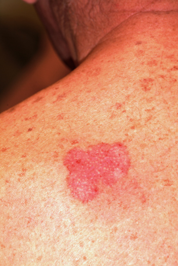
A 50-year-old man has had a nonhealing but otherwise asymptomatic lesion on his back for more than three years. The lesion has slowly grown, despite the use of a number of treatments, including OTC and prescription antifungal creams and OTC steroid creams. The consensus from his primary care providers is that this must represent some form of fungal infection, although they are at a loss to explain why it has not responded to treatment for that condition. The patient presents to dermatology because his wife insisted on referral to a specialist when the lesion finally became focally eroded around its margins. Further history taking reveals that there are no pets or children at home, there is no personal history of atopy, and the patient has never worked with livestock. For 30 years, he worked outdoors for the power company, but he is now assigned to a desk job. He is not immunosuppressed, and his only medication is for hypertension. During the physical examination, you observe an arciform, faintly and focally eroded border surrounding a 5-cm pink patch on the patient’s left upper back. Closer inspection reveals prominent pilosebaceous units confined by well-defined borders, with a background of uniformly atrophic, glassy-looking telangiectatic skin. No scale is seen on or around the lesion. Elsewhere, you see a moderate number of solar lentigines across the patient’s shoulders and note that his posterior neck has a deeply lined appearance. The rest of his trunk and face have similar lesions, although none is especially worrisome. To confirm your clinical suspicions, you perform a shave biopsy on the central portion of the lesion, which shows superficial basal cell carcinoma (BCC). Treatment options are then discussed at length with the patient.
