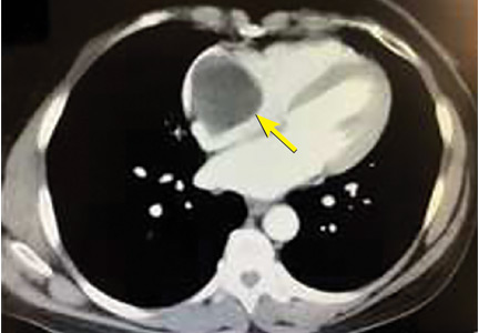
A right atrial mass

Article Type
Changed
Mon, 07/01/2019 - 08:53
Display Headline
A right atrial mass

Figure 1. CT of the chest. An arrow points to the mass in the right atrium.
Figure 1. Computed tomography of the chest. An arrow points to the mass in the right atrium.
A 55-year-old woman with hypertension, hyperthyroidism, and endometrial adenocarcinoma treated 5 years earlier presented with occasional shortness of breath and dizziness. To rule out pulmonary embolism, computed tomography of the chest was done and showed a high-density lesion in the right atrium of the heart, measuring up to 4.5 cm (Figure 1).

Figure 2. Transthoracic echocardiography.
Transthoracic echocardiography confirmed a large round echogenic right atrial mass measuring 4.5 by 4.3 cm (Figure 2). The mass appeared to encroach into the tricuspid valve orifice, but there was no evidence of functional obstruction or stenosis.

Figure 3. Transesophageal echocardiography.
Transesophageal echocardiography showed a large globular echogenic mass measuring 5.5 by 4.6 cm with a stalk originating from the right atrial appendage (Figures 3 and 4). The mass was minimally mobile and did not obstruct the superior vena cava or the tricuspid orifice.

Figure 4. Transesophageal echocardiography.
Given her history, the intracardiac mass strongly suggested metastatic adenocarcinoma. Other possibilities in the differential diagnosis were thrombus, myxoma, lipoma, angiosarcoma, and lymphoma.1,2

Figure 5. Angiography with pigtail catheter.
The cardiovascular team was consulted, and they recommended proceeding with open heart surgery and resection of the mass. In preparation for the surgery, cardiac catheterization was performed, and did not show any significant coronary artery disease. However, using a pigtail catheter, right atrial angiography demonstrated a large, round, mobile mass attached to the wall of the right atrium (Figure 5).

Figure 6. Operative photograph.
The patient underwent surgery, and the tumor was resected (Figures 6 and 7). Pathologic study confirmed the diagnosis of cardiac benign myxoma. The patient recovered and was eventually discharged to her home.

PRIMARY HEART TUMORS ARE RARE

Figure 7. Gross section.
Metastatic cardiac tumors are more common than primary cardiac tumors. On autopsy, between 0.01% and 0.1% of people in the general population have primary cardiac tumors, while 0.7% to 3.5% have metastatic tumors.3 About 75% of primary cardiac tumors are benign. In autopsies of patients with malignant neoplasms, cardiac metastasis has been found in 6% to 20%.4 Further, right-sided masses typically raise the concern of metastatic disease, whereas benign masses are more commonly found on the left side.

The most common neoplasms that metastasize to the heart are malignant melanoma, lymphoma, leukemia, breast, and lung cancers. The layers of the heart affected by malignant neoplasms in order of frequency from highest to lowest are the pericardium, epicardium, myocardium, and endocardium.3

MYXOMA: A PRIMARY CARDIAC TUMOR

The most common type of primary cardiac tumor is myxoma. Most—75% to 80%—occur in the left atrium, while 15% to 20% occur in the right atrium.5 Right atrial myxomas are usually found in the intraatrial septum at the border of the fossa ovalis.6 Myxomas can occur at any age, but are most common in women between the third and sixth decades.2

The cause of atrial myxomas is currently unknown. Most cases are sporadic. However, 10% are familial, with an autosomal-dominant pattern.7

The clinical symptoms of right atrial myxoma depend on the tumor’s size, location, and mobility and on the patient’s physical activity and body position.4 Common presenting symptoms include shortness of breath, pulmonary edema, cough, hemoptysis, and fatigue. Thirty percent of patients present with constitutional symptoms.4

Auscultation may reveal a characteristic “tumor plop” early in diastole.4,7 About 35% of patients have laboratory abnormalities such as elevations in erythrocyte sedimentation rate, C-reactive protein, and globulin levels and anemia. Our patient did not.

Embolization occurs in about 10% of cases of right-sided myxoma and can result in pulmonary artery embolism or a stroke. Pulmonary artery embolism occurs with myxoma embolization to the lungs. Strokes can occur in patients who have a patent foramen ovale or atrial septal defect, through which embolism to the systemic arterial circulation can occur.

The primary treatment for myxoma is complete resection of the tumor and its base with wide safety margins. This is particularly important to prevent recurrence of the myxoma and need for repeat operations, with their risk of surgical complications.9

References
  1. Dujardin KS, Click RL, Oh JK. The role of intraoperative transesophageal echocardiography in patients undergoing cardiac mass removal. J Am Soc Echocardiogr 2000; 13(12):1080–1083. pmid:11119275
  2. Jang KH, Shin DH, Lee C, Jang JK, Cheong S, Yoo SY. Left atrial mass with stalk: thrombus or myxoma? J Cardiovasc Ultrasound 2010; 18(4):154–156. doi:10.4250/jcu.2010.18.4.154
  3. Goldberg AD, Blankstein R, Padera RF. Tumors metastatic to the heart. Circulation 2013; 128(16):1790–1794. doi:10.1161/CIRCULATIONAHA.112.000790
  4. Aggarwal SK, Barik R, Sarma TC, et al. Clinical presentation and investigation findings in cardiac myxomas: new insights from the developing world. Am Heart J 2007; 154(6):1102–1107. doi:10.1016/j.ahj.2007.07.032
  5. Diaz A, Di Salvo C, Lawrence D, Hayward M. Left atrial and right ventricular myxoma: an uncommon presentation of a rare tumour. Interact Cardiovasc Thorac Surg 2011; 12(4):622–623. doi:10.1510/icvts.2010.255661
  6. Reynen K. Cardiac myxomas. N Engl J Med 1995; 333(24):1610–1617. doi:10.1056/NEJM199512143332407
  7. Kolluru A, Desai D, Cohen GI. The etiology of atrial myxoma tumor plop. J Am Coll Cardiol 2011; 57(21):e371. doi:10.1016/j.jacc.2010.09.085
  8. Kassab R, Wehbe L, Badaoui G, el Asmar B, Jebara V, Ashoush R. Recurrent cerebrovascular accident: unusual and isolated manifestation of myxoma of the left atrium. J Med Liban 1999; 47(4):246–250. French. pmid:10641454
  9. Guhathakurta S, Riordan JP. Surgical treatment of right atrial myxoma. Tex Heart Inst J 2000; 27(1):61–63. pmid:10830633
Article PDF
Author and Disclosure Information

Zeel Shah, MD
Wayne State University School of Medicine, Detroit, MI

M. Chadi Alraies, MD
Department of Cardiovascular Medicine, Wayne State University School of Medicine/Detroit Medical Center, Detroit, MI

Ameer Harp, MD
Wayne State University School of Medicine/Detroit Medical Center, Detroit, MI

Salam Khalil, MD
Wayne State University School of Medicine/Detroit Medical Center, Detroit, MI

Amir Kaki, MD
Wayne State University School of Medicine and Ascension St. John Hospital, Detroit, MI

Mahir Elder, MD
Wayne State University School of Medicine and Beaumont Hospital, Detroit, MI

Address: M. Chadi Alraies, MD, Wayne State University, Detroit Medical Center, 311 Mack Avenue, Detroit, MI 48201; alraies@hotmail.com

Issue
Cleveland Clinic Journal of Medicine - 86(7)
Publications
Topics
Page Number
445-447
Legacy Keywords
atrial mass, atrial tumor, heart tumor, cardiac tumor, computed tomography, CT, echocardiography, TTE, TEE, angiography, adenocarcinoma, myxoma, tumor plop, cardiac imaging, Zeel Shah, Chadi Alraies, Ameer Harp, Salam Khalil, Amir Kaki, Mahir Elder
Sections
Author and Disclosure Information

Zeel Shah, MD
Wayne State University School of Medicine, Detroit, MI

M. Chadi Alraies, MD
Department of Cardiovascular Medicine, Wayne State University School of Medicine/Detroit Medical Center, Detroit, MI

Ameer Harp, MD
Wayne State University School of Medicine/Detroit Medical Center, Detroit, MI

Salam Khalil, MD
Wayne State University School of Medicine/Detroit Medical Center, Detroit, MI

Amir Kaki, MD
Wayne State University School of Medicine and Ascension St. John Hospital, Detroit, MI

Mahir Elder, MD
Wayne State University School of Medicine and Beaumont Hospital, Detroit, MI

Address: M. Chadi Alraies, MD, Wayne State University, Detroit Medical Center, 311 Mack Avenue, Detroit, MI 48201; alraies@hotmail.com

Article PDF
Article PDF
Related Articles

Figure 1. CT of the chest. An arrow points to the mass in the right atrium.
Figure 1. Computed tomography of the chest. An arrow points to the mass in the right atrium.
A 55-year-old woman with hypertension, hyperthyroidism, and endometrial adenocarcinoma treated 5 years earlier presented with occasional shortness of breath and dizziness. To rule out pulmonary embolism, computed tomography of the chest was done and showed a high-density lesion in the right atrium of the heart, measuring up to 4.5 cm (Figure 1).

Figure 2. Transthoracic echocardiography.
Transthoracic echocardiography confirmed a large round echogenic right atrial mass measuring 4.5 by 4.3 cm (Figure 2). The mass appeared to encroach into the tricuspid valve orifice, but there was no evidence of functional obstruction or stenosis.

Figure 3. Transesophageal echocardiography.
Transesophageal echocardiography showed a large globular echogenic mass measuring 5.5 by 4.6 cm with a stalk originating from the right atrial appendage (Figures 3 and 4). The mass was minimally mobile and did not obstruct the superior vena cava or the tricuspid orifice.

Figure 4. Transesophageal echocardiography.
Given her history, the intracardiac mass strongly suggested metastatic adenocarcinoma. Other possibilities in the differential diagnosis were thrombus, myxoma, lipoma, angiosarcoma, and lymphoma.1,2

Figure 5. Angiography with pigtail catheter.
The cardiovascular team was consulted, and they recommended proceeding with open heart surgery and resection of the mass. In preparation for the surgery, cardiac catheterization was performed, and did not show any significant coronary artery disease. However, using a pigtail catheter, right atrial angiography demonstrated a large, round, mobile mass attached to the wall of the right atrium (Figure 5).

Figure 6. Operative photograph.
The patient underwent surgery, and the tumor was resected (Figures 6 and 7). Pathologic study confirmed the diagnosis of cardiac benign myxoma. The patient recovered and was eventually discharged to her home.

PRIMARY HEART TUMORS ARE RARE

Figure 7. Gross section.
Metastatic cardiac tumors are more common than primary cardiac tumors. On autopsy, between 0.01% and 0.1% of people in the general population have primary cardiac tumors, while 0.7% to 3.5% have metastatic tumors.3 About 75% of primary cardiac tumors are benign. In autopsies of patients with malignant neoplasms, cardiac metastasis has been found in 6% to 20%.4 Further, right-sided masses typically raise the concern of metastatic disease, whereas benign masses are more commonly found on the left side.

The most common neoplasms that metastasize to the heart are malignant melanoma, lymphoma, leukemia, breast, and lung cancers. The layers of the heart affected by malignant neoplasms in order of frequency from highest to lowest are the pericardium, epicardium, myocardium, and endocardium.3

MYXOMA: A PRIMARY CARDIAC TUMOR

The most common type of primary cardiac tumor is myxoma. Most—75% to 80%—occur in the left atrium, while 15% to 20% occur in the right atrium.5 Right atrial myxomas are usually found in the intraatrial septum at the border of the fossa ovalis.6 Myxomas can occur at any age, but are most common in women between the third and sixth decades.2

The cause of atrial myxomas is currently unknown. Most cases are sporadic. However, 10% are familial, with an autosomal-dominant pattern.7

The clinical symptoms of right atrial myxoma depend on the tumor’s size, location, and mobility and on the patient’s physical activity and body position.4 Common presenting symptoms include shortness of breath, pulmonary edema, cough, hemoptysis, and fatigue. Thirty percent of patients present with constitutional symptoms.4

Auscultation may reveal a characteristic “tumor plop” early in diastole.4,7 About 35% of patients have laboratory abnormalities such as elevations in erythrocyte sedimentation rate, C-reactive protein, and globulin levels and anemia. Our patient did not.

Embolization occurs in about 10% of cases of right-sided myxoma and can result in pulmonary artery embolism or a stroke. Pulmonary artery embolism occurs with myxoma embolization to the lungs. Strokes can occur in patients who have a patent foramen ovale or atrial septal defect, through which embolism to the systemic arterial circulation can occur.

The primary treatment for myxoma is complete resection of the tumor and its base with wide safety margins. This is particularly important to prevent recurrence of the myxoma and need for repeat operations, with their risk of surgical complications.9

References
  1. Dujardin KS, Click RL, Oh JK. The role of intraoperative transesophageal echocardiography in patients undergoing cardiac mass removal. J Am Soc Echocardiogr 2000; 13(12):1080–1083. pmid:11119275
  2. Jang KH, Shin DH, Lee C, Jang JK, Cheong S, Yoo SY. Left atrial mass with stalk: thrombus or myxoma? J Cardiovasc Ultrasound 2010; 18(4):154–156. doi:10.4250/jcu.2010.18.4.154
  3. Goldberg AD, Blankstein R, Padera RF. Tumors metastatic to the heart. Circulation 2013; 128(16):1790–1794. doi:10.1161/CIRCULATIONAHA.112.000790
  4. Aggarwal SK, Barik R, Sarma TC, et al. Clinical presentation and investigation findings in cardiac myxomas: new insights from the developing world. Am Heart J 2007; 154(6):1102–1107. doi:10.1016/j.ahj.2007.07.032
  5. Diaz A, Di Salvo C, Lawrence D, Hayward M. Left atrial and right ventricular myxoma: an uncommon presentation of a rare tumour. Interact Cardiovasc Thorac Surg 2011; 12(4):622–623. doi:10.1510/icvts.2010.255661
  6. Reynen K. Cardiac myxomas. N Engl J Med 1995; 333(24):1610–1617. doi:10.1056/NEJM199512143332407
  7. Kolluru A, Desai D, Cohen GI. The etiology of atrial myxoma tumor plop. J Am Coll Cardiol 2011; 57(21):e371. doi:10.1016/j.jacc.2010.09.085
  8. Kassab R, Wehbe L, Badaoui G, el Asmar B, Jebara V, Ashoush R. Recurrent cerebrovascular accident: unusual and isolated manifestation of myxoma of the left atrium. J Med Liban 1999; 47(4):246–250. French. pmid:10641454
  9. Guhathakurta S, Riordan JP. Surgical treatment of right atrial myxoma. Tex Heart Inst J 2000; 27(1):61–63. pmid:10830633
Issue
Cleveland Clinic Journal of Medicine - 86(7)
Issue
Cleveland Clinic Journal of Medicine - 86(7)
Page Number
445-447
Page Number
445-447
Publications
Publications
Topics
Article Type
Display Headline
A right atrial mass
Display Headline
A right atrial mass
Legacy Keywords
atrial mass, atrial tumor, heart tumor, cardiac tumor, computed tomography, CT, echocardiography, TTE, TEE, angiography, adenocarcinoma, myxoma, tumor plop, cardiac imaging, Zeel Shah, Chadi Alraies, Ameer Harp, Salam Khalil, Amir Kaki, Mahir Elder
Legacy Keywords
atrial mass, atrial tumor, heart tumor, cardiac tumor, computed tomography, CT, echocardiography, TTE, TEE, angiography, adenocarcinoma, myxoma, tumor plop, cardiac imaging, Zeel Shah, Chadi Alraies, Ameer Harp, Salam Khalil, Amir Kaki, Mahir Elder
Sections
Disallow All Ads
Content Gating
No Gating (article Unlocked/Free)
Alternative CME
Disqus Comments
Default
Gate On Date
Tue, 06/25/2019 - 15:45
Un-Gate On Date
Tue, 06/25/2019 - 15:45
Use ProPublica
CFC Schedule Remove Status
Tue, 06/25/2019 - 15:45
Hide sidebar & use full width
render the right sidebar.
Article PDF Media