User login
VIDEO: Bottom line on maternal infections and cerebral palsy
VANCOUVER, B.C. – Intra- or extra-amniotic fluid infections during pregnancy elevate the risk of having a child with cerebral palsy, according to analysis of 6 million California birth records – although the findings shows an association, not causality.
Researchers showed that pregnant women who were hospitalized with a diagnosis of chorioamnionitis had a fourfold increase in the risk of having a child with cerebral palsy (CP), while genitourinary and respiratory infections increased that risk twofold.
"I think this is a very important study, and it took us a step further in trying to understand what could cause cerebral palsy," said Dr. Lee M. Sanders, associate professor of pediatrics and codirector of the Center for Policy, Outcomes, and Prevention at Stanford (Calif.) University.
Because the study shows an association only, however, "it’s important not to cause alarm," cautioned Dr. Sanders, who was not involved in the study.
In a video interview, Dr. Sanders explains what’s known so far about this association, the implications of the study’s findings, and the research that’s already underway.
The video associated with this article is no longer available on this site. Please view all of our videos on the MDedge YouTube channel
On Twitter @naseemmiller
AT THE PAS ANNUAL MEETING
Maternal infections associated with increased risk of cerebral palsy
VANCOUVER, B.C. – Intra- or extra-amniotic fluid infections during pregnancy are associated with an increased risk of having a child with cerebral palsy, according to analysis of six million California birth records.
Researchers found that pregnant women who were hospitalized with diagnosis of chorioamnionitis had a fourfold increase in risk of having a child with cerebral palsy (CP), while genitourinary and respiratory infections increased that risk by twofold, each.
"I think this is a very important study, and it took us a step further in trying to understand what could cause cerebral palsy," said Dr. Lee M. Sanders, associate professor of pediatrics and codirector of the Center for Policy, Outcomes, and Prevention at Stanford (Calif.) University. But because the study shows an association only, "it’s important not to cause alarm," said Dr. Sanders, who was not involved in the study.
Dr. Joshua Bear of the University of California, San Francisco, and his colleagues analyzed the California birth records from 1991 to 2001, in addition to records of all children receiving services for CP from the California Department of Developmental Services through 2006.
There were close to 8,500 CP cases, or 1.4 per 1,000 live births, which is at the lower end of some of the reported statistics, Dr. Bear said.
Analysis of infection diagnoses showed that 6% of the women had unaffected births, while 15.3% had children with CP, with a relative risk of 2.7. Among the latter group, 7.6% had chorioamnionitis, 5.2% had other genitourinary infections, and 3.5% had respiratory infections.
Among women hospitalized with chorioamnionitis, there was a fourfold increase in risk of having CP in preterm infants and twofold increase in term births. Most women hospitalized with chorioamnionitis gave birth at that hospitalization, said Dr. Bear.
For other genitourinary infections, there was a 1.4-fold increase in the risk of CP for prenatal hospitalization, and 1.9-fold increase for birth hospitalization.
Respiratory infections showed a similar trend, with a twofold increased risk for prenatal hospitalization and 2.6-fold increase risk for birth hospitalization.
Increased risk of CP with other genitourinary and respiratory infections, even during prenatal hospitalization, suggest that "these infections may be directly or indirectly contributing to harm on the developing fetal brain and not be simply another complication during delivery," Dr. Bear said during his presentation at the annual meeting of Pediatric Academic Societies.
Analysis of sociodemographic risk factors showed that women younger than 18 and older than 35 were more likely to have a child with CP. Black women were at a higher risk of CP, compared with white and Hispanic women, and Asian women were at lower risk. Years of education was inversely associated with the risk of CP. Also, male infants were at a slightly higher risk of CP than were their female counterparts.
Yet when researchers adjusted for maternal age, race, education, socioeconomic status, and infant sex, the association between infections and increased risk of CP remained the same.
Dr. Bear and his colleagues also stratified the data by presence of birth asphyxia, to find clues if it could be a cause for CP. The results showed a large drop in rate ratios of chorioamnionitis, from 4 to 1.5 when asphyxia was diagnosed at birth, but the rates for other genitourinary and respiratory infections remained the same. "This suggests that there’s some interaction between chorioamnionitis and presence of birth asphyxia, although the nature of that interaction is unclear." said Dr. Bear.
The study had several limitations, including the use of ICD-9 codes, researchers’ inability to account for comorbid conditions related to hospitalization, and being limited to hospital-based diagnoses.
The findings, said Dr. Bear, highlighted the need for further research in several areas, including the association between CP and respiratory infections, and whether risk of CP can be decreased by preventing or treating the infections.
The findings can also "help us with public health interventions, such as better rates of influenza vaccinations among pregnant women," said Dr. Sanders.
Dr. Bear and Dr. Sanders had no relevant disclosures.
On Twitter @naseemmiller
AT THE PAS ANNUAL MEETING
Key clinical finding: The association between maternal infections and increased risk of CP could be a call for public health interventions, such as higher rate of flu vaccination for pregnant women.
Major finding: Pregnant women who were hospitalized with diagnosis of chorioamnionitis had a fourfold increase in risk of having a child with cerebral palsy (CP), while genitourinary and respiratory infections increased that risk by twofold, each.
Data source: Analysis of California birth records from 1991 to 2001, and records of all children receiving services for CP from the California Department of Developmental Services through 2006.
Disclosures: Dr. Bear and Dr. Sanders had no relevant disclosures.
VIDEO: A cardiologist updates her view on AHA/ACC risk estimator
In November 2013, the American College of Cardiology and the American Heart Association published new U.S. cholesterol management guidelines, including a risk estimator.
The guidelines were released during the American Heart Association’s scientific sessions, and they stirred up some controversy. At the time, one of the attendees, Dr. Eugenia Gianos, the director of Preventive Cardiology fellowship at NYU Langone Medical Center, New York, was hesitant to use the new risk estimator.
However, in the past few months, Dr. Gianos said she has had the opportunity to apply these guidelines clinically, and has reviewed the recently released National Lipid Association’s draft "Recommendations for Patient-Centered Management of Dyslipidemia."
In a video interview, she shares what changed her view and what she has changed in her practice that could help other clinicians.
The video associated with this article is no longer available on this site. Please view all of our videos on the MDedge YouTube channel
On Twitter @naseemmiller
Prospective studies needed to compare MS in African Americans and whites
DALLAS – There is a need for prospective, multicenter studies to examine the differences between African Americans and whites with multiple sclerosis and their response to therapy, according to Dr. Omar Khan.
The impact of education, disease awareness, and access to health care as covariates must also be included in future studies, "otherwise, this question will continue to come up, to find out whether these factors do or don’t have a bearing on disease severity and outcomes," said Dr. Khan, professor and chair of neurology at Wayne State University, Detroit, during a presentation at a meeting of the Consortium of Multiple Sclerosis Centers and the Americas Committee for Treatment and Research in Multiple Sclerosis.
Another point to consider, especially in regard to trials evaluating response to disease-modifying therapies, is that African Americans continue to be a small proportion of participants in these trials, he said.
Up until recent decades, it was believed that multiple sclerosis didn’t affect African Americans. Even today, some resources, such as the National MS Society's website, try to bust that myth first before delving into other facts about multiple sclerosis.
Nearly 10 years ago, Dr. Bruce Cree and his colleagues presented a study about the clinical characteristics of multiple sclerosis in African Americans, compared with whites, and they showed that African American patients were more likely to have a more aggressive disease course, and a greater likelihood of developing opticospinal MS and transverse myelitis (Neurology 2004;63:2039-45).
The study showed that the median time from onset to diagnosis was 1 year in African Americans, compared with 2 years in whites. Also, the median time from onset to treatment was 4 years in African American patients, compared with 5 years in whites. The former group also had a significantly shorter median time to a Kurtzke Expanded Disability Status Scale (EDSS) score of greater than or equal to 6 and 7, compared with whites.
These findings have been consistently shown in later trials.
In 2004, a group in France showed that the MS course was more aggressive in North African than in European patients (Neurology 2007;68:29-32). Researchers found that the mean age of onset was younger in North Africans, compared with Europeans. They also found that the median times to EDSS scores of 4 and 6 were both significantly shorter in the former group.
"You’re going to see this theme in other studies," said Dr. Khan. "Clearly there wasn’t a delay in diagnosing the disease based on these studies."
Then in 2008, a study in France showed that North African migrants with MS appear to be as responsive as other MS patients are to disease-modifying therapies, although they received the treatment sooner than their white counterparts and had more frequent use of immunosuppressive drugs (Mult. Scler. 2008;14:933-9). The study showed that the mean time to an EDSS score of 6 was 6 years in North Africans, which was almost 10 years sooner than in white patients.
In 2009, in a retrospective study of pediatric-onset MS, Dr. Khan and his colleagues yet again found "a more aggressive disease phenotype among American blacks with adult-onset MS," and called for larger multicenter studies, which Dr. Khan continues to advocate for (Pediatr. Neurol. 2009;40:31-3). The group found an earlier onset of disease to initiation of disease-modifying therapy among African American patients, compared with whites, although the difference didn’t reach statistical significance. The relapse rate was also higher among the former group.
Another study on pediatric-onset MS showed that African American patients may be at higher risk for adverse cognitive impact on language and complex attention (Neurology 2010;75:2097-102).
Dr. Khan also pointed to several studies quantifying central nervous system tissue injury in African Americans with MS.
A 2012 study showed that African Americans had a significantly greater lesion volume, compared with white patients, but the brain volumes weren’t significantly different between the two groups. Researchers concluded that "a higher lesion accumulation, rather than a pronounced brain volume decrease, might explain the early progress to ambulatory assistance of [African Americans] with MS" (PLoS One 2012;7:e43061).
"I don’t think we should be arguing that there is a clear indication that African Americans have greater disability, CNS tissue injury, and possibly suboptimal response to therapy," compared with whites, Dr. Khan said.
Other quantitative studies have suggested differences in the disease biology. "And they provide more credence to these observations, despite the fact that almost all of them have been cross-sectional or retrospective," he said, stressing the need for prospectively designed multicenter studies.
"And I think it’s equally important to keep in mind [that] the impact of education, health awareness, and access to health care as covariates at a bare minimum must be included in these studies," Dr. Khan said.
His group conducted a study in 2012 to find out whether there was a difference between African Americans and whites with MS in terms of level of education, disease awareness, and disease severity, but they didn’t find any statistically significant differences.
Additional study is needed, he said.
Dr. Khan said he had no relevant financial disclosures.
On Twitter @naseemmiller
EXPERT ANALYSIS AT THE CMSC/ACTRIMS ANNUAL MEETING
When switching from natalizumab to teriflunomide, timing is key
DALLAS – Teriflunomide could be a safe and effective option when patients with multiple sclerosis need to be switched from natalizumab, especially when the oral treatment starts 1 month after the last natalizumab treatment, according to a small, retrospective study.
Multiple studies have shown that natalizumab (Tysabri) is a risk factor for progressive multifocal leukoencephalopathy (PML). "So, there’s always the question of when do we switch," said Dr. Francisco J. Gomez, coauthor of the study and a physician at the MS Center of Northeastern New York in Latham.

"If you don’t switch people [from natalizumab] earlier in the disease, you might end up giving them worsened outcomes," Dr. Gomez said at a meeting of the Consortium of Multiple Sclerosis Centers and the Americas Committee for Treatment and Research in Multiple Sclerosis.
At the same time, he noted, "you’re limited on how to switch. So you’re caught between a rock and a hard place. You want to switch them, but you don’t want to lose the only opportunity you have in controlling their disease."
Dr. Gomez and his colleagues looked at the safety and efficacy of teriflunomide (Aubagio), which was approved by theFood and Drug Administration in 2012 and has been shown to cut the annual MS relapse rate by more than a third.
The investigators used the center’s database to conduct a 12-month retrospective study of 30 patients who switched to teriflunomide after receiving natalizumab for more than a year and were all clinically stable. Most patients began the new treatment within 4 weeks of their last natalizumab treatment.
Cranial MRI and a clinical exam were conducted at the time of last natalizumab, and at 3 and 6 months after natalizumab treatment, to check for possible PML and new MS activity.
Of the 30 patients, 11 were triple positive for PML, meaning that they had received more than 24 natalizumab treatments, had prior immunosuppression, and were positive for anti–John Cunningham virus antibody. Fifteen were positive for anti-JCV antibody, and four were negative for anti-JCV antibody.
The mean number of natalizumab treatments was 38, ranging from 14 to 81. The patients’ mean age was 50 years, and 80% were female. Their mean duration of treatment with teriflunomide after natalizumab discontinuation was 13 months, ranging from 6 to 13.
Of all patients, 23 are still on teriflunomide, Dr. Gomez said. None have developed PML. Two have moved away and were lost to follow-up.
Five patients discontinued the treatment, one of whom developed significant exacerbation, including severe optic neuritis 4 weeks after starting on teriflunomide. That patient also had a 3-month delay between last natalizumab dose and the initiation of teriflunomide, compared with the rest, who started within a month of the last natalizumab treatment.
So, "a reduced washout period after natalizumab may be important to lessen breakthrough disease," Dr. Gomez and his colleagues said in the poster presented at the meeting.
Two others discontinued treatment because of persistent diarrhea and abdominal cramps, and one patient withdrew due to hair thinning. Minor diarrhea and transient hair thinning occurred in seven and five patients, respectively.
Despite the clinical exacerbations, "MRI scans haven’t shown disease, so we feel" that the disease is under control, Dr. Gomez said.
He advised physicians "to make the switch as soon as possible, no more than 4 weeks from the last natalizumab infusion, and then monitor the patient closely for potential MS relapse and PML for the next 6 months. Also, we have only been looking at patients switching to teriflunomide who have been stable while on natalizumab. If the patient has had exacerbations while on natalizumab, we usually go to rituximab and perhaps, in the future, alemtuzumab."
Dr. Gomez reported having no disclosures.
On Twitter @naseemmiller
DALLAS – Teriflunomide could be a safe and effective option when patients with multiple sclerosis need to be switched from natalizumab, especially when the oral treatment starts 1 month after the last natalizumab treatment, according to a small, retrospective study.
Multiple studies have shown that natalizumab (Tysabri) is a risk factor for progressive multifocal leukoencephalopathy (PML). "So, there’s always the question of when do we switch," said Dr. Francisco J. Gomez, coauthor of the study and a physician at the MS Center of Northeastern New York in Latham.
"If you don’t switch people [from natalizumab] earlier in the disease, you might end up giving them worsened outcomes," Dr. Gomez said at a meeting of the Consortium of Multiple Sclerosis Centers and the Americas Committee for Treatment and Research in Multiple Sclerosis.
At the same time, he noted, "you’re limited on how to switch. So you’re caught between a rock and a hard place. You want to switch them, but you don’t want to lose the only opportunity you have in controlling their disease."
Dr. Gomez and his colleagues looked at the safety and efficacy of teriflunomide (Aubagio), which was approved by theFood and Drug Administration in 2012 and has been shown to cut the annual MS relapse rate by more than a third.
The investigators used the center’s database to conduct a 12-month retrospective study of 30 patients who switched to teriflunomide after receiving natalizumab for more than a year and were all clinically stable. Most patients began the new treatment within 4 weeks of their last natalizumab treatment.
Cranial MRI and a clinical exam were conducted at the time of last natalizumab, and at 3 and 6 months after natalizumab treatment, to check for possible PML and new MS activity.
Of the 30 patients, 11 were triple positive for PML, meaning that they had received more than 24 natalizumab treatments, had prior immunosuppression, and were positive for anti–John Cunningham virus antibody. Fifteen were positive for anti-JCV antibody, and four were negative for anti-JCV antibody.
The mean number of natalizumab treatments was 38, ranging from 14 to 81. The patients’ mean age was 50 years, and 80% were female. Their mean duration of treatment with teriflunomide after natalizumab discontinuation was 13 months, ranging from 6 to 13.
Of all patients, 23 are still on teriflunomide, Dr. Gomez said. None have developed PML. Two have moved away and were lost to follow-up.
Five patients discontinued the treatment, one of whom developed significant exacerbation, including severe optic neuritis 4 weeks after starting on teriflunomide. That patient also had a 3-month delay between last natalizumab dose and the initiation of teriflunomide, compared with the rest, who started within a month of the last natalizumab treatment.
So, "a reduced washout period after natalizumab may be important to lessen breakthrough disease," Dr. Gomez and his colleagues said in the poster presented at the meeting.
Two others discontinued treatment because of persistent diarrhea and abdominal cramps, and one patient withdrew due to hair thinning. Minor diarrhea and transient hair thinning occurred in seven and five patients, respectively.
Despite the clinical exacerbations, "MRI scans haven’t shown disease, so we feel" that the disease is under control, Dr. Gomez said.
He advised physicians "to make the switch as soon as possible, no more than 4 weeks from the last natalizumab infusion, and then monitor the patient closely for potential MS relapse and PML for the next 6 months. Also, we have only been looking at patients switching to teriflunomide who have been stable while on natalizumab. If the patient has had exacerbations while on natalizumab, we usually go to rituximab and perhaps, in the future, alemtuzumab."
Dr. Gomez reported having no disclosures.
On Twitter @naseemmiller
AT THE CMSC/ACTRIMS ANNUAL MEETING
Key clinical point: Make the switch as soon as possible – no more than 4 weeks from the last natalizumab infusion – and then monitor the patient closely.
Major finding: Despite clinical exacerbations in five patients, MRI scans remained stable.
Data source: A 12-month retrospective study involving 30 patients who switched to teriflunomide after receiving natalizumab for more than a year.
Disclosures: Dr. Gomez reported having no disclosures.

Don’t Dread Pediatric Dermatology
ORLANDO – There are a few simple things you can do when you have a pediatric dermatology patient to create a better office experience for the little ones, their parents, and yourself. To start, greet the child first and by name, advises Dr. Lawrence A. Schachner, a pediatric dermatologist and professor at the University of Miami.
Some of his other top tips:
• Don’t block the door.
• Make small talk to bond with them. For instance, tell them that you like their shoes, or ask them what they do for fun. (This can also be a significant indicator for depression, Dr. Schachner said.)
• Try to shake hands with your pinky and their hand, or what Dr. Schachner calls, "the Schachner Shake." This trick also lets you examine the child’s extremities, he said.
• To examine their abdomen and back, ask them if they’ve "brought their belly button with them or left it home," said Dr. Schachner, during a presentation at the annual meeting of the Florida Society of Dermatology and Dermatologic Surgery.
• Have fun with some of the tools you need to use. For instance, use the Wood’s lamp to make the parent blue. And if you have to use liquid nitrogen, don’t come at the child with a canister, Dr. Schachner advised.
• Speak kid: Instead of topical anesthesia, use a term like "magic cream." Instead of punch biopsy, say "cookie cutter."
• And use little distractions – what Dr. Schachner calls distraction anesthesia – when you need to use needles or perform a surgical procedure. For injections, pinch the skin and inject the tented area. Or use a shot blocker, like the Buzzy, for intralesional injections, he advised.
• And finally, don’t forget to smile!
Check out our interview for more of Dr. Schachner’s tips on distraction anesthesia, and other pediatric pearls.
Dr. Schachner is an investigator for several companies, including Astellas, Ferndale Labs, and Novartis. He’s a consultant for Beiersdorf, Lexington, and TopMD.
On Twitter @naseemmiller
AT FSDDS 2014
AUDIO: Don’t dread pediatric dermatology
ORLANDO – There are a few simple things you can do when you have a pediatric dermatology patient to create a better office experience for the little ones, their parents, and yourself. To start, greet the child first and by name, advises Dr. Lawrence A. Schachner, a pediatric dermatologist and professor at the University of Miami.
Some of his other top tips:
• Don’t block the door.
• Make small talk to bond with them. For instance, tell them that you like their shoes, or ask them what they do for fun. (This can also be a significant indicator for depression, Dr. Schachner said.)
• Try to shake hands with your pinky and their hand, or what Dr. Schachner calls, "the Schachner Shake." This trick also lets you examine the child’s extremities, he said.
• To examine their abdomen and back, ask them if they’ve "brought their belly button with them or left it home," said Dr. Schachner, during a presentation at the annual meeting of the Florida Society of Dermatology and Dermatologic Surgery.
• Have fun with some of the tools you need to use. For instance, use the Wood’s lamp to make the parent blue. And if you have to use liquid nitrogen, don’t come at the child with a canister, Dr. Schachner advised.
• Speak kid: Instead of topical anesthesia, use a term like "magic cream." Instead of punch biopsy, say "cookie cutter."
• And use little distractions – what Dr. Schachner calls distraction anesthesia – when you need to use needles or perform a surgical procedure. For injections, pinch the skin and inject the tented area. Or use a shot blocker, like the Buzzy, for intralesional injections, he advised.
• And finally, don’t forget to smile!
Check out our interview for more of Dr. Schachner’s tips on distraction anesthesia, and other pediatric pearls.
Dr. Schachner is an investigator for several companies, including Astellas, Ferndale Labs, and Novartis. He’s a consultant for Beiersdorf, Lexington, and TopMD.
On Twitter @naseemmiller
AT FSDDS 2014
Tubby toddlers: Don’t delay the tough conversation
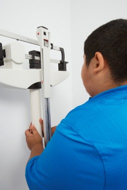
VANCOUVER, B.C. – Very few overweight and obese children outgrow their at-risk weight, and the window to address the problem is fairly narrow.
Dr. Raquel G. Hernandez, assistant professor of pediatrics at Johns Hopkins University, and her colleagues looked at obesity resilience, seeking to identify the prevalence of favorable growth patterns, such as healthy weight maintenance and return to healthy weight in school-aged children.
"There are pervasive expectations among parents that their children will magically outgrow their weight risk, and unfortunately, I think this applies to some providers as well," Dr. Hernandez said at the annual meeting of the Pediatric Academic Societies.

The researchers analyzed data on more than 9,000 children in the Early Childhood Longitudinal Study database, looking at their growth during the elementary school years. Measurements included body mass index (BMI), race/ethnicity, gender, and socioeconomic status and were compared for kindergarten, first, third, and fifth grade.
At baseline, nearly 26% of children were overweight or obese, while most (70%) were classified as healthy weight (BMI from the fifth through the 85th percentile). The rest were underweight. The majority of the cohort maintained healthy weight, but less than one in five (17%) of the overweight or obese children were able to return to healthy weight (BMI between the 5th and 85th percentile) over the study period.
In this unpublished study, Hispanic boys and girls were less likely to return to healthy weight, compared with children of other races/ethnicities. Boys of all races/ethnicities and socioeconomic statuses were more likely to be persistently overweight or obese. For girls, middle and low socioeconomic status was associated with lower likelihood of returning to healthy body weight.
"Most surprisingly, in contrast to pervasive expectations, greatest incidence of outgrowth occurred in overweight kids during kindergarten to first grade," said Dr. Hernandez, who is also associate director of medical education at All Children’s Hospital Johns Hopkins Medicine in St. Petersburg, Fla.
Less than 5% of children returned to healthy weight after third grade, she said.
"It’s time to open [the conversation about weight], and also parents should be open about having that conversation, knowing that outgrowth doesn’t really happen as frequently as we thought it would," Dr. Hernandez said in an interview.
The study had several limitations, she said. A portion of children weren’t included in the analysis because of missing relevant data, leading to potential for selection bias. Also, the criteria for return to healthy weight and healthy weight maintenance might have led to exclusion of children who made favorable transitions, the authors said.
"Curbing the pediatric obesity epidemic will depend on exploring all growth trends in children, including the potential for obesity-resilient growth patterns," said Dr. Hernandez. Future work should seek ways to help "clinicians to more deliberately message healthy weight maintenance and explore how these patterns can be more robustly discussed among children who are at risk, especially Hispanic children and children who have low socioeconomic status," she added.
Dr. Hernandez reported no financial disclosures. The research was funded by an institutional grant from the All Children’s Hospital Johns Hopkins Foundation.
On Twitter @naseemmiller
VANCOUVER, B.C. – Very few overweight and obese children outgrow their at-risk weight, and the window to address the problem is fairly narrow.
Dr. Raquel G. Hernandez, assistant professor of pediatrics at Johns Hopkins University, and her colleagues looked at obesity resilience, seeking to identify the prevalence of favorable growth patterns, such as healthy weight maintenance and return to healthy weight in school-aged children.
"There are pervasive expectations among parents that their children will magically outgrow their weight risk, and unfortunately, I think this applies to some providers as well," Dr. Hernandez said at the annual meeting of the Pediatric Academic Societies.
The researchers analyzed data on more than 9,000 children in the Early Childhood Longitudinal Study database, looking at their growth during the elementary school years. Measurements included body mass index (BMI), race/ethnicity, gender, and socioeconomic status and were compared for kindergarten, first, third, and fifth grade.
At baseline, nearly 26% of children were overweight or obese, while most (70%) were classified as healthy weight (BMI from the fifth through the 85th percentile). The rest were underweight. The majority of the cohort maintained healthy weight, but less than one in five (17%) of the overweight or obese children were able to return to healthy weight (BMI between the 5th and 85th percentile) over the study period.
In this unpublished study, Hispanic boys and girls were less likely to return to healthy weight, compared with children of other races/ethnicities. Boys of all races/ethnicities and socioeconomic statuses were more likely to be persistently overweight or obese. For girls, middle and low socioeconomic status was associated with lower likelihood of returning to healthy body weight.
"Most surprisingly, in contrast to pervasive expectations, greatest incidence of outgrowth occurred in overweight kids during kindergarten to first grade," said Dr. Hernandez, who is also associate director of medical education at All Children’s Hospital Johns Hopkins Medicine in St. Petersburg, Fla.
Less than 5% of children returned to healthy weight after third grade, she said.
"It’s time to open [the conversation about weight], and also parents should be open about having that conversation, knowing that outgrowth doesn’t really happen as frequently as we thought it would," Dr. Hernandez said in an interview.
The study had several limitations, she said. A portion of children weren’t included in the analysis because of missing relevant data, leading to potential for selection bias. Also, the criteria for return to healthy weight and healthy weight maintenance might have led to exclusion of children who made favorable transitions, the authors said.
"Curbing the pediatric obesity epidemic will depend on exploring all growth trends in children, including the potential for obesity-resilient growth patterns," said Dr. Hernandez. Future work should seek ways to help "clinicians to more deliberately message healthy weight maintenance and explore how these patterns can be more robustly discussed among children who are at risk, especially Hispanic children and children who have low socioeconomic status," she added.
Dr. Hernandez reported no financial disclosures. The research was funded by an institutional grant from the All Children’s Hospital Johns Hopkins Foundation.
On Twitter @naseemmiller
AT THE PAS ANNUAL MEETING
Key clinical point: Intervene before the first grade to help overweight or obese children achieve a healthy weight.
Major finding: Only 17% of children outgrew their at-risk weight by fifth grade.
Data source: Longitudinal analysis of the Early Childhood Longitudinal Study database.
Disclosures: Dr. Hernandez reported no financial disclosures. The research was funded by an institutional grant from the All Children’s Hospital Johns Hopkins Foundation.
VIDEO: How to talk to parents about kids’ obesity
VANCOUVER, B.C. – Many overweight and obese children don’t outgrow their risk as they get older – and if they do, it’s usually between kindergarten and first grade, according to a longitudinal study that followed kindergartners to fifth grade.
But how should physicians open up the conversation with parents about their children’s obesity risks?
In this video interview at the annual meeting of the Pediatric Academic Societies, Dr. Raquel G. Hernandez, the study’s lead investigator who is with the department of pediatrics at the Johns Hopkins University School and is the associate director of medical education at All Children’s Hospital Johns Hopkins Medicine, St. Petersburg, Fla., shares her practice tips for talking with parents.
On Twitter @naseemmiller
AT THE PAS ANNUAL MEETING
Aquatic therapy deemed beneficial for MS patients
DALLAS – Aquatic exercise can be beneficial for individuals with multiple sclerosis, and it can be considered as an adjunct to other therapies, according to a systematic review and a small randomized, controlled trial presented by the same group at a meeting of the Consortium of Multiple Sclerosis Centers and the Americas Committee for Treatment and Research in Multiple Sclerosis.
In the systematic review, all 11 studies had positive outcomes in patients with multiple sclerosis (MS), and none identified any exacerbation or adverse changes in neurologic status, reported Yasser Salem, Ph.D., of the University of North Texas Health Science Center’s Physical Therapy Program, Fort Worth.
Dr. Salem and his colleagues also conducted a single-blind randomized, controlled trial of 20 patients and found better overall scores in all outcome measures in individuals with MS who received aquatic therapy, compared with those who didn’t.
Aquatic therapy has started receiving attention in recent years and is "now recommended by many physicians, therapists, and the National Multiple Sclerosis Society [NMSS] as an adjunct treatment for individuals with multiple sclerosis," Dr. Salem said in an interview.
The therapy uses the unique aspects and natural properties of water and is generally a well-tolerated form of exercise, Dr. Salem said. "There is no specific contraindication to aquatic therapy other than general contraindications associated with exercise and immersion in water such as fever and severe cardiac diseases," he said.
For their systematic review, Dr. Salem and his colleagues analyzed 11 studies that totaled 141 participants with MS. They said this is the first time such a systematic review has been done.
Of the 11 studies, 3 were randomized, controlled trials; 5 had a single-subject design; and 3 were case studies. The length of treatment ranged from 5 to 12 weeks, and the frequency ranged from 1 to 3 times per week. Most studies didn’t report the duration of the workout sessions, but the longest duration was 1 hour. The exercises included stretching, strength training, aerobics, endurance, balance, and general exercises.
All studies indicated that individuals with MS benefited from aquatic therapy, including increases in mobility and muscle strength, improved quality of life, cardiovascular fitness, and grip strength.
"There is a need for more studies with longer-term follow-up to determine if any gains are retained" on a long-term basis, Dr. Salem and his associates wrote in their unpublished study, which was presented as a poster.
Their single-blind randomized, controlled trial included 20 people with chronic MS, and examined the effects of a 10-week aquatic training program on mobility function, strength, fatigue, and quality of life.
Ten participants were in the exercise group and 10 in the control group. The training included strength, balance, walking, and aerobic training, originally designed by the NMSS and implemented at several local community settings sponsored by the local chapter of NMSS, according to the authors.
Participants were assessed 1 week before and after the training by a blinded investigator. Primary outcomes included a self-paced 10-m walk test, timed Up & Go (TUG) test, Berg Balance Scale, grip strength measured by a hand-held dynamometer, Modified Fatigue Impact Scale, Quality of Life using the MS Quality of Life 54 test (MSQOL-54), and a survey and interview.
The average attendance at the training sessions was 78%. No MS-related exacerbations were reported. "Participants reported better overall scores in all outcome measures, compared with participants in the control group," the authors wrote in the poster. The majority were also extremely satisfied with the program.
Currently, there’s not enough evidence to show whether aquatic therapy is superior to land therapy or whether it can substitute for land therapy. But Dr. Salem said that based on the current literature, it can be an adjunct to other forms of therapy. "When properly performed, aquatic therapy is a gentle and invigorating exercise with little or no adverse effects," he said.
Dr. Salem highly recommends the American Physical Therapy Association’s (APTA’s) Aquatic Physical Therapy Section website, aquaticpt.org, and its "Find an Aquatic PT Clinic" feature to search for places that offer aquatic therapy in your state.
In addition, some local support groups offer free aquatic classes for people with MS. For example, some local offices of the NMSS sponsor aquatic exercise programs designed for individuals with MS. An MS Navigator, available at 1-800-344-4867, can be used to find out if other resources are available in your area.
Dr. Salem reported having no financial disclosures.
On Twitter @naseemmiller
AT THE CMSC/ACTRIMS ANNUAL MEETING
Key clinical point: An aquatic exercise regimen may benefit many areas of life for patients with MS without adverse effects.
Major finding: In a review, 11 studies showed positive results for MS patients who received aquatic therapy. A small randomized, controlled trial also showed benefits.
Data source: Systematic review of 11 studies and a single-blind randomized, controlled study of 20 patients.
Disclosures: Dr. Salem reported having no financial disclosures.
