User login
Little-used fitness measure could be key to exercise results
A new study out of Brigham Young University, Provo, Utah, suggests doctors could take that initiative to the next level, prescribing exercise plans that result in a specific health outcome; say, lowering your blood pressure or losing weight.
“The findings of this study, and others, suggest that we should be able to more consistently and accurately prescribe exercise like medicine,” says senior study author Jayson Gifford, PhD, an exercise sciences professor at BYU.
These exercise prescriptions would be tailored to patients based on a largely ignored fitness measure called critical power, or maximum steady state – the fastest you can go while maintaining a pace you can sustain for a long time.
By crafting workouts around critical power instead of the more frequently used VO2 max (maximum effort), we could more accurately predict health outcomes, just as we can with medicine, the researchers wrote in the Journal of Applied Physiology.
“We’ve known for centuries that exercise is part of the way to develop a healthy and long life,” says Jordan Metzl, MD, a sports medicine doctor at the Hospital for Special Surgery in New York and author of The Exercise Cure. “But it’s only in the past 70 years that we’ve recognized the medicinal value of exercise.”
Dr. Metzl, who was not involved in the study, helped develop an annual seminar at Cornell Medical School teaching medical students ways to prescribe exercise that go beyond the “30 minutes per day” cookie-cutter advice. Still, doctors and other health care professionals often struggle to prescribe exercise to prevent or treat disease. And a recent study from Oxford found that when doctors do give weight loss advice, it’s often vague and hard for patients to use.
“The drug of movement is one of the safest and most effective forms of preventive health,” says Dr. Metzl. “We need to get the medical community fully engaged in prescribing exercise for their patients.”
This study suggests that a focus on critical power could be key in making that happen.
What the research found
In the study, 22 adults completed 8 weeks of either moderate-intensity training or high-intensity interval training (HIIT). The intensity levels specified in both plans were based on VO2 max. So, the people in the study trained at given percentages of their VO2 max.
Both groups saw improvements in endurance, but results varied greatly from person to person. Those mixed results could be explained by individual differences in critical power.
“Improvement was much more correlated with the percentage of critical powers the individuals worked at rather than the percentage of their VO2max, like exercise physiologists have thought for years,” says lead study author Jessica Collins, a researcher at Brigham Young University.
Not only that but several subjects who did not improve their VO2 max did see an increase in critical power and endurance.
“People tend to only focus on VO2 max,” Dr. Gifford says. “Many might see the lack of increase in VO2 max for some people and conclude that the training was ineffective. I personally believe that a lot of potentially useful therapies have been ruled out because of an almost exclusive focus on VO2 max.”
Turns out, critical power varies a lot from person to person, even among those with similar VO2 maxes.
“Let’s say you and Jessica had the same VO2 max,” explains Dr. Gifford. “If we had you both going at 70% of [your VO2 max], it could be above your maximum steady state, which would make it really hard for you. And it could be below her maximum steady state, which would make it easy for her.”
This means you are each stressing your body differently, and that stress is what triggers improvements in fitness and endurance.
“Below critical power, the metabolic stressors are well-managed and maintained at elevated-but-steady levels,” Dr. Gifford says. “Above critical power, the metabolic stressors are produced so fast that they cannot be controlled, and consistently accrue until reaching very high levels that cause failure.”
Knowing your critical power means you can predict how those stressors will build up, and you can tailor an exercise program that provides just the right stressor “dose” for you, Dr. Gifford says.
Such programs could be used for rehab patients recovering from a heart attack or from lung disease, Dr. Gifford suggests. Or they could help older adults improve endurance and physical function, Ms. Collins notes.
But first, researchers must confirm these results by programming workouts based on people’s critical power and seeing how much different measures improve.
How to find your critical power
Critical power is not new, but exercise physiologists and medical professionals have largely ignored it because it’s not easy to measure.
“People generally train off VO2 max or maximum heart rate, which is even less precise,” Dr. Gifford says.
Finding people’s critical power in the study involved multiple timed trials and calculating the relationship between speed/power and time, Dr. Gifford explains.
But for a rough measure of your critical power, you could use an app that measures functional threshold power (FTP), something Dr. Gifford refers to as the “Walmart version” of critical power. “It’s not exactly the same, but it’s close,” he says. (The app Strava features FTP as well as a pretty sophisticated power analysis.)
Or skip the tech and go by feel. If you’re below your critical power, “it’s going to be challenging, but you’ll feel under control,” Dr. Gifford says. Above your critical power, “your breathing and heart rate will continuously climb until you fail in about 2 to 15 minutes, depending on how far above you are.” Still, you don’t need to know your critical power to start exercising, Ms. Collins notes.
“The beauty of exercise is that it is such a powerful drug that you can see benefits without fine-tuning the workout this way,” Dr. Gifford explains. “I would hate for this to become a barrier to exercising. The important thing is to do something.”
A version of this article first appeared on WebMD.com.
FROM THE JOURNAL OF APPLIED PHYSIOLOGY
Why do women get Alzheimer’s disease more often than men? Study offers clue
. A new study published online in Cell may help explain the gender gap – and offer clues to new treatments for helping patients of both sexes fight back.
Researchers zeroed in on a gene named USP11, found on the X chromosome. People assigned female at birth have two X chromosomes, while people assigned male at birth have one X and one Y. So while all males have one copy of USP11, females have two.
The body’s trash collection system
In the normal course of events, the brain creates waste that must be removed lest it becomes toxic. One waste product is the protein tau. Too little tau can damage nerve cells, explained researchers David Kang, PhD, and Jung-A “Alexa” Woo, PhD, who led the study. But too much becomes toxic and can lead to neurodegenerative diseases such as Alzheimer’s disease. In fact, new research suggests that testing for changes in tau may someday help doctors diagnose Alzheimer’s disease earlier.
To manage tau, the brain uses a regulatory protein called ubiquitin to “tag” or signal the body that extra tau should be removed.
USP11’s job is to give instructions to make an enzyme that removes the ubiquitin tag to maintain balance. But if too much of the enzyme is present, too much tau gets untagged – and not enough of it gets cleared.
“Our study showed USP11 is higher in females than males in both humans and in mice,” Dr. Kang said. “That’s already true before the onset of dementia. But once someone has Alzheimer’s disease, USP11 is much higher – regardless of sex.”
The study adds to a growing body of evidence that shows that women may be more vulnerable than men to higher levels of tau, possibly explaining why women are affected by the disease more often than men.
But what if there was a way to “turn off” or deactivate the USP11 gene? Might that help prevent Alzheimer’s disease? And could it be done safely?
What happened when the gene was eliminated?
To examine these questions, researchers used a method of gene manipulation to completely delete the USP11 gene in mice. They then examined the mice for changes. The result? The mice seemed fine.
“The mice bred well. Their brains looked fine,” Dr. Woo said.
It would not be possible – or ethical – to remove a gene from humans. But when a medical condition makes a certain gene unhelpful, that gene can be partially blocked or expression of the gene can be reduced with medication. In fact, medications targeting enzymes are common. Examples include statins for cardiovascular disease or HIV treatments that inhibit protease enzymes.
“If we are able to identify some type of medicine that would inhibit USP11, our study suggests it would be well tolerated and benefit women,” Dr. Woo said.
Dr. Kang also cautions that the process for creating such a therapy takes at least 10-15 years. The researchers said they’d like to shorten the timeline and plan to study currently approved FDA medications to see if any might work to target USP11 gene activity – and hopefully bring forth a new treatment for Alzheimer’s disease sooner.
A version of this article first appeared on WebMD.com.
FROM CELL
The next blood pressure breakthrough: temporary tattoos
As scientists work on wearable technology that promises to revolutionize health care, researchers from the University of Texas at Austin and Texas A&M University, College Station, are reporting a big win in the pursuit of one highly popular target: a noninvasive solution for continuous blood pressure monitoring at home.
Not only that, but this development comes in the surprising form of a temporary tattoo. That’s right: Just like the kind that children like to wear.
the researchers report in their new study.
“With this new technology, we are going to have an opportunity to understand how our blood pressure fluctuates during the day. We will be able to quantify how stress is impacting us,” says Roozbeh Jafari, PhD, a professor of biomedical engineering, electrical engineering, and computer science at Texas A&M, College Station, and a coauthor of the study.
Revealing the whole picture, not just dots
At-home blood pressure monitors have been around for many years now. They work just like the blood pressure machines doctors use at their office: You place your arm inside a cuff, press a button, feel a squeeze on your arm, and get a reading.
While results from this method are accurate, they are also just a moment in time. Our blood pressure can vary greatly throughout the day – especially among people who have labile hypertension, where blood pressure changes from one extreme to the other. So, looking at point-in-time readings is a bit like focusing on a few dots inside of a pointillism painting – one might miss the bigger picture.
Doctors may also find continuous monitoring useful for getting rid of false readings from “white coat syndrome.” Basically, this means a person’s blood pressure rises due to the anxiety of being in a doctor’s office but is not true hypertension.
Bottom line: The ability to monitor a person’s blood pressure continuously for hours or even days can provide clearer, and more accurate, insights into a person’s health.
How do health monitoring tattoos work?
Electronic tattoos for health monitoring are not completely new. John A. Rogers, PhD, of Northwestern University, Chicago, first put forth the idea of monitoring through temporary tattoos 12 years ago. Some concepts, such as UV monitoring tattoos, had already been adopted by scientists and put on the market. But the existing models weren’t suitable for monitoring blood pressure, according to Deji Akinwande, PhD, a professor of electrical and computer engineering at the University of Texas at Austin and another coauthor of the study.
“[UV monitoring tattoos] are very thick,” he says. “They create too much movement when used to measure blood pressure because they slide around.”
So, the Texas-based research team worked to develop an option that was slimmer and more stable.
“The key ingredient within e-tattoos is graphene,” says Dr. Akinwande.
Graphene is carbon that’s similar to what’s inside your graphite pencil. The material is conductive, meaning it can conduct small electrical currents through the skin. For blood pressure monitoring, graphene promotes bioelectrical impedance analysis (BIA), which is like the technology used in smart scales that measure body fat.
With e-tattoos, the thin layers of graphene stick to the skin and do not slide around, getting rid of “artifacts,” or bad data. The graphene e-tattoos can be worn on the skin for about a week – or roughly as long as the temporary tattoos kids love.
Once the graphene captures the raw data, a machine learning algorithm interprets the information and provides results in units used for measuring blood pressure: millimeters of mercury (mmHg), commonly referred to as blood pressure “points.”
How accurate are the results? The tests measured blood pressure within 0.2 ± 5.8 mmHg (systolic), 0.2 ± 4.5 mmHg (diastolic), and 0.1 ± 5.3 mmHg (mean arterial pressure). In other words: If this were a basketball player shooting baskets, the great majority of shots taken would be swishes and occasionally a few would hit the rim. That means good accuracy.
When will e-tattoos be available?
The teams of Dr. Jafari and Dr. Akinwande are working on a second generation of their e-tattoo that they expect to be available in the next 5 years.
The upgrade they envision will be smaller and compatible with smartwatches and phones that use Bluetooth technology and near-field communication (NFC) to transfer data and give it power. With these updates, e-tattoos for continuous blood pressure monitoring will be ready for clinical trials and mainstream use soon after.
“Everyone can benefit from knowing their blood pressure recordings,” Dr. Akinwande says. “It is not just for people at risk for hypertension but for others to proactively monitor their health, for stress and other factors.”
A version of this article first appeared on WebMD.com.
Will you have cardiac arrest? New tech may predict if and when
Deaths from COVID-19 may have caught more attention lately, but heart disease remains the leading cause of death in the United States.
More than 300,000 Americans will die this year of sudden cardiac arrest (also called sudden cardiac death, or SCD), when the heart abruptly stops working.
These events happen suddenly and often without warning, making them nearly impossible to predict. But that may be changing, thanks to 3D imaging and artificial intelligence (AI) technology under study at Johns Hopkins University, Baltimore.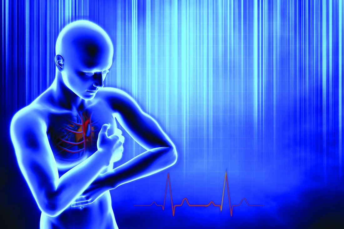
There, researchers are working to create more accurate and personalized models of the heart – and not just any heart, your heart, if you have heart disease.
“Right now, a clinician can only say whether a patient is at risk or not at risk for sudden death,” says Dan Popescu, PhD, a Johns Hopkins research scientist and first author of a new study on AI’s ability to predict sudden cardiac arrest. “With this new technology, you can have much more nuanced predictions of probability of an event over time.”
Put another way: With AI, clinicians may be able not only to predict if someone is at risk for sudden cardiac arrest, but also when it is most likely to happen. They can do this using a much clearer and more personalized look at the electrical “wiring” of your heart.
Your heart, the conductor
Your heart isn’t just a metronome responsible for keeping a steady stream of blood pumping to tissues with every beat. It’s also a conductor through which vital energy flows.
To make the heart beat, electrical impulses flow from the top to the bottom of the organ. Healthy heart cells relay this electricity seamlessly. But in a heart damaged by inflammation or a past heart attack, scar tissue will block the energy flow.
When an electrical impulse encounters a scarred area, the signal can become erratic, disrupting the set top-to-bottom path and causing irregular heartbeats (arrhythmias), which increase someone’s danger of sudden cardiac death.
Seeing the heart in 3D
Today’s tests offer some insights into the heart’s makeup. For example, MRI scans can reveal damaged areas. PET scans can show inflammation. And EKGs can record the heart’s electrical signals from beat to beat.
But all these technologies offer only a snapshot, showing heart health at a moment in time. They can’t predict the future. That’s why scientists at Johns Hopkins are going further to develop 3D digital replicas of a person’s heart, known as computational heart models.
Computational models are computer-simulated replicas that combine mathematics, physics, and computer science. These models have been around for a long time and are used in many fields, ranging from manufacturing to economics.
In heart medicine, these models are populated with digital “cells,” which imitate living cells and can be programmed with different electrical properties, depending on whether they are healthy or diseased.
“Currently available imaging and testing (MRIs, PETs, EKGs) give some representation of the scarring, but you cannot translate that to what is going to happen over time,” says Natalia Trayanova, PhD, of the Johns Hopkins department of biomedical engineering.
“With computational heart models, we create a dynamic digital image of the heart. We can then give the digital image an electrical stimulus and assess how the heart is able to respond. Then you can better predict what is going to happen.”
The computerized 3D models also mean better, more accurate treatment for heart conditions.
For example, a common treatment for a type of arrhythmia known as atrial fibrillation is ablation, or burning some heart tissue. Ablation stops the erratic electrical impulses causing the arrhythmia, but it can also damage otherwise healthy heart cells.
A personalized computational heart model could allow doctors to see more accurately what areas should and shouldn’t be treated for a specific patient.
Using deep learning AI to predict health outcomes
Dr. Trayanova’s colleague Dr. Popescu is applying deep learning and AI to do more with computerized heart models to predict the future.
In a recent paper in Nature Cardiovascular Research, the research team showed their algorithm assessed the health of 269 patients and was able to predict the chance of sudden cardiac arrest up to 10 years in advance.
“This is really the first time ever, as far as we know, where deep learning technology has been proven to analyze scarring of the heart in a successful way,” Dr. Popescu says.
Dr. Popescu and Dr. Trayanova say the AI algorithm gathers information from the 3D computational heart models with patient data like MRIs, ethnicity, age, lifestyle, and other clinical information. Analyzing all these data can produce accurate and consistent estimates about how long patients might live if they are at risk for sudden death.
“You can’t afford to be wrong. If you are wrong, you can actually impact a patient’s quality of life dramatically,” Dr. Popescu says. “Having clinicians use this technology in the decision-making process will provide confidence in a better diagnosis and prognosis.”
While the current study was specifically about patients with a particular type of heart disease, Dr. Popescu says his algorithm can also be trained to assess other health conditions.
So when might you see this being used outside of a research study? Dr. Trayanova predicts 3D imaging of heart models could be available in 2 years, but first the technique must be tested in more clinical trials – some of which are happening right now.
Adding AI to the heart models will require more studies and Food and Drug Administration approval, so the timeline is less clear. But perhaps the biggest hurdle is that after approval the technologies would need to be adopted and used by clinicians and caregivers.
“The much harder question to answer is, ‘When will doctors be perfectly comfortable with AI tools?’ And I don’t know the answer,” Dr. Popescu says. “How to use AI as an aid in the decision-making process is something that’s not currently taught.”
A version of this article first appeared on WebMD.com.