User login
Bariatric surgery for type 2 diabetes: Weighing the impact for obese patients
Evidence is mounting for the use of bariatric surgery to treat type 2 diabetes mellitus in patients whose body mass index (BMI) is 35 kg/m2 or higher. In obese patients who also have type 2 diabetes, bariatric surgery sends it into remission (defined as normoglycemic control without the need for diabetic medications) in more than three-fourths of cases, with higher rates with the Roux-en-Y gastric bypass procedure than with the laparoscopic adjustable gastric banding procedure.
However, data on the effects of this surgery on type 2 diabetes come primarily from observational studies that lacked appropriate control groups, and the relative benefit of bariatric surgery vs aggressive medical antidiabetic therapy is not yet known. Needed are randomized trials comparing the two types of therapy (and the various types of bariatric surgery) in diabetic patients with less-severe obesity.
Further, why would bariatric surgery help with diabetes, and why would one procedure do it better than another? To be honest, we are not sure, but evidence points not only to weight loss but also to better insulin sensitivity and to alterations in levels of hormones secreted by the gut that increase insulin secretion.
OBESITY PROMOTES DIABETES; WEIGHT LOSS COUNTERACTS IT
Type 2 diabetes mellitus is a complex metabolic disease characterized by insulin resistance and progressive failure of pancreatic beta cells, resulting in hyperglycemia.1,2
Obesity, a potent risk factor for type 2 diabetes, contributes to its development by inducing insulin resistance and inflammation, which in turn impair glucose regulation.3,4 Fat deposits in the abdomen, muscles, and liver contribute to elevations of circulating free fatty acids and adipocyte-derived cytokines that mediate insulin resistance and inflammatory pathways.5
In the Diabetes Prevention Program,6 modest weight loss (5% to 10% of body weight) through diet and exercise reduced the incidence of type 2 diabetes, and in the ongoing Action for Health in Diabetes (Look AHEAD) study of the National Institutes of Health, it improved glucose homeostasis.7,8
The current medical approach to type 2 diabetes includes advising the patient to lose weight through lifestyle modification, and prescribing drugs that restore glycemic control by reducing insulin resistance (biguanides, glitazones) and improving insulin secretion (incretin mimetics and analogues and sulfonylureas). 9,10
However, several factors make type 2 diabetes challenging to treat in obese people. Patients who lose weight via behavioral changes and weight-loss drugs tend to gain the weight back. Antidiabetic drugs pose the risk of hypoglycemia. Moreover, although many new classes of drugs have been developed to treat type 2 diabetes, most patients fail to achieve the American Diabetes Association goal for glycemic control, ie, a hemoglobin A1c level lower than 7%.11
BARIATRIC PROCEDURES AND THEIR EFFECT ON DIABETES CONTROL
After bariatric surgery, patients lose more weight than with traditional weight-loss methods—up to 25% of their total body weight. Furthermore, of those with type 2 diabetes, 87% achieve at least better glucose control and need fewer antidiabetic medications,12 and an average of 78% achieve normal glycemic control without taking any antidiabetic medications at all.12,13
But not all bariatric procedures have the same effect on weight and diabetes: certain procedures have a greater effect.
The two major types are classified as gastric restrictive procedures and intestinal bypass procedures. The classification was initially based on the presumed mechanism of weight loss.
Gastric restrictive procedures (laparoscopic adjustable gastric banding, sleeve gastrectomy, vertical gastroplasty) limit gastric volume and, hence, restrict the intake of calories by inducing satiety. Afterward, patients lose approximately 10% to 20% of their total body weight.
Furthermore, multiple studies, including a randomized controlled trial14 (more about this below), have shown remission of type 2 diabetes with laparoscopic adjustable gastric banding but not with conventional medical therapy. The effect is primarily mediated by weight loss and improved insulin sensitivity, both of which occur several months following surgery. Of note, however: in this trial,14 all the patients had diabetes of short duration, less than 2 years.
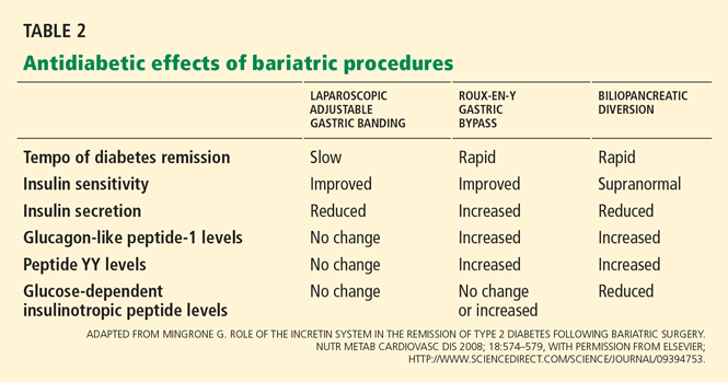
Hence, different procedures have different effects on diabetes.12 The speed at which type 2 diabetes goes into remission differs with restrictive vs malabsorptive procedures. After Roux-en-Y gastric bypass and biliopancreatic diversion, diabetes remits within days, even before the patient has lost much weight.15 This does not happen after gastric restrictive procedures.12,16
Observational studies of the effect of Roux-en-Y surgery on diabetes
Several observational studies have evaluated the benefit of Roux-en-Y surgery for patients with type 2 diabetes mellitus.
Pories et al15 followed 608 severely obese patients, of whom 165 (27%) had type 2 diabetes or impaired glucose tolerance.
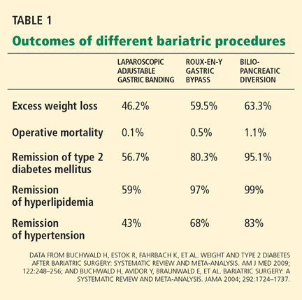
At a mean follow-up of 7.6 years after surgery, 83% of the diabetic patients were off their antidiabetic drugs, and 99% of those with impaired glucose tolerance were normoglycemic, with normal fasting glucose and hemoglobin A1c levels. Marked improvements in hyperlipidemia, hypertension, fertility, osteoarthritis, and obstructive sleep apnea were also noted.
Schauer et al17 observed similar results in 1,160 morbidly obese patients, of whom 240 (21%) had type 2 diabetes or impaired fasting glucose.
After laparoscopic Roux-en-Y gastric bypass surgery, fasting glucose and hemoglobin A1c levels returned to normal levels in 83% of cases and were markedly improved in the remaining 17%. Significantly (80%) fewer patients needed oral antidiabetic agents or insulin (79% fewer). Patients most likely to achieve complete remission of diabetes were those with the shortest duration of diabetes (< 5 years), the mildest severity of diabetes (diet-controlled), and the greatest weight loss after surgery. The rate of diabetes remission in patients who had been diabetic for 5 years or less was 95%, compared with 75% in those who had been diabetic for 6 to 10 years and 54% in those who had been diabetic for more than 10 years (P < .001).
The Swedish Obese Subjects (SOS) study18 prospectively followed 1,703 patients, of whom 118 had type 2 diabetes, for 10 years after various bariatric surgery procedures (primarily vertical gastroplasty). In a control group that received medical therapy, 77 patients had type 2 diabetes. Medical therapy was ill-defined with respect to aggressiveness and adherence to intervention with lifestyle and pharmacotherapy.
At 2 years, the surgical group had lost a mean of 28 kg, glycemic control had improved in the diabetic patients, and many of them had been able to stop taking oral hypoglycemic drugs or insulin. In contrast, the need for these agents increased in the medically treated patients. The proportion treated by diet alone rose from 59% to 73% in the surgical group, but declined from 55% to 34% in the nonsurgical group.13
In these studies, surgery also reduced the risk of progressing from impaired glucose tolerance to type 2 diabetes; the risk was 30 times lower in the study by Pories et al.15 In the SOS study,18 the frequency of diabetes was 30 times lower at 2 years and five times lower at 8 years after surgery.
Studies of biliopancreatic diversion
Data on the effects of biliopancreatic diversion, a primarily malabsorptive procedure, are limited to European studies.
Scopinaro et al19,20 reported long-term follow-up data on 312 patients with type 2 diabetes who underwent biliopancreatic diversion; 310 patients (99%) achieved normal fasting glucose values by 1 year after surgery. At 10 years after surgery, 98% of the patients were still in complete remission of diabetes, defined as normal glucose values without the use of antidiabetic medications.
Others have noted similar findings.21,22
Limitations of the studies
Although these data seem encouraging, these studies had major limitations.
The patients were mostly white women with severe obesity, ie, a BMI greater than 40 kg/m2, which is not representative of patients with type 2 diabetes in the community. Only about 20% had glucose intolerance or overt type 2 diabetes mellitus. Would other groups benefit, particularly men and those with lesssevere obesity?
Moreover, these studies were observational, with no randomized control groups. Many reports consisted of large case series. It is not clear how specific bariatric procedures were chosen or what criteria were used for performing bariatric surgery. A lack of complete follow-up data is also a concern.
Needed are large randomized trials evaluating the effects of various bariatric procedures in a less obese cohort with type 2 diabetes, ie, typical patients seen in the community. Moreover, surgery has not been compared directly with more vigorous medical weight-loss strategies, such as those used in the Diabetes Prevention Project6 and the Look AHEAD trial.7,8
A randomized controlled trial of gastric banding
The only randomized controlled trial to date that compared standard medical diabetes therapy with bariatric surgery was conducted by Dixon et al.14
Sixty patients with type 2 diabetes (duration < 2 years and mean hemoglobin A1c 7.7%) were randomized either to receive medical management as defined by the American Diabetes Association guidelines or to undergo laparoscopic adjustable gastric banding.
At 2 years, the rate of remission (defined as hemoglobin A1c < 6.2% and a normal fasting glucose level) was 13% in the medical treatment group vs 73% in the surgery group (P < .001). Patients receiving medical treatment had lost a mean of 1.7% of their body weight, vs 20.7% in the surgical patients (P < .001). Weight loss was strongly associated with remission of type 2 diabetes after surgery.
This study was controversial in that the medical intervention in this trial was not as aggressive as in the Diabetes Prevention Project and Look AHEAD trials.
INDICATIONS FOR BARIATRIC SURGERY IN PATIENTS WITH DIABETES
According to guidelines from the National Institutes of Health,23 the current indications for bariatric surgery include a BMI of 40 kg/m2 or higher, or a BMI between 35 and 40 kg/m2 with at least two obesity-related comorbidities. Diabetes is considered a key comorbidity that justifies the risk of surgery. The guidelines suggest that bariatric surgery be discussed with all severely obese patients (BMI > 35 kg/m2) with type 2 diabetes who have not been able to lose weight with other weight-control approaches.
Since type 2 diabetes mellitus is a progressive disease characterized by relentless deterioration of beta-cell function, many endocrinologists favor aggressive weight-loss approaches early in the course of the disease. We believe that bariatric surgery should be considered early, as it may help preserve pancreatic betacell function and slow the progression of microvascular and macrovascular complications.
HOW DOES BARIATRIC SURGERY IMPROVE TYPE 2 DIABETES?
Hypothesis 1: Weight loss increases insulin sensitivity
The enforced caloric restriction, negative energy balance, and weight loss after bariatric surgery reduce insulin resistance. Consequently, the beta cells can rest because they don’t need to produce as much insulin. These effects have been observed after both gastric restrictive procedures and gastric bypass procedures.
Hypothesis 2: Less lipotoxicity, inflammation
Another theory is that bariatric surgery lessens insulin resistance by reducing “lipotoxicity,” a condition related to dysregulated fatty acid flux, lipid metabolites in tissues, and direct and indirect effects of hormones secreted by adipocytes.
The strongest evidence for this theory comes from Bikman et al,26 who found that insulin sensitivity increased after Roux-en-Y surgery more than expected from weight loss alone. One year after surgery, even though they remained anthropometrically obese (BMI > 30 kg/m2), the patients had insulin sensitivity levels similar to those in a control group of lean people (BMI < 25 kg/m2).
Insulin sensitivity begins to improve within 1 week of intestinal bypass procedures,15,27 suggesting that these procedures are doing something more than simply forcing weight loss via caloric restriction, as gastric restrictive procedures do.
Hypothesis 3: An effect on gut hormones

The “hindgut hypothesis” raised by Cummings et al24 suggests that accelerated transit of concentrated nutrients (particularly glucose) to the distal intestine results in increased production of insulinotropic and appetite-controlling substances, which account for the reversal of hyperglycemia and obesity.
In contrast, the “foregut hypothesis” raised by Rubino et al28 suggests that nutrient interactions in the duodenum are diabetogenic and, hence, bypassing the duodenum would reverse this defect. Their conclusions come from experiments in rodents that underwent jejunoileal bypass and subsequent refeeding through the bypassed intestine.
GUT HORMONES AND OTHER PEPTIDES ALTERED BY BARIATRIC SURGERY
Incretin hormones: GLP-1, GIP
Gastrointestinal hormones that increase insulin release after a meal are known as incretins. Of interest, they have this effect only when glucose is ingested orally—not when it is infused intravenously.29,30
Glucagon-like peptide 1 (GLP-1) and glucose-dependent insulinotropic peptide (GIP) account for 50% to 60% of nutrient-related insulin secretion. In addition to stimulating insulin, GLP-1 suppresses glucagon and slows gastric emptying, which delays digestion and reduces postprandial glycemia. GLP-1 also acts on the hypothalamus to induce satiety.
Laferrère et al31 and others32,33 documented robust increases in postprandial levels of GLP-1 within 4 weeks after Roux-en-Y surgery. GLP-1 levels did not increase with comparable weight loss induced by diet.
Rubino et al28,34 documented similar findings that occurred prior to marked weight loss, suggesting that the benefit of Roux-en-Y surgery on remission of diabetes may not be completely attributable to reduced caloric intake and weight loss. Insulin secretion is generally reduced after gastric restrictive procedures (eg, laparoscopic adjustable gastric banding) and biliopancreatic diversion,35 and is increased after Roux-en-Y gastric bypass.32,33,36
Noninsulinotropic peptides: Ghrelin, peptide YY
Noninsulinotropic gut peptides that are altered after Roux-en-Y surgery include ghrelin and peptide YY.
Ghrelin, a hormone derived from the gastric fundus, stimulates appetite. Ghrelin concentrations are lower after Roux-en-Y surgery, indicating that suppression of hunger signals helps sustain weight loss. In contrast, ghrelin levels increase with diet-induced weight loss.37 However, the data on ghrelin levels at various times after bariatric surgical procedures are not consistent.33,38
Peptide YY, like GLP-1, is secreted by L cells of the distal small intestine and is responsible for increasing satiety and delaying gastric emptying after meals. Numerous studies have consistently documented increases in postprandial peptide YY and GLP-1 levels after gastric bypass.32,33,39–41
ACUTE EFFECTS OF BARIATRIC SURGERY ON INSULIN SECRETION, SENSITIVITY
Bariatric surgery alters both insulin secretion and insulin sensitivity, thus improving glucose regulation.
The relationship between insulin secretion and sensitivity is a hyperbolic curve, so that any change in insulin sensitivity is balanced by a reciprocal and proportionate change in insulin secretion. The development of type 2 diabetes is characterized by a reduction in insulin secretion (decompensation) relative to the severity of insulin resistance.
In the first 6 weeks after Roux-en-Y gastric bypass or biliopancreatic diversion, insulin sensitivity improves while insulin secretion increases disproportionately, associated with a robust increase in GLP-1, and resulting in normal glucose homeostasis.16,31,42
In contrast, patients who lose weight by dieting or undergoing gastric restrictive procedures show a modest increase in insulin sensitivity and a compensatory reduction in insulin secretion, termed “beta-cell rest.”16,31,42
RISKS OF BARIATRIC SURGERY
Short-term risks
An important concern about using bariatric surgery to treat type 2 diabetes is the risk of morbidity and death associated with these procedures.
Buchwald et al13 performed a meta-analysis of 136 bariatric studies that included 22,094 patients. The 30-day operative death rates were 1.1% with biliopancreatic diversion, 0.5% with Roux-en-Y surgery, and 0.1% with restrictive procedures.
Laparoscopic adjustable gastric banding is considered the safest of the current bariatric procedures. It does not involve bowel anastomosis, and the risks of major hemorrhage, gastric perforation, and pulmonary embolism are less than 1%. Late complications requiring reoperation include band slippage or prolapse (5%–10%) and band erosion (1%–3%). The entire intestinal tract is left intact, so subsequent nutritional deficiencies are rare.43
Roux-en-Y gastric bypass carries an overall risk of major complications of 10% to 15%. Anastomotic leak (1%–5%), pulmonary embolism (< 1%), and hemorrhage (1%–4%) can be life-threatening but are rare if the staff are experienced. Late complications such as ulcer or stricture formation at the gastrojejunostomy site occur in 5% to 10% of cases and are managed nonoperatively.
Nutritional deficiencies
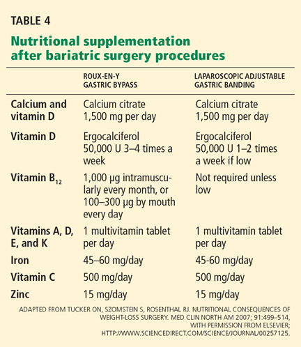
Protein-calorie malnutrition is recognized by signs such as edema, hypoalbuminemia, anemia, and hair loss. To minimize this problem after Roux-en-Y surgery, we suggest that patients take in 60 to 80 g of protein and 700 to 800 kcal a day.
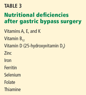
Vitamin deficiencies can lead to Wernicke encephalopathy (due to thiamine deficiency), peripheral neuropathy (due to vitamin B12 deficiency),45,46 and metabolic bone disease (due to long-term deficiencies of vitamin D and calcium). Often, vitamin deficiencies are present before surgery and require prompt supplementation to avoid exacerbation of these deficiencies afterward.
Biliopancreatic diversion procedures are performed at relatively few centers worldwide, largely because of the massive amounts of protein, fat, and carbohydrate malabsorption they cause. Long-term deficiencies of fat-soluble vitamins, iron, calcium, and vitamins B12 and D have been reported in one-third to one-half of patients undergoing these procedures, and nutritional supplementation is mandatory.43 Protein-calorie malnutrition occurs in 7% of cases, and 2% of patients require operative revision to lengthen the common channel.
In rare cases, severe hypoglycemia has been noted after Roux-en-Y surgery and is associated with prandial hyperinsulinemia related to elevated GLP-1 levels.36,47 Neuroglycopenia and seizures have been reported in severe cases. Initial treatment of hypoglycemia involves dietary modification targeting carbohydrate restriction, the use of alpha glucosidase inhibitors such as acarbose (Precose), and referral to an endocrinologist for further management.
Long-term death rates
Death rates after bariatric surgery must be weighed against the long-term cardiovascular risks of continued obesity and type 2 diabetes.
Strong evidence now exists that bariatric surgery increases life expectancy48 and that this is largely attributable to reduction in cardiovascular risk factors such as diabetes and cancer. Recent studies have found that the long-term death rate is 32% to 73% lower for patients undergoing bariatric surgery than in matched controls who do not undergo surgery.49 A decrease in the death rate related to diabetes has played an important role in these results.
Acknowledgments: We acknowledge support from the National Institutes of Health, Multidisciplinary Clinical Research Career Development Programs Grant 5K12RR023264 (SRK), National Center for Research Resources, CTSA 1UL1RR024989, and research grants from Ethicon Endo-Surgery (PS,SRK).
- DeFronzo RA. Pathogenesis of type 2 diabetes mellitus. Med Clin North Am 2004; 88:787–835.
- Kashyap SR, Defronzo RA. The insulin resistance syndrome: physiological considerations. Diab Vasc Dis Res 2007; 4:13–19.
- Mokdad AH, Ford ES, Bowman BA, et al. Prevalence of obesity, diabetes, and obesity-related health risk factors, 2001. JAMA 2003; 289:76–79.
- Unger RH. Minireview: weapons of lean body mass destruction: the role of ectopic lipids in the metabolic syndrome. Endocrinology 2003; 144:5159–5165.
- Itani SI, Ruderman NB, Schmieder F, Boden G. Lipid-induced insulin resistance in human muscle is associated with changes in diacylglycerol, protein kinase C, and IkappaB-alpha. Diabetes 2002; 51:2005–2011.
- Diabetes Prevention Program Research Group. Reduction in the incidence of type 2 diabetes with lifestyle intervention or metformin. N Engl J Med 2002; 346:393–403.
- Look AHEAD Research Group; Pi-Sunyer X, Blackburn G, Brancati FL, et al. Reduction in weight and cardiovascular disease risk factors in individuals with type 2 diabetes: one-year results of the look AHEAD trial. Diabetes Care 2007; 30:1374–1383.
- Look AHEAD Research Group; Wadden TA, West DS, Delahanty L, et al. The Look AHEAD study: a description of the lifestyle intervention and the evidence supporting it. Obesity (Silver Spring) 2006; 14:737–752.
- Nathan DM. Clinical practice. Initial management of glycemia in type 2 diabetes mellitus. N Engl J Med 2002; 347:1342–1349.
- Nathan DM, Buse JB, Davidson MB, et al. Management of hyperglycemia in type 2 diabetes: a consensus algorithm for the initiation and adjustment of therapy: update regarding thiazolidinediones: a consensus statement from the American Diabetes Association and the European Association for the Study of Diabetes. Diabetes Care 2008; 31:173–175.
- Spann SJ, Nutting PA, Galliher JM, et al. Management of type 2 diabetes in the primary care setting: a practice-based research network study. Ann Fam Med 2006; 4:23–31.
- Buchwald H, Estok R, Fahrbach K, et al. Weight and type 2 diabetes after bariatric surgery: systematic review and meta-analysis. Am J Med 2009; 122:248–256.
- Buchwald H, Avidor Y, Braunwald E, et al. Bariatric surgery: a systematic review and meta-analysis. JAMA 2004; 292:1724–1737.
- Dixon JB, O’Brien PE, Playfair J, et al. Adjustable gastric banding and conventional therapy for type 2 diabetes. JAMA 2008; 299:316–323.
- Pories WJ, Swanson MS, MacDonald KG, et al. Who would have thought it? An operation proves to be the most effective therapy for adult-onset diabetes mellitus. Ann Surg 1995; 222:339–350.
- Kashyap SR, Daud S, Kelly KR, et al. Acute effects of gastric bypass versus gastric restrictive surgery on beta-cell function and insulinotropic hormones in severely obese patients with type 2 diabetes. Int J Obes (Lond) 2009; epub ahead of print
- Schauer PR, Burguera B, Ikramuddin S, et al. Effect of laparoscopic Roux-en Y gastric bypass on type 2 diabetes mellitus. Ann Surg 2003; 238:467–484.
- Sjöström L, Lindroos AK, Peltonen M, et al; Swedish Obese Subjects Study Scientific Group. Lifestyle, diabetes, and cardiovascular risk factors 10 years after bariatric surgery. N Engl J Med 2004; 351:2683–2693.
- Scopinaro N, Marinari GM, Camerini GB, Papadia FS, Adami GF. Specific effects of biliopancreatic diversion on the major components of metabolic syndrome: a long-term follow-up study. Diabetes Care 2005; 28:2406–2411.
- Scopinaro N, Papadia F, Marinari G, Camerini G, Adami G. Long-term control of type 2 diabetes mellitus and the other major components of the metabolic syndrome after biliopancreatic diversion in patients with BMI < 35 kg/m2. Obes Surg 2007; 17:185–192.
- Alexandrides TK, Skroubis G, Kalfarentzos F. Resolution of diabetes mellitus and metabolic syndrome following Roux-en-Y gastric bypass and a variant of biliopancreatic diversion in patients with morbid obesity. Obes Surg 2007; 17:176–184.
- Chiellini C, Rubino F, Castagneto M, Nanni G, Mingrone G. The effect of bilio-pancreatic diversion on type 2 diabetes in patients with BMI < 35 kg/m2. Diabetologia 2009; 52:1027–1030.
- Consensus Development Conference Panel. NIH conference. Gastrointestinal surgery for severe obesity. Ann Intern Med 1991; 115:956–961.
- Cummings DE, Overduin J, Foster-Schubert KE. Gastric bypass for obesity: mechanisms of weight loss and diabetes resolution. J Clin Endocrinol Metab 2004; 89:2608–2615.
- Cummings DE, Flum DR. Gastrointestinal surgery as a treatment for diabetes. JAMA 2008; 299:341–343.
- Bikman BT, Zheng D, Pories WJ, et al. Mechanism for improved insulin sensitivity after gastric bypass surgery. J Clin Endocrinol Metab 2008; 93:4656–4663.
- Guidone C, Manco M, Valera-Mora E, et al. Mechanisms of recovery from type 2 diabetes after malabsorptive bariatric surgery. Diabetes 2006; 55:2025–2031.
- Rubino F, Forgione A, Cummings DE, et al. The mechanism of diabetes control after gastrointestinal bypass surgery reveals a role of the proximal small intestine in the pathophysiology of type 2 diabetes. Ann Surg 2006; 244:741–749.
- Vilsbøll T, Krarup T, Madsbad S, Holst JJ. Both GLP-1 and GIP are insulinotropic at basal and postprandial glucose levels and contribute nearly equally to the incretin effect of a meal in healthy subjects. Regul Pept 2003; 114:115–121.
- Vollmer K, Holst JJ, Baller B, et al. Predictors of incretin concentrations in subjects with normal, impaired, and diabetic glucose tolerance. Diabetes 2008; 57:678–687.
- Laferrère B, Teixeira J, McGinty J, et al. Effect of weight loss by gastric bypass surgery versus hypocaloric diet on glucose and incretin levels in patients with type 2 diabetes. J Clin Endocrinol Metab 2008; 93:2479–2485.
- Korner J, Bessler M, Inabnet W, Taveras C, Holst JJ. Exaggerated glucagon-like peptide-1 and blunted glucose-dependent insulinotropic peptide secretion are associated with Roux-en-Y gastric bypass but not adjustable gastric banding. Surg Obes Relat Dis 2007; 3:597–601.
- le Roux CW, Aylwin SJ, Batterham RL, et al. Gut hormone profiles following bariatric surgery favor an anorectic state, facilitate weight loss, and improve metabolic parameters. Ann Surg 2006; 243:108–114.
- Rubino F, Gagner M, Gentileschi P, et al. The early effect of the Roux-en-Y gastric bypass on hormones involved in body weight regulation and glucose metabolism. Ann Surg 2004; 240:236–242.
- Salinari S, Bertuzzi A, Asnaghi S, Guidone C, Manco M, Mingrone G. First-phase insulin secretion restoration and differential response to glucose load depending on the route of administration in type 2 diabetic subjects after bariatric surgery. Diabetes Care 2009; 32:375–380.
- Goldfine AB, Mun EC, Devine E, et al. Patients with neuroglycopenia after gastric bypass surgery have exaggerated incretin and insulin secretory responses to a mixed meal. J Clin Endocrinol Metab 2007; 92:4678–4685.
- Cummings DE, Weigle DS, Frayo RS, et al. Plasma ghrelin levels after diet-induced weight loss or gastric bypass surgery. N Engl J Med 2002; 346:1623–1630.
- Chandarana K, Drew ME, Emmanuel J, et al. Subject standardization, acclimatization, and sample processing affect gut hormone levels and appetite in humans. Gastroenterology 2009; 136:2115–2126.
- Korner J, Inabnet W, Febres G, et al. Prospective study of gut hormone and metabolic changes after adjustable gastric banding and Roux-en-Y gastric bypass. Int J Obes (Lond) 2009; 33:786–795.
- Boey D, Sainsbury A, Herzog H. The role of peptide YY in regulating glucose homeostasis. Peptides 2007; 28:390–395.
- Hanusch-Enserer U, Ghatei MA, Cauza E, Bloom SR, Prager R, Roden M. Relation of fasting plasma peptide YY to glucose metabolism and cardiovascular risk factors after restrictive bariatric surgery. Wien Klin Wochenschr 2007; 119:291–296.
- Laferrère B, Heshka S, Wang K, et al. Incretin levels and effect are markedly enhanced 1 month after Roux-en-Y gastric bypass surgery in obese patients with type 2 diabetes. Diabetes Care 2007; 30:1709–1716.
- Tucker ON, Szomstein S, Rosenthal RJ. Nutritional consequences of weight-loss surgery. Med Clin North Am 2007; 91:499–514.
- Davies DJ, Baxter JM, Baxter JN. Nutritional deficiencies after bariatric surgery. Obes Surg 2007; 17:1150–1158.
- Angstadt JD, Bodziner RA. Peripheral polyneuropathy from thiamine deficiency following laparoscopic Roux-en-Y gastric bypass. Obes Surg 2005; 15:890–892.
- Ritz P, Becouarn G, Douay O, Sallé A, Topart P, Rohmer V. Gastric bypass is not associated with protein malnutrition in morbidly obese patients. Obes Surg 2009; 19:840–844.
- Service GJ, Thompson GB, Service FJ, Andrews JC, Collazo-Clavell ML, Lloyd RV. Hyperinsulinemic hypoglycemia with nesidioblastosis after gastric-bypass surgery. N Engl J Med 2005; 353:249–254.
- Sjöström L, Narbro K, Sjöström CD, et al;Swedish Obese Subjects Study. Effects of bariatric surgery on mortality in Swedish obese subjects. N Engl J Med 2007; 357:741–752.
- Adams TD, Gress RE, Smith SC, et al. Long-term mortality after gastric bypass surgery. N Engl J Med 2007; 357:753–761.
Evidence is mounting for the use of bariatric surgery to treat type 2 diabetes mellitus in patients whose body mass index (BMI) is 35 kg/m2 or higher. In obese patients who also have type 2 diabetes, bariatric surgery sends it into remission (defined as normoglycemic control without the need for diabetic medications) in more than three-fourths of cases, with higher rates with the Roux-en-Y gastric bypass procedure than with the laparoscopic adjustable gastric banding procedure.
However, data on the effects of this surgery on type 2 diabetes come primarily from observational studies that lacked appropriate control groups, and the relative benefit of bariatric surgery vs aggressive medical antidiabetic therapy is not yet known. Needed are randomized trials comparing the two types of therapy (and the various types of bariatric surgery) in diabetic patients with less-severe obesity.
Further, why would bariatric surgery help with diabetes, and why would one procedure do it better than another? To be honest, we are not sure, but evidence points not only to weight loss but also to better insulin sensitivity and to alterations in levels of hormones secreted by the gut that increase insulin secretion.
OBESITY PROMOTES DIABETES; WEIGHT LOSS COUNTERACTS IT
Type 2 diabetes mellitus is a complex metabolic disease characterized by insulin resistance and progressive failure of pancreatic beta cells, resulting in hyperglycemia.1,2
Obesity, a potent risk factor for type 2 diabetes, contributes to its development by inducing insulin resistance and inflammation, which in turn impair glucose regulation.3,4 Fat deposits in the abdomen, muscles, and liver contribute to elevations of circulating free fatty acids and adipocyte-derived cytokines that mediate insulin resistance and inflammatory pathways.5
In the Diabetes Prevention Program,6 modest weight loss (5% to 10% of body weight) through diet and exercise reduced the incidence of type 2 diabetes, and in the ongoing Action for Health in Diabetes (Look AHEAD) study of the National Institutes of Health, it improved glucose homeostasis.7,8
The current medical approach to type 2 diabetes includes advising the patient to lose weight through lifestyle modification, and prescribing drugs that restore glycemic control by reducing insulin resistance (biguanides, glitazones) and improving insulin secretion (incretin mimetics and analogues and sulfonylureas). 9,10
However, several factors make type 2 diabetes challenging to treat in obese people. Patients who lose weight via behavioral changes and weight-loss drugs tend to gain the weight back. Antidiabetic drugs pose the risk of hypoglycemia. Moreover, although many new classes of drugs have been developed to treat type 2 diabetes, most patients fail to achieve the American Diabetes Association goal for glycemic control, ie, a hemoglobin A1c level lower than 7%.11
BARIATRIC PROCEDURES AND THEIR EFFECT ON DIABETES CONTROL
After bariatric surgery, patients lose more weight than with traditional weight-loss methods—up to 25% of their total body weight. Furthermore, of those with type 2 diabetes, 87% achieve at least better glucose control and need fewer antidiabetic medications,12 and an average of 78% achieve normal glycemic control without taking any antidiabetic medications at all.12,13
But not all bariatric procedures have the same effect on weight and diabetes: certain procedures have a greater effect.
The two major types are classified as gastric restrictive procedures and intestinal bypass procedures. The classification was initially based on the presumed mechanism of weight loss.
Gastric restrictive procedures (laparoscopic adjustable gastric banding, sleeve gastrectomy, vertical gastroplasty) limit gastric volume and, hence, restrict the intake of calories by inducing satiety. Afterward, patients lose approximately 10% to 20% of their total body weight.
Furthermore, multiple studies, including a randomized controlled trial14 (more about this below), have shown remission of type 2 diabetes with laparoscopic adjustable gastric banding but not with conventional medical therapy. The effect is primarily mediated by weight loss and improved insulin sensitivity, both of which occur several months following surgery. Of note, however: in this trial,14 all the patients had diabetes of short duration, less than 2 years.
Hence, different procedures have different effects on diabetes.12 The speed at which type 2 diabetes goes into remission differs with restrictive vs malabsorptive procedures. After Roux-en-Y gastric bypass and biliopancreatic diversion, diabetes remits within days, even before the patient has lost much weight.15 This does not happen after gastric restrictive procedures.12,16
Observational studies of the effect of Roux-en-Y surgery on diabetes
Several observational studies have evaluated the benefit of Roux-en-Y surgery for patients with type 2 diabetes mellitus.
Pories et al15 followed 608 severely obese patients, of whom 165 (27%) had type 2 diabetes or impaired glucose tolerance.
At a mean follow-up of 7.6 years after surgery, 83% of the diabetic patients were off their antidiabetic drugs, and 99% of those with impaired glucose tolerance were normoglycemic, with normal fasting glucose and hemoglobin A1c levels. Marked improvements in hyperlipidemia, hypertension, fertility, osteoarthritis, and obstructive sleep apnea were also noted.
Schauer et al17 observed similar results in 1,160 morbidly obese patients, of whom 240 (21%) had type 2 diabetes or impaired fasting glucose.
After laparoscopic Roux-en-Y gastric bypass surgery, fasting glucose and hemoglobin A1c levels returned to normal levels in 83% of cases and were markedly improved in the remaining 17%. Significantly (80%) fewer patients needed oral antidiabetic agents or insulin (79% fewer). Patients most likely to achieve complete remission of diabetes were those with the shortest duration of diabetes (< 5 years), the mildest severity of diabetes (diet-controlled), and the greatest weight loss after surgery. The rate of diabetes remission in patients who had been diabetic for 5 years or less was 95%, compared with 75% in those who had been diabetic for 6 to 10 years and 54% in those who had been diabetic for more than 10 years (P < .001).
The Swedish Obese Subjects (SOS) study18 prospectively followed 1,703 patients, of whom 118 had type 2 diabetes, for 10 years after various bariatric surgery procedures (primarily vertical gastroplasty). In a control group that received medical therapy, 77 patients had type 2 diabetes. Medical therapy was ill-defined with respect to aggressiveness and adherence to intervention with lifestyle and pharmacotherapy.
At 2 years, the surgical group had lost a mean of 28 kg, glycemic control had improved in the diabetic patients, and many of them had been able to stop taking oral hypoglycemic drugs or insulin. In contrast, the need for these agents increased in the medically treated patients. The proportion treated by diet alone rose from 59% to 73% in the surgical group, but declined from 55% to 34% in the nonsurgical group.13
In these studies, surgery also reduced the risk of progressing from impaired glucose tolerance to type 2 diabetes; the risk was 30 times lower in the study by Pories et al.15 In the SOS study,18 the frequency of diabetes was 30 times lower at 2 years and five times lower at 8 years after surgery.
Studies of biliopancreatic diversion
Data on the effects of biliopancreatic diversion, a primarily malabsorptive procedure, are limited to European studies.
Scopinaro et al19,20 reported long-term follow-up data on 312 patients with type 2 diabetes who underwent biliopancreatic diversion; 310 patients (99%) achieved normal fasting glucose values by 1 year after surgery. At 10 years after surgery, 98% of the patients were still in complete remission of diabetes, defined as normal glucose values without the use of antidiabetic medications.
Others have noted similar findings.21,22
Limitations of the studies
Although these data seem encouraging, these studies had major limitations.
The patients were mostly white women with severe obesity, ie, a BMI greater than 40 kg/m2, which is not representative of patients with type 2 diabetes in the community. Only about 20% had glucose intolerance or overt type 2 diabetes mellitus. Would other groups benefit, particularly men and those with lesssevere obesity?
Moreover, these studies were observational, with no randomized control groups. Many reports consisted of large case series. It is not clear how specific bariatric procedures were chosen or what criteria were used for performing bariatric surgery. A lack of complete follow-up data is also a concern.
Needed are large randomized trials evaluating the effects of various bariatric procedures in a less obese cohort with type 2 diabetes, ie, typical patients seen in the community. Moreover, surgery has not been compared directly with more vigorous medical weight-loss strategies, such as those used in the Diabetes Prevention Project6 and the Look AHEAD trial.7,8
A randomized controlled trial of gastric banding
The only randomized controlled trial to date that compared standard medical diabetes therapy with bariatric surgery was conducted by Dixon et al.14
Sixty patients with type 2 diabetes (duration < 2 years and mean hemoglobin A1c 7.7%) were randomized either to receive medical management as defined by the American Diabetes Association guidelines or to undergo laparoscopic adjustable gastric banding.
At 2 years, the rate of remission (defined as hemoglobin A1c < 6.2% and a normal fasting glucose level) was 13% in the medical treatment group vs 73% in the surgery group (P < .001). Patients receiving medical treatment had lost a mean of 1.7% of their body weight, vs 20.7% in the surgical patients (P < .001). Weight loss was strongly associated with remission of type 2 diabetes after surgery.
This study was controversial in that the medical intervention in this trial was not as aggressive as in the Diabetes Prevention Project and Look AHEAD trials.
INDICATIONS FOR BARIATRIC SURGERY IN PATIENTS WITH DIABETES
According to guidelines from the National Institutes of Health,23 the current indications for bariatric surgery include a BMI of 40 kg/m2 or higher, or a BMI between 35 and 40 kg/m2 with at least two obesity-related comorbidities. Diabetes is considered a key comorbidity that justifies the risk of surgery. The guidelines suggest that bariatric surgery be discussed with all severely obese patients (BMI > 35 kg/m2) with type 2 diabetes who have not been able to lose weight with other weight-control approaches.
Since type 2 diabetes mellitus is a progressive disease characterized by relentless deterioration of beta-cell function, many endocrinologists favor aggressive weight-loss approaches early in the course of the disease. We believe that bariatric surgery should be considered early, as it may help preserve pancreatic betacell function and slow the progression of microvascular and macrovascular complications.
HOW DOES BARIATRIC SURGERY IMPROVE TYPE 2 DIABETES?
Hypothesis 1: Weight loss increases insulin sensitivity
The enforced caloric restriction, negative energy balance, and weight loss after bariatric surgery reduce insulin resistance. Consequently, the beta cells can rest because they don’t need to produce as much insulin. These effects have been observed after both gastric restrictive procedures and gastric bypass procedures.
Hypothesis 2: Less lipotoxicity, inflammation
Another theory is that bariatric surgery lessens insulin resistance by reducing “lipotoxicity,” a condition related to dysregulated fatty acid flux, lipid metabolites in tissues, and direct and indirect effects of hormones secreted by adipocytes.
The strongest evidence for this theory comes from Bikman et al,26 who found that insulin sensitivity increased after Roux-en-Y surgery more than expected from weight loss alone. One year after surgery, even though they remained anthropometrically obese (BMI > 30 kg/m2), the patients had insulin sensitivity levels similar to those in a control group of lean people (BMI < 25 kg/m2).
Insulin sensitivity begins to improve within 1 week of intestinal bypass procedures,15,27 suggesting that these procedures are doing something more than simply forcing weight loss via caloric restriction, as gastric restrictive procedures do.
Hypothesis 3: An effect on gut hormones
The “hindgut hypothesis” raised by Cummings et al24 suggests that accelerated transit of concentrated nutrients (particularly glucose) to the distal intestine results in increased production of insulinotropic and appetite-controlling substances, which account for the reversal of hyperglycemia and obesity.
In contrast, the “foregut hypothesis” raised by Rubino et al28 suggests that nutrient interactions in the duodenum are diabetogenic and, hence, bypassing the duodenum would reverse this defect. Their conclusions come from experiments in rodents that underwent jejunoileal bypass and subsequent refeeding through the bypassed intestine.
GUT HORMONES AND OTHER PEPTIDES ALTERED BY BARIATRIC SURGERY
Incretin hormones: GLP-1, GIP
Gastrointestinal hormones that increase insulin release after a meal are known as incretins. Of interest, they have this effect only when glucose is ingested orally—not when it is infused intravenously.29,30
Glucagon-like peptide 1 (GLP-1) and glucose-dependent insulinotropic peptide (GIP) account for 50% to 60% of nutrient-related insulin secretion. In addition to stimulating insulin, GLP-1 suppresses glucagon and slows gastric emptying, which delays digestion and reduces postprandial glycemia. GLP-1 also acts on the hypothalamus to induce satiety.
Laferrère et al31 and others32,33 documented robust increases in postprandial levels of GLP-1 within 4 weeks after Roux-en-Y surgery. GLP-1 levels did not increase with comparable weight loss induced by diet.
Rubino et al28,34 documented similar findings that occurred prior to marked weight loss, suggesting that the benefit of Roux-en-Y surgery on remission of diabetes may not be completely attributable to reduced caloric intake and weight loss. Insulin secretion is generally reduced after gastric restrictive procedures (eg, laparoscopic adjustable gastric banding) and biliopancreatic diversion,35 and is increased after Roux-en-Y gastric bypass.32,33,36
Noninsulinotropic peptides: Ghrelin, peptide YY
Noninsulinotropic gut peptides that are altered after Roux-en-Y surgery include ghrelin and peptide YY.
Ghrelin, a hormone derived from the gastric fundus, stimulates appetite. Ghrelin concentrations are lower after Roux-en-Y surgery, indicating that suppression of hunger signals helps sustain weight loss. In contrast, ghrelin levels increase with diet-induced weight loss.37 However, the data on ghrelin levels at various times after bariatric surgical procedures are not consistent.33,38
Peptide YY, like GLP-1, is secreted by L cells of the distal small intestine and is responsible for increasing satiety and delaying gastric emptying after meals. Numerous studies have consistently documented increases in postprandial peptide YY and GLP-1 levels after gastric bypass.32,33,39–41
ACUTE EFFECTS OF BARIATRIC SURGERY ON INSULIN SECRETION, SENSITIVITY
Bariatric surgery alters both insulin secretion and insulin sensitivity, thus improving glucose regulation.
The relationship between insulin secretion and sensitivity is a hyperbolic curve, so that any change in insulin sensitivity is balanced by a reciprocal and proportionate change in insulin secretion. The development of type 2 diabetes is characterized by a reduction in insulin secretion (decompensation) relative to the severity of insulin resistance.
In the first 6 weeks after Roux-en-Y gastric bypass or biliopancreatic diversion, insulin sensitivity improves while insulin secretion increases disproportionately, associated with a robust increase in GLP-1, and resulting in normal glucose homeostasis.16,31,42
In contrast, patients who lose weight by dieting or undergoing gastric restrictive procedures show a modest increase in insulin sensitivity and a compensatory reduction in insulin secretion, termed “beta-cell rest.”16,31,42
RISKS OF BARIATRIC SURGERY
Short-term risks
An important concern about using bariatric surgery to treat type 2 diabetes is the risk of morbidity and death associated with these procedures.
Buchwald et al13 performed a meta-analysis of 136 bariatric studies that included 22,094 patients. The 30-day operative death rates were 1.1% with biliopancreatic diversion, 0.5% with Roux-en-Y surgery, and 0.1% with restrictive procedures.
Laparoscopic adjustable gastric banding is considered the safest of the current bariatric procedures. It does not involve bowel anastomosis, and the risks of major hemorrhage, gastric perforation, and pulmonary embolism are less than 1%. Late complications requiring reoperation include band slippage or prolapse (5%–10%) and band erosion (1%–3%). The entire intestinal tract is left intact, so subsequent nutritional deficiencies are rare.43
Roux-en-Y gastric bypass carries an overall risk of major complications of 10% to 15%. Anastomotic leak (1%–5%), pulmonary embolism (< 1%), and hemorrhage (1%–4%) can be life-threatening but are rare if the staff are experienced. Late complications such as ulcer or stricture formation at the gastrojejunostomy site occur in 5% to 10% of cases and are managed nonoperatively.
Nutritional deficiencies
Protein-calorie malnutrition is recognized by signs such as edema, hypoalbuminemia, anemia, and hair loss. To minimize this problem after Roux-en-Y surgery, we suggest that patients take in 60 to 80 g of protein and 700 to 800 kcal a day.
Vitamin deficiencies can lead to Wernicke encephalopathy (due to thiamine deficiency), peripheral neuropathy (due to vitamin B12 deficiency),45,46 and metabolic bone disease (due to long-term deficiencies of vitamin D and calcium). Often, vitamin deficiencies are present before surgery and require prompt supplementation to avoid exacerbation of these deficiencies afterward.
Biliopancreatic diversion procedures are performed at relatively few centers worldwide, largely because of the massive amounts of protein, fat, and carbohydrate malabsorption they cause. Long-term deficiencies of fat-soluble vitamins, iron, calcium, and vitamins B12 and D have been reported in one-third to one-half of patients undergoing these procedures, and nutritional supplementation is mandatory.43 Protein-calorie malnutrition occurs in 7% of cases, and 2% of patients require operative revision to lengthen the common channel.
In rare cases, severe hypoglycemia has been noted after Roux-en-Y surgery and is associated with prandial hyperinsulinemia related to elevated GLP-1 levels.36,47 Neuroglycopenia and seizures have been reported in severe cases. Initial treatment of hypoglycemia involves dietary modification targeting carbohydrate restriction, the use of alpha glucosidase inhibitors such as acarbose (Precose), and referral to an endocrinologist for further management.
Long-term death rates
Death rates after bariatric surgery must be weighed against the long-term cardiovascular risks of continued obesity and type 2 diabetes.
Strong evidence now exists that bariatric surgery increases life expectancy48 and that this is largely attributable to reduction in cardiovascular risk factors such as diabetes and cancer. Recent studies have found that the long-term death rate is 32% to 73% lower for patients undergoing bariatric surgery than in matched controls who do not undergo surgery.49 A decrease in the death rate related to diabetes has played an important role in these results.
Acknowledgments: We acknowledge support from the National Institutes of Health, Multidisciplinary Clinical Research Career Development Programs Grant 5K12RR023264 (SRK), National Center for Research Resources, CTSA 1UL1RR024989, and research grants from Ethicon Endo-Surgery (PS,SRK).
- DeFronzo RA. Pathogenesis of type 2 diabetes mellitus. Med Clin North Am 2004; 88:787–835.
- Kashyap SR, Defronzo RA. The insulin resistance syndrome: physiological considerations. Diab Vasc Dis Res 2007; 4:13–19.
- Mokdad AH, Ford ES, Bowman BA, et al. Prevalence of obesity, diabetes, and obesity-related health risk factors, 2001. JAMA 2003; 289:76–79.
- Unger RH. Minireview: weapons of lean body mass destruction: the role of ectopic lipids in the metabolic syndrome. Endocrinology 2003; 144:5159–5165.
- Itani SI, Ruderman NB, Schmieder F, Boden G. Lipid-induced insulin resistance in human muscle is associated with changes in diacylglycerol, protein kinase C, and IkappaB-alpha. Diabetes 2002; 51:2005–2011.
- Diabetes Prevention Program Research Group. Reduction in the incidence of type 2 diabetes with lifestyle intervention or metformin. N Engl J Med 2002; 346:393–403.
- Look AHEAD Research Group; Pi-Sunyer X, Blackburn G, Brancati FL, et al. Reduction in weight and cardiovascular disease risk factors in individuals with type 2 diabetes: one-year results of the look AHEAD trial. Diabetes Care 2007; 30:1374–1383.
- Look AHEAD Research Group; Wadden TA, West DS, Delahanty L, et al. The Look AHEAD study: a description of the lifestyle intervention and the evidence supporting it. Obesity (Silver Spring) 2006; 14:737–752.
- Nathan DM. Clinical practice. Initial management of glycemia in type 2 diabetes mellitus. N Engl J Med 2002; 347:1342–1349.
- Nathan DM, Buse JB, Davidson MB, et al. Management of hyperglycemia in type 2 diabetes: a consensus algorithm for the initiation and adjustment of therapy: update regarding thiazolidinediones: a consensus statement from the American Diabetes Association and the European Association for the Study of Diabetes. Diabetes Care 2008; 31:173–175.
- Spann SJ, Nutting PA, Galliher JM, et al. Management of type 2 diabetes in the primary care setting: a practice-based research network study. Ann Fam Med 2006; 4:23–31.
- Buchwald H, Estok R, Fahrbach K, et al. Weight and type 2 diabetes after bariatric surgery: systematic review and meta-analysis. Am J Med 2009; 122:248–256.
- Buchwald H, Avidor Y, Braunwald E, et al. Bariatric surgery: a systematic review and meta-analysis. JAMA 2004; 292:1724–1737.
- Dixon JB, O’Brien PE, Playfair J, et al. Adjustable gastric banding and conventional therapy for type 2 diabetes. JAMA 2008; 299:316–323.
- Pories WJ, Swanson MS, MacDonald KG, et al. Who would have thought it? An operation proves to be the most effective therapy for adult-onset diabetes mellitus. Ann Surg 1995; 222:339–350.
- Kashyap SR, Daud S, Kelly KR, et al. Acute effects of gastric bypass versus gastric restrictive surgery on beta-cell function and insulinotropic hormones in severely obese patients with type 2 diabetes. Int J Obes (Lond) 2009; epub ahead of print
- Schauer PR, Burguera B, Ikramuddin S, et al. Effect of laparoscopic Roux-en Y gastric bypass on type 2 diabetes mellitus. Ann Surg 2003; 238:467–484.
- Sjöström L, Lindroos AK, Peltonen M, et al; Swedish Obese Subjects Study Scientific Group. Lifestyle, diabetes, and cardiovascular risk factors 10 years after bariatric surgery. N Engl J Med 2004; 351:2683–2693.
- Scopinaro N, Marinari GM, Camerini GB, Papadia FS, Adami GF. Specific effects of biliopancreatic diversion on the major components of metabolic syndrome: a long-term follow-up study. Diabetes Care 2005; 28:2406–2411.
- Scopinaro N, Papadia F, Marinari G, Camerini G, Adami G. Long-term control of type 2 diabetes mellitus and the other major components of the metabolic syndrome after biliopancreatic diversion in patients with BMI < 35 kg/m2. Obes Surg 2007; 17:185–192.
- Alexandrides TK, Skroubis G, Kalfarentzos F. Resolution of diabetes mellitus and metabolic syndrome following Roux-en-Y gastric bypass and a variant of biliopancreatic diversion in patients with morbid obesity. Obes Surg 2007; 17:176–184.
- Chiellini C, Rubino F, Castagneto M, Nanni G, Mingrone G. The effect of bilio-pancreatic diversion on type 2 diabetes in patients with BMI < 35 kg/m2. Diabetologia 2009; 52:1027–1030.
- Consensus Development Conference Panel. NIH conference. Gastrointestinal surgery for severe obesity. Ann Intern Med 1991; 115:956–961.
- Cummings DE, Overduin J, Foster-Schubert KE. Gastric bypass for obesity: mechanisms of weight loss and diabetes resolution. J Clin Endocrinol Metab 2004; 89:2608–2615.
- Cummings DE, Flum DR. Gastrointestinal surgery as a treatment for diabetes. JAMA 2008; 299:341–343.
- Bikman BT, Zheng D, Pories WJ, et al. Mechanism for improved insulin sensitivity after gastric bypass surgery. J Clin Endocrinol Metab 2008; 93:4656–4663.
- Guidone C, Manco M, Valera-Mora E, et al. Mechanisms of recovery from type 2 diabetes after malabsorptive bariatric surgery. Diabetes 2006; 55:2025–2031.
- Rubino F, Forgione A, Cummings DE, et al. The mechanism of diabetes control after gastrointestinal bypass surgery reveals a role of the proximal small intestine in the pathophysiology of type 2 diabetes. Ann Surg 2006; 244:741–749.
- Vilsbøll T, Krarup T, Madsbad S, Holst JJ. Both GLP-1 and GIP are insulinotropic at basal and postprandial glucose levels and contribute nearly equally to the incretin effect of a meal in healthy subjects. Regul Pept 2003; 114:115–121.
- Vollmer K, Holst JJ, Baller B, et al. Predictors of incretin concentrations in subjects with normal, impaired, and diabetic glucose tolerance. Diabetes 2008; 57:678–687.
- Laferrère B, Teixeira J, McGinty J, et al. Effect of weight loss by gastric bypass surgery versus hypocaloric diet on glucose and incretin levels in patients with type 2 diabetes. J Clin Endocrinol Metab 2008; 93:2479–2485.
- Korner J, Bessler M, Inabnet W, Taveras C, Holst JJ. Exaggerated glucagon-like peptide-1 and blunted glucose-dependent insulinotropic peptide secretion are associated with Roux-en-Y gastric bypass but not adjustable gastric banding. Surg Obes Relat Dis 2007; 3:597–601.
- le Roux CW, Aylwin SJ, Batterham RL, et al. Gut hormone profiles following bariatric surgery favor an anorectic state, facilitate weight loss, and improve metabolic parameters. Ann Surg 2006; 243:108–114.
- Rubino F, Gagner M, Gentileschi P, et al. The early effect of the Roux-en-Y gastric bypass on hormones involved in body weight regulation and glucose metabolism. Ann Surg 2004; 240:236–242.
- Salinari S, Bertuzzi A, Asnaghi S, Guidone C, Manco M, Mingrone G. First-phase insulin secretion restoration and differential response to glucose load depending on the route of administration in type 2 diabetic subjects after bariatric surgery. Diabetes Care 2009; 32:375–380.
- Goldfine AB, Mun EC, Devine E, et al. Patients with neuroglycopenia after gastric bypass surgery have exaggerated incretin and insulin secretory responses to a mixed meal. J Clin Endocrinol Metab 2007; 92:4678–4685.
- Cummings DE, Weigle DS, Frayo RS, et al. Plasma ghrelin levels after diet-induced weight loss or gastric bypass surgery. N Engl J Med 2002; 346:1623–1630.
- Chandarana K, Drew ME, Emmanuel J, et al. Subject standardization, acclimatization, and sample processing affect gut hormone levels and appetite in humans. Gastroenterology 2009; 136:2115–2126.
- Korner J, Inabnet W, Febres G, et al. Prospective study of gut hormone and metabolic changes after adjustable gastric banding and Roux-en-Y gastric bypass. Int J Obes (Lond) 2009; 33:786–795.
- Boey D, Sainsbury A, Herzog H. The role of peptide YY in regulating glucose homeostasis. Peptides 2007; 28:390–395.
- Hanusch-Enserer U, Ghatei MA, Cauza E, Bloom SR, Prager R, Roden M. Relation of fasting plasma peptide YY to glucose metabolism and cardiovascular risk factors after restrictive bariatric surgery. Wien Klin Wochenschr 2007; 119:291–296.
- Laferrère B, Heshka S, Wang K, et al. Incretin levels and effect are markedly enhanced 1 month after Roux-en-Y gastric bypass surgery in obese patients with type 2 diabetes. Diabetes Care 2007; 30:1709–1716.
- Tucker ON, Szomstein S, Rosenthal RJ. Nutritional consequences of weight-loss surgery. Med Clin North Am 2007; 91:499–514.
- Davies DJ, Baxter JM, Baxter JN. Nutritional deficiencies after bariatric surgery. Obes Surg 2007; 17:1150–1158.
- Angstadt JD, Bodziner RA. Peripheral polyneuropathy from thiamine deficiency following laparoscopic Roux-en-Y gastric bypass. Obes Surg 2005; 15:890–892.
- Ritz P, Becouarn G, Douay O, Sallé A, Topart P, Rohmer V. Gastric bypass is not associated with protein malnutrition in morbidly obese patients. Obes Surg 2009; 19:840–844.
- Service GJ, Thompson GB, Service FJ, Andrews JC, Collazo-Clavell ML, Lloyd RV. Hyperinsulinemic hypoglycemia with nesidioblastosis after gastric-bypass surgery. N Engl J Med 2005; 353:249–254.
- Sjöström L, Narbro K, Sjöström CD, et al;Swedish Obese Subjects Study. Effects of bariatric surgery on mortality in Swedish obese subjects. N Engl J Med 2007; 357:741–752.
- Adams TD, Gress RE, Smith SC, et al. Long-term mortality after gastric bypass surgery. N Engl J Med 2007; 357:753–761.
KEY POINTS
- After Roux-en-Y gastric bypass and biliopancreatic diversion, normoglycemia is restored within days, even before the patient has lost much weight.
- Alterations in postprandial levels of intestine-derived hormones (glucagon-like peptide 1, peptide YY, and ghrelin) contribute to the robust metabolic benefits of intestinal bypass procedures.
- Nutritional deficiencies are common after bariatric surgery, and long-term follow-up is mandatory for surveillance of metabolic status.
- Although curing diabetes cannot yet be considered a goal of bariatric surgery, it may be a serendipitous benefit.

Perioperative management of bariatric surgery patients: Focus on metabolic bone disease
A 56-year-old woman who underwent Roux-en-y bariatric surgery because of morbid obesity 6 years ago presents to her primary care physician with vague complaints of fatigue, myalgias, arthralgias, and weakness that have slowly been getting worse. Before surgery she weighed 340 pounds (154 kg), and in the first 2 years afterward she lost 160 pounds (72.5 kg). She is postmenopausal, has no history of fractures, nephrolithiasis, or thyroid disease, and does not smoke or consume alcohol. She gives herself monthly intramuscular vitamin B12 injections, and takes a multivitamin tablet, calcium carbonate 500 mg, and vitamin D 400 IU daily.
After her surgery she returned for her first two postoperative appointments, but because she was feeling well, was losing weight, and had returned to work full-time, she cancelled all subsequent appointments with the surgeon, bariatrician, and dietitian.
On physical examination, the patient’s weight is stable at 187 pounds (84.5 kg), her height is 165.1 cm, and her body mass index is 31. Her head, eyes, ears, nose, throat, heart, lungs, and abdomen are normal. Her upper legs are weak, requiring her to use her arms in rising from a chair, and she feels discomfort when the proximal muscles of her arms and legs are palpated. She has mild osteoarthritis of the hands and knees. Her neurologic examination is normal.
Pertinent laboratory data:
- Calcium 8.1 mg/dL (reference range 8.5–10.5)
- Albumin 3.7 g/dL (3.4–4.7)
- Magnesium 1.9 mg/dL (1.7–2.6)
- Phosphorus 2.7 mg/dL (2.4–4.5)
- Alkaline phosphatase 240 U/L (40–150)
- Intact parathyroid hormone 215 pg/mL (10–60)
- 25-hydroxyvitamin D < 7.0 ng/mL (31–80)
- 24-hour urine volume 2,310 mL
- Urine creatinine normal
- Urine calcium 25.4 mg/24 hours (100–300).
Dual-energy x-ray absorptiometry (DXA) data, lumbar spine:
- Bone mineral density 0.933 g/cm2
- T score –2.0
- Z score –0.8.
Left total hip:
- Bone mineral density 0.628 g/cm2
- T score –2.6
- Z score –2.4.
METABOLIC BONE DISEASE: A CASE IN POINT
This is a classic presentation of metabolic bone disease in a bariatric surgery patient lost to follow-up. Many patients have non-specific and vague symptoms for many months or years that are often incorrectly diagnosed as fibromyalgia, rheumatoid arthritis, polymyalgia rheumatica, Paget disease, or depression.1 They typically have low serum and urine calcium levels, very low or undetectable 25-hydroxyvitamin D levels, high alkaline phosphatase levels, secondary hyperparathyroidism, and a clinical picture consistent with both osteomalacia and osteoporosis.1
This case underscores the importance of monitoring nutrients and biochemical markers at baseline and on an ongoing basis to detect early indicators of malabsorption and ultimately prevent the development of metabolic bone disease and fragility fracture, with its risks of disability and even death. It also illustrates the essential role that primary care physicians play in the continuing care of these patients.
THE OBESITY-BONE CONNECTION
Although we used to think that morbid obesity protected against metabolic bone disease, in fact, vitamin D and calcium deficiencies and elevated parathyroid hormone (PTH) levels are common in extremely obese people, placing them at risk of low bone mass.2–6 More than 60% of candidates for weight-loss surgery are deficient in vitamin D,5,7 and 25% to 48% have elevated PTH levels.5,6
And that is before bariatric surgery: afterward, severely restricted oral intake and significant weight loss, coupled with a procedure that bypasses the major site of calcium absorption, place many patients at extremely high risk.5,8
After combination restrictive and malabsorptive procedures (eg, the popular Rouxen-y procedure, in which the stomach is reduced in size—”restricted”—and the proximal duodenum is bypassed so that less food is absorbed), as patients lose weight their PTH levels rise and 25-hydroxyvitamin D levels decrease, although corrected calcium levels usually remain within normal limits.3,9 Secondary hyperparathyroidism has been documented as soon as 8 weeks after bariatric surgery, and osteomalacia after gastric bypass surgery is not uncommon.1,7,10–13
Exclusively restrictive procedures such as gastric banding, formerly presumed not to alter bone metabolism, now also appear to place patients at risk of metabolic bone disease due to inadequate intake of calcium and vitamin D in the immediate postoperative period.10
Numerous reported cases further illustrate the ever-present risk of metabolic bone disease in this population if adequate supplementation of calcium and vitamin D is not given. In these cases, significant bone disease occurred from 8 weeks to 32 years after bariatric surgery, often with devastating consequences.1,4,8,11–14
Voluntary weight loss, involuntary bone loss
When overweight or obese people lose weight—whether by dieting or by bariatric surgery—they also lose bone: a voluntary loss of approximately 10% of body weight results in a loss of 1% to 2% of bone at all sites. This loss appears to vary among populations: premenopausal women younger than 45 years may be able to lose a moderate amount of weight without a significant increase in fracture risk, while a study of overweight men found that a 7% weight loss resulted in a 1% bone loss.15
The percentage of bone lost correlates strongly with how fast the weight is lost. A recent study found that losing 0.7 kg/week was more detrimental to bone than a slower loss of 0.3 kg/week, due to the activation of the calcium-PTH axis.16
After bariatric surgery, many patients rapidly lose 50 kg—some even lose 100 kg or more. This rapid weight loss, combined with severely restricted oral intake, decreased calcium absorption, and vitamin D deficiency places these patients at extremely high risk of rapidly developing metabolic bone disease.3,8,9 In one large study, metabolic bone disease developed in more than 70% of patients who underwent a malabsorptive procedure, while in a second study, markers of bone resorption were elevated as soon as 8 weeks after bariatric surgery, regardless of whether the patient underwent a malabsorptive or restrictive bariatric procedure.13 Yet another study found that 48% of patients had a statistically significant bone mineral reduction of more than 3% 12 months after undergoing gastric banding.10
ESSENTIAL NUTRIENTS FOR BONE HEALTH
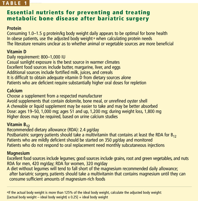
Protein
Dietary protein is needed to maintain bone structure, and although there is a link between high protein intake, calciuria, and fracture risk, the potentially harmful effects appear to be ameliorated when high protein intake is coupled with adequate calcium.17–20 This fact is of particular importance after bariatric surgery because once the patient can consume enough fluids to maintain hemodynamic stability, he or she is given a relatively high-protein diet to prevent protein malnutrition.21
Inadequate protein intake also has a detrimental effect on bone; therefore, it is essential to assess postoperative protein intake.22 Rizzoli and Bonjour23 noted that markers of bone turnover were higher with a low-protein diet (0.7 g protein per kg body weight) than with a diet containing 2.1 g protein per kg. In two trials examining graded levels of protein ingestion (0.7, 0.8, 0.9, and 1.0 g protein per kg body weight), decreased calcium absorption and an acute rise in PTH were noted by day 4 of the 0.7- and 0.8-g/kg diets but not during the 0.9- or 1.0-g/kg diets.24,25 And a systematic review of protein and bone health concluded that diets containing 1.0 to 1.5 g protein/kg are best for bone health.26 This is particularly worrisome, since the current recommended dietary allowance for protein is only 0.8 g/kg, which may be insufficient to promote calcium homeostasis.26,27
Vitamin D
Vitamin D is essential for calcium absorption, stimulation of osteoblast activity, and normal bone mineralization throughout the life span.27 Dietary vitamin D is mainly absorbed by passive diffusion in the proximal and mid small intestine in a process that is highly dependent on bile salts.28–30 Dietary sources of vitamin D are clinically important because exposure to ultraviolet B radiation is often insufficient, especially in northern latitudes.27
Up to 84% of morbidly obese patients have vitamin D deficiency.2,3,5,2,29,31 The mechanism of vitamin D deficiency and secondary hyperparathyroidism in the morbidly obese remains unclear, although one study concluded they were likely due to sequestration of vitamin D in adipose tissue and subsequent limited bioavailability.32
Correction of vitamin D deficiency requires more than just an over-the-counter multivitamin, but standard multivitamins also contain vitamin A, so taking more than one tablet a day increases the risk of vitamin A excess.33 Repletion can often be safely achieved orally by giving 50,000 IU of vitamin D weekly for 8 weeks, followed by a maintenance dose of one 50,000 IU tablet every 2 weeks. If a repeat serum level shows suboptimal repletion (less than 32 ng/mL), an additional 8-week course is recommended.29 For patients who cannot tolerate or adequately absorb oral supplements, exposure to sunlight is still the best source of vitamin D and is an effective alternative.29,33
Calcium
Dietary calcium deficiency is a well-established risk factor for osteoporosis and fragility fractures. Therefore, supplemental calcium should be prescribed for patients who do not meet their defined need.34,35 Of note: a normal serum calcium level does not imply adequate calcium intake or absorption. Calcium homeostasis is tightly regulated and is maintained by a combination of gut absorption, bone resorption, and renal reabsorption. If dietary intake is inadequate, calcium is resorbed from the bone.
The duodenum is the major site of active calcium uptake, while the rest of the small intestine and the colon appear to absorb some calcium passively. When the physiologic need for calcium is increased, active transport appears to take place throughout the duodenum, the ileum, and, to a lesser degree, the jejunum and the colon.36
In the normal gastrointestinal tract, 20% to 60% of dietary calcium is absorbed.36–38 Patients who have lost absorptive surface area (eg, after Roux-en-y bariatric surgery) need to have their calcium intake optimized. However, optimal dosing based on the type of surgical procedure is currently undefined.
Judicious monitoring for compliance and adequate absorption is recommended. Some patients will stop taking their calcium supplement due to gastrointestinal side effects such as gas, bloating, or constipation. And for some patients, a standard calcium supplement may be insufficient to promote adequate calcium absorption. Measuring urinary calcium in a 24-hour sample can help in assessing the adequacy of calcium intake: abnormally low urine calcium in the presence of normal renal function suggests inadequate absorption. For patients reporting gastrointestinal side effects or those with a history of calcium oxalate renal stones, calcium citrate supplements are better tolerated, alter urine acidity, and often prevent further stone formation.
Vitamin B12 (cobalamin)
Vitamin B12 deficiency is associated with increased fracture risk, and it may be an important modifiable risk factor for osteoporosis.39–41 After surgery, malabsorption of vitamin B12 is commonly the result of altered gut function in the gastric pouch or sleeve, but malabsorption also occurs when more than 60 to 100 cm of terminal ileum has been bypassed.42
Vitamin B12 supplementation is recommended for all patients after bariatric surgery, because deficiency is common.42 Patients with relatively mild malabsorption can maintain their B12 level by taking 350 μg orally; however, many patients require lifelong subcutaneous injections.7,39,42–45
Magnesium
Magnesium appears to affect bone remodeling and strength, to have a positive association with hip bone mineral density, and to play an important role in calcium and bone metabolism.
Magnesium is absorbed in the distal small intestine by carrier-mediated and paracellular routes.46 When the distal small intestine is bypassed, magnesium deficiency occurs as a result of reduced absorption and chelation with unabsorbed fatty acids in the bowel lumen.42 Chronic hypomagnesemia impairs PTH secretion, resulting in altered calcium metabolism, hypocalcemia, and vitamin D abnormalities, further decreasing jejunal magnesium absorption.26,42,47
Few well-designed studies have investigated the effect of magnesium intake on bone health, and although there is evidence that postmenopausal women may benefit from magnesium supplementation, studies of magnesium supplementation after bariatric surgery are lacking.47,48
A prevailing misconception promoted by manufacturers of calcium-magnesium supplements and others is that magnesium is necessary for calcium absorption and efficacy. In fact, magnesium deficiency typically must be severe to impair calcium absorption. With usual dietary intake of magnesium and normal serum magnesium levels, no such relationship exists.49–53
THE ROLE OF DXA IN THE CARE OF THE BARIATRIC SURGERY PATIENT
DXA is the gold standard for measuring bone density. The results are reported as a T score and as a Z score.
The T score is the bone density in an area of interest expressed in standard deviations from the mean value of a reference database of young adults. The World Health Organization defines normal as a T score greater than or equal to –1, low bone mass (previously called osteopenia) as a score between –1 and –2.5, and osteoporosis as a score of less than or equal to –2.5. (If a fragility fracture has occurred, “established” or “severe” osteoporosis is present.) Of note: these criteria only apply to DXA of the posterior-anterior spine, femoral neck, and the proximal (33%) radius in post-menopausal women and men over the age of 50 years.54,55 The International Society of Clinical Densitometry has extended the criteria to include total hip measurements.56
The Z score should be used instead of the T score for premenopausal women and men younger than 50 years.56 The Z score is the patient’s bone mineral density expressed in standard deviations from the mean in a reference population matched for sex and age. A Z score greater than –2.0 is “within the expected range for age,” and –2.0 or lower is “below the expected range for age.” There are separate guidelines for DXA reporting in the diagnosis of metabolic bone disease in people younger than 20 years, and this topic is beyond the scope of this article.
Who should undergo DXA?
According to the International Society of Clinical Densitometry, bone density testing is indicated in the general population in women 65 years of age and older, postmenopausal women younger than 65 with risk factors, men 70 and older, adults with fragility fractures, adults taking a medication or having a disease or condition associated with low bone mass or bone loss, any patient being treated for low bone mass (to monitor the treatment effect), and any person in whom evidence of bone loss would affect treatment decisions.58
The National Osteoporosis Foundation recommends initiating therapy to reduce fracture risk in postmenopausal women with a central DXA T score below –2 in the absence of risk factors, and in women with T scores below –1.5 if one or more risk factors is present.34 Therefore, in view of the known risks, the likely need for interventions before surgery, and the ability to prevent the illness and death associated with metabolic bone disease, we recommend that all bariatric surgery patients undergo DXA at baseline as part of the preoperative evaluation.
Improvements in DXA technology
Newer DXA machines can accommodate patients weighing up to 450 pounds (the limit with older machines was 275 pounds for central measurements). In addition to measuring bone density, they also can map the distribution of fat in the body—patients with an android (apple-shaped) distribution are at higher risk of cardiovascular disease than those with a gynecoid (pear-shaped) distribution.59–63 For those patients who cannot be accommodated on a DXA table, DXA of the forearm can be used to assess bone density and fracture risk.
How often should DXA be repeated?
The estimated monitoring time interval is derived from the statistically defined least significant change divided by the anticipated change in bone density over time.64 When estimating the monitoring time interval for changes in body composition, the rate of weight loss and the psychological impact on the patient must be taken into consideration.
In general, DXA testing more frequently than every 2 years remains controversial unless one is initiating, monitoring, or changing therapy or monitoring conditions associated with rapid bone loss such as glucocorticoid therapy. In the bariatric surgery population, however, there is convincing evidence that significant changes may be detected after 12 months that would influence clinical decisions, particularly in the year immediately after surgery.8,9,13,21,27,65
Anabolic and antiresorptive bone drugs
Prescribed medications for the prevention and treatment of osteoporosis should also be an integral part of the treatment plan for at-risk morbidly obese patients. But the decision to prescribe an antiresorptive or bone-forming medication must take into consideration the patient’s risk-benefit profile, including the likelihood of gastrointestinal side effects and his or her ability and willingness to follow specific dosing instructions. Intravenous preparations are now available for patients who cannot absorb or tolerate oral antiresorptive medications. However, specific recommendations about the use of anabolic or antiresorptive bone medications in perioperative bariatric patients have yet to be elucidated.
RECOMMENDATIONS
Although a variety of recommendations have been published, there are no established guidelines for perioperative screening, risk stratification, or management of metabolic bone disease in bariatric surgery patients.7,44,65–67 And the literature remains inconclusive on key issues such as when to start supplements, which biochemical indices should be checked before surgery, whether baseline and annual DXA should be done, and whether antiresorptive agents such as bisphosphonates should be used prophylactically during rapid weight loss.
However, numerous studies and case reports cited here and elsewhere further underscore the ever-present risk of metabolic bone disease in this patient population, and the need for meticulous perioperative and long-term monitoring.44,65–67
With these caveats in mind, we offer our recommendations (Table 3).
Preoperative assessment
We recommend obtaining baseline biochemical indices, including albumin, 25-hydroxy-vitamin D, calcium, magnesium, phosphorus, alkaline phosphatase, folate, vitamin B12, thyroid-stimulating hormone, and PTH levels, and DXA in all bariatric surgery candidates. These indices should be used to assess for primary and secondary metabolic bone disease, to enable prompt presurgical interventions, and to guide the clinician in selecting appropriate postoperative interventions and surveillance.
We recommend starting a multivitamin with minerals at the first preoperative visit. A calcium supplement that provides calcium and vitamin D appropriate to the patient’s age and sex is also recommended until surgery. After surgery, and while rapid weight loss is occurring, a minimum of 1,800 mg of calcium and 800 to 1,000 IU of vitamin D is recommended, keeping in mind that the required level of supplemental vitamin D during periods of rapid weight loss remains unclear.68,69
However, before prescribing supplementation, one should thoroughly review the patient’s nutrition history, including the use of homeopathic medications, herbal preparations, and supplements. Many over-the-counter and over-the-Internet supplements are touted as being good for bone health, and some may indeed be beneficial, but others can be detrimental and need to be discontinued.27,47 Furthermore, in a patient with a severely restricted stomach capacity, it is important to ensure that less efficacious supplements do not compromise the intake of essential fluids, protein, and prescribed medications.
Immediate postoperative period
Hospitalization and surgery result in nutrient deficiencies. In bariatric surgery patients, particularly those who have preoperative nutritional deficiencies, repletion in the immediate postoperative period is believed to be of benefit. Therefore, in the immediate postoperative period we recommend infusing a standard-dose multivitamin with minerals daily along with adequate intravenous hydration until the patient can resume oral feeding. Once the patient can tolerate liquids, presurgical supplementation needs to be resumed, preferably in a liquid or chewable form to facilitate tolerance and absorption.
Short-term and long-term follow-up
Follow-up visits with a bariatric specialist should start 4 weeks after surgery and should be repeated every 3 to 4 months for the first year. If the patient continues to do well, annual visits may be sufficient thereafter. Compliance with supplements should be checked as indicated, as should nutritional indices. DXA should be repeated every 1 to 2 years, depending on the patient’s risk profile.
WHAT SHOULD BE DONE FOR OUR PATIENT?
Initial treatment for the patient described at the beginning of this article should include vitamin D repletion with cholecalciferol 50,000 IU, calcium supplements of at least 1,200 mg daily, and addressing of modifiable risk factors for fracture, including the risk of falling due to her proximal weakness. Her laboratory studies should be repeated in 6 to 12 weeks, with calcium and vitamin D supplement dosages adjusted on the basis of her response. Once the serum calcium level has normalized, we would consider the use of a bisphosphonate. DXA should be repeated in 1 to 2 years to monitor the effectiveness of the prescribed interventions.
- De Prisco C, Levine SN. Metabolic bone disease after gastric bypass surgery for obesity. Am J Med Sci 2005; 329:57–61.
- Ybarra J, Sanchez-Harnandez J, Gich I, et al. Unchanged hypovitaminosis D and secondary hyperparathyroidism in morbid obesity after bariatric surgery. Obes Surg 2005; 15:330–335.
- Hamoui N, Anthone G, Crookes F. Calcium metabolism in the morbidly obese. Obes Surg 2004; 14:9–12.
- Parikh SJ, Edelman M, Uwaifo GI, et al. Gastric bypass surgery for morbid obesity leads to an increase in bone turnover and a decrease in bone mass. J Clin Endocrinol Metab 2004; 89:1196–1199.
- Carlin AM, Rao DS, Meslemani AM, et al. Prevalence of vitamin D depletion among morbidly obese patients seeking gastric bypass surgery. Surg Obes Rel Dis 2006; 2:98–103.
- Puzziferri N, Blankenship J, Wolfe BM. Surgical treatment of obesity. Endocrine 2006; 29:11–19.
- Mason EM, Jalagani H, Vinik AI. Metabolic complications of bariatric surgery: diagnosis and management issues. Gastroenterol Clin North Am 2006 34:25–33.
- Haria DM, Sibonga JD, Taylor HC. Hypocalcemia, hypovitaminosis D osteopathy, osteopenia, and secondary hyperparathyroidism 32 years after jejunoileal bypass. Endocr Pract 2005; 11:335–340.
- Newbery L, Dolan K, Hatzifotis M, et al. Calcium and vitamin D depletion and elevated parathyroid hormone following biliopancreatic diversion. Obes Surg 2003; 13:893–895.
- Pugnale N, Guisti V, Suter M, et al. Bone metabolism and risk of secondary hyperparathyroidism 12 months after gastric banding in obese pre-menopausal women. Int J Obesity 2003; 27:110–116.
- Goldner WS, O’Dorisio TM, Dillon JS, Mason EE. Severe metabolic bone disease as a long-term complication of obesity surgery. Obes Surg 2002; 12:685–692.
- Atreja A, Abacan C, Licata A. A 51-year-old woman with debilitating cramps 12 years after bariatric surgery. Cleve Clin J Med 2003; 70:417–426.
- Collazo-Clavell ML, Jimenez A, Hodgson SF, et al. Osteomalacia after Rouxen-Y gastric bypass. Endocr Pract 2004; 10:287–288.
- Dennisson E. Osteoporosis. In:Pinchera A, Bertagna X, Fischer J, editors. Endocrinology and Metabolism. London: McGraw-Hill International (UK) Ltd; 2001:271–282.
- Shapses SA, Cifuentes M. Body weight/composition and weight change: effects on bone health. In:Holick MF, Dawson-Hughes B, editors. Nutrition and Bone Health New Jersey: Humana Press; 2004:549–573.
- Shapses SA, Cifuentes M, Sherrell R, et al. Rate of weight loss influences calcium absorption. J Bone Miner Res 2002; 17:S471.
- Munger RG, Cerhan JR, Chiu BC. Prospective study of dietary protein intake and risk of hip fracture in postmenopausal women. Am J Clin Nutr 1999; 69:147–152.
- Weikert C, Walter D, Hoffmann K, et al. The relation between dietary protein, calcium and bone health in women: results from the EPIC-Potsdam cohort. Ann Nutr Metab 2005; 49:312–318.
- Whiting SJ, Boyle JL, Thompson A. Dietary protein, phosphorus and potassium are beneficial to bone mineral density in adult men consuming adequate dietary calcium. J Am Col Nutr 2002; 21:402–409.
- Teegarden D, Lyle RM, McCabe GP, et al. Dietary calcium, protein, and phosphorus are related to bone mineral density and content in young women. Am J Clin Nutr 1998; 68:749–754.
- Bloomberg RD, Fleishman A, Nalle JE, et al. Nutritional deficiencies following bariatric surgery: what have we learned? Obes Surg 2005; 15:145–154.
- Greenspan SL, Resnick NM. Geriatric endocrinology. In:Greenspan FS, Gardner DG, editors. Basic and Clinical Endocrinology 7 New York: McGraw-Hill; 2004:842–866.
- Rizzoli R, Bonjour JP. Dietary protein and bone health. J Bone Miner Res 2004; 19:527–531.
- Kerstetter J, Svastisalee C, Caseria D, et al. A threshold for low-protein-diet-induced elevations in parathyroid hormone. Am J Clin Nutr 2000; 72:168–173.
- Giannini S, Nobile M, Sartori L, et al. Acute effects of moderate dietary protein restriction in patients with idiopathic hypercalciuria and calcium nephrolithiasis. Am J Clin Nutr 1999; 69:267–271.
- Ilich JZ, Kerstetter JE. Nutrition in bone health revisited: a story beyond calcium. J Am Coll Nutr 2000; 19:715–737.
- Williams S, Seidner D. Metabolic bone disease in gastrointestinal illness. Gastroenterol Clin North Am 2007; 36 1:161–190.
- Shoback D, Marcus R, Bilke D. Metabolic bone disease. In:Greenspan FS, Gardner DG, editors. Basic and Clinical Endocrinology 7. New York: McGraw-Hill; 2004:295–361.
- Holick MF. Vitamin D. In:Shils ME, Olsen JA, Shine M, et al, editors. Modern Nutrition in Health and Disease 9. Philadelphia: Lippincott Williams & Wilkins; 1999:329–346.
- Rosen CJ. Vitamin D and bone health in adults and the elderly. In:Holick MF. Vitamin D: Physiology, Molecular Biology, and Clinical Applications. New Jersey: Humana Press; 1999:287–306.
- Buffington C, Walker B, Cowan GS, et al. Vitamin D deficiency in the morbidly obese. Obes Surg 1993; 3:421–424.
- Wortsman J, Matsuoka L, Chen TC, et al. Decreased bioavailability of vitamin D in obesity. Am J Clin Nutr 2000; 72:690–693.
- Holick MF, Garabedian M. Vitamin D: photobiology, metabolism, mechanism of action, and clinical applications. Primer on the Metabolic Bone Diseases and Disorders of Mineral Metabolism 6. Washington DC. American Society for Bone and Mineral Research, 2006:106–114.
- National Osteoporosis Foundation. Physician’s Guide to Prevention and Treatment of Osteoporosis. http://www.nof.org/physguide. Washington D.C. 1998.
- National Academy of Sciences Executive Summary: Dietary Reference Intakes for Calcium, Phosphorus, Magnesium, Vitamin D, and Fluoride. Washington DC. 1997.
- Favus MJ, Bushinsky DA, Lemann J. Regulation of calcium, magnesium and phosphate metabolism. Primer on the Metabolic Bone Diseases and Disorders of Mineral Metabolism 6. Washington DC. American Society for Bone and Mineral Research, 2006:76–83.
- Weaver CM, Heaney RP. Calcium. In:Shils ME, Olsen JA, Shine M, et al, editors. Modern Nutrition in Health and Disease 9. Philadelphia: Lippincott Williams & Wilkins, 1999:141–155.
- Gueguen L, Oiubtukkart A. The bioavailability of dietary calcium. J Am Coll Nutr 2000; 19:119S–136S.
- Tucker KL, Hannan MT, Qiao N, et al. Low plasma vitamin B12 is associated with lower BMD: the Framingham osteoporosis study. J Bone Miner Res 2005; 20:152–158.
- Goerss JB, Kim CH, Atkinson EJ, et al. Risk of fractures in patients with pernicious anemia. J Bone Miner Res 1992; 7:573–579.
- Eastell R, Vieira NE, Yergey AL, et al. Pernicious anemia is a risk factor for osteoporosis. Clin Sci 1992; 82:681–685.
- Nightingale JMD, Woodward JM. Guidelines for management of patients with a short bowel. Gut 2006; 55:1–12.
- Rhode BM, Tamin H, Gilfix BM, et al. Treatment of vitamin B12 deficiency after gastric surgery for severe obesity. Obes Surg 1995; 5:154–158.
- Brethauer SA, Chand C, Schauer PR. Risks and benefits of bariatric surgery: current evidence. Cleve Clin J Med 2006; 73:993–1007.
- Weir DG, Scott JW. Vitamin B12 “cobalamin”. In:Shils ME, Olsen JA, Shine M, et al, editors. Modern Nutrition in Health and Disease 9. Philadelphia: Lippincott Williams & Wilkins; 1999:447–458.
- Shils ME. Magnesium. In:Shils ME, Olsen JA, Shine M, et al, editors. Modern Nutrition in Health and Disease 9. Philadelphia: Lippincott Williams & Wilkins; 1999:169–192.
- National Osteoporosis Foundation. Osteoporosis Clinical Updates: Over-the-counter products & osteoporosis: Case discussions. 2002; Vol IIIIssue 2. Washington DC.
- Stendig-Lindenberg G, Tepper R, Leicher I. Trabecular bone density in a two year controlled trial of personal magnesium in osteoporosis. Magnes Res 1993:155–163.
- Heaney RP. Sodium, potassium, phosphorus, and magnesium. In:Holick MF, Dawson-Hughes B, editors. Nutrition and Bone Health. New Jersey: Humana Press; 2004:327–344.
- Spencer H, Fuller H, Norris C, et al. Effect of magnesium on the intestinal absorption of calcium in man. J Am Coll Nutr 1994; 13:483–492.
- Chapuy MC, Arlot ME, Duboeuf F, et al. Vitamin D and calcium to prevent hip fractures in elderly women. N Engl J Med 1992; 327:1637–1642.
- Dawson-Hughes B, Harris SS, Krall EA, et al. Effect of calcium and vitamin D supplementation on bone density in men and women 65 years of age and older. N Engl J Med 1997; 337:670–676.
- Rude RK, Olerich M. Magnesium deficiency: possible role in osteoporosis associated with gluten-sensitive enteropathy. Osteoporos Int 1996; 6:453–461.
- Report of a WHO Study Group. Assessment of fracture risk and its application to screening for postmenopausal osteoporosis. 1994; 843:1–129.
- Kanis JA, Melton LJ, Christiansen C, et al. The diagnosis of osteoporosis. J Bone Miner Res 1994; 9:1137–1141.
- Official Positions of the International Society for Clinical Densitometry. West Hartford, CT. September, 2005. http://www.iscd.org/Visitors/positions/OfficialPositionsText.cfm.
- Licata A. Diagnosing primary osteoporosis: It’s more than a T score. Cleve Clin J Med 2006; 73:473–476.
- Writing group for the ISCD Position Development Conference. Indications and reporting for dual-energy x-ray absorptiometry. J Clin Densitom 2004; 7:37–44.
- Park YW, Heymsfield SB, Gallagher D. Are dual-energy X-ray absorptiometry regional estimates associated with visceral adipose tissue mass? Int J Obes Relat Metab Disord 2002; 26:978–983.
- Glickman SG, Marn CS, Supiano MA, et al. Validity and reliability of dual-energy x-ray absorptiometry for the assessment of abdominal adiposity. J Appl Physiol 2004; 97:509–514.
- Hull HR, Hester CN, Fields DA. The effect of the holiday season on body weight and composition in college students. Nutr Metab (Lond) 2006; 3:44–51.
- Aubertin-Leheudre M, Goulet EDB, Khalil A, et al. Effect of sarcopenia on cardiovascular disease risk factors in obese post-menopausal women. Obesity 2006; 14:2277–2283.
- Lubrano C, Cornoldi A, Pili M, et al. Reduction of risk factors for cardiovascular diseases in morbid-obese patients following biliary-intestinal bypass: 3 years’ follow-up. Int J Obes Relat Metab Disord 2004; 28:1600–1606.
- Bonnick SL, Johnston CC, Kleerekoper M, et al. Importance of precision in bone density measurements. J Clin Densitom 2001; 4:105–110.
- Mason EM, Jalagani H, Vinik AI. Metabolic complications of bariatric surgery: diagnosis and management issues. Gastroenterol Clin North Am 2006 34:25–33.
- Hensrud DD, McMahon MM. Bariatric surgery in adults with extreme (not morbid) obesity. Mayo Clin Proc 2006; 81 suppl:S3–S4.
- McGlinch BP, Que FG, Nelson JL, et al. Perioperative care of patients undergoing bariatric surgery. Mayo Clin Proc 2006; 81 10, suppl:S25–S33.
- Ricci TA, Chowdhury HA, Heymsfield SB, et al. Calcium supplementation suppresses bone turnover during weight reduction in postmenopausal women. J Bone Miner Res 1998; 13:1045–1050.
- Jensen LB, Kollerup G, Quaade F, et al. Bone mineral changes in obese women during moderate weight loss with and without calcium supplementation. J Bone Miner Res 2001; 16:141–147.
A 56-year-old woman who underwent Roux-en-y bariatric surgery because of morbid obesity 6 years ago presents to her primary care physician with vague complaints of fatigue, myalgias, arthralgias, and weakness that have slowly been getting worse. Before surgery she weighed 340 pounds (154 kg), and in the first 2 years afterward she lost 160 pounds (72.5 kg). She is postmenopausal, has no history of fractures, nephrolithiasis, or thyroid disease, and does not smoke or consume alcohol. She gives herself monthly intramuscular vitamin B12 injections, and takes a multivitamin tablet, calcium carbonate 500 mg, and vitamin D 400 IU daily.
After her surgery she returned for her first two postoperative appointments, but because she was feeling well, was losing weight, and had returned to work full-time, she cancelled all subsequent appointments with the surgeon, bariatrician, and dietitian.
On physical examination, the patient’s weight is stable at 187 pounds (84.5 kg), her height is 165.1 cm, and her body mass index is 31. Her head, eyes, ears, nose, throat, heart, lungs, and abdomen are normal. Her upper legs are weak, requiring her to use her arms in rising from a chair, and she feels discomfort when the proximal muscles of her arms and legs are palpated. She has mild osteoarthritis of the hands and knees. Her neurologic examination is normal.
Pertinent laboratory data:
- Calcium 8.1 mg/dL (reference range 8.5–10.5)
- Albumin 3.7 g/dL (3.4–4.7)
- Magnesium 1.9 mg/dL (1.7–2.6)
- Phosphorus 2.7 mg/dL (2.4–4.5)
- Alkaline phosphatase 240 U/L (40–150)
- Intact parathyroid hormone 215 pg/mL (10–60)
- 25-hydroxyvitamin D < 7.0 ng/mL (31–80)
- 24-hour urine volume 2,310 mL
- Urine creatinine normal
- Urine calcium 25.4 mg/24 hours (100–300).
Dual-energy x-ray absorptiometry (DXA) data, lumbar spine:
- Bone mineral density 0.933 g/cm2
- T score –2.0
- Z score –0.8.
Left total hip:
- Bone mineral density 0.628 g/cm2
- T score –2.6
- Z score –2.4.
METABOLIC BONE DISEASE: A CASE IN POINT
This is a classic presentation of metabolic bone disease in a bariatric surgery patient lost to follow-up. Many patients have non-specific and vague symptoms for many months or years that are often incorrectly diagnosed as fibromyalgia, rheumatoid arthritis, polymyalgia rheumatica, Paget disease, or depression.1 They typically have low serum and urine calcium levels, very low or undetectable 25-hydroxyvitamin D levels, high alkaline phosphatase levels, secondary hyperparathyroidism, and a clinical picture consistent with both osteomalacia and osteoporosis.1
This case underscores the importance of monitoring nutrients and biochemical markers at baseline and on an ongoing basis to detect early indicators of malabsorption and ultimately prevent the development of metabolic bone disease and fragility fracture, with its risks of disability and even death. It also illustrates the essential role that primary care physicians play in the continuing care of these patients.
THE OBESITY-BONE CONNECTION
Although we used to think that morbid obesity protected against metabolic bone disease, in fact, vitamin D and calcium deficiencies and elevated parathyroid hormone (PTH) levels are common in extremely obese people, placing them at risk of low bone mass.2–6 More than 60% of candidates for weight-loss surgery are deficient in vitamin D,5,7 and 25% to 48% have elevated PTH levels.5,6
And that is before bariatric surgery: afterward, severely restricted oral intake and significant weight loss, coupled with a procedure that bypasses the major site of calcium absorption, place many patients at extremely high risk.5,8
After combination restrictive and malabsorptive procedures (eg, the popular Rouxen-y procedure, in which the stomach is reduced in size—”restricted”—and the proximal duodenum is bypassed so that less food is absorbed), as patients lose weight their PTH levels rise and 25-hydroxyvitamin D levels decrease, although corrected calcium levels usually remain within normal limits.3,9 Secondary hyperparathyroidism has been documented as soon as 8 weeks after bariatric surgery, and osteomalacia after gastric bypass surgery is not uncommon.1,7,10–13
Exclusively restrictive procedures such as gastric banding, formerly presumed not to alter bone metabolism, now also appear to place patients at risk of metabolic bone disease due to inadequate intake of calcium and vitamin D in the immediate postoperative period.10
Numerous reported cases further illustrate the ever-present risk of metabolic bone disease in this population if adequate supplementation of calcium and vitamin D is not given. In these cases, significant bone disease occurred from 8 weeks to 32 years after bariatric surgery, often with devastating consequences.1,4,8,11–14
Voluntary weight loss, involuntary bone loss
When overweight or obese people lose weight—whether by dieting or by bariatric surgery—they also lose bone: a voluntary loss of approximately 10% of body weight results in a loss of 1% to 2% of bone at all sites. This loss appears to vary among populations: premenopausal women younger than 45 years may be able to lose a moderate amount of weight without a significant increase in fracture risk, while a study of overweight men found that a 7% weight loss resulted in a 1% bone loss.15
The percentage of bone lost correlates strongly with how fast the weight is lost. A recent study found that losing 0.7 kg/week was more detrimental to bone than a slower loss of 0.3 kg/week, due to the activation of the calcium-PTH axis.16
After bariatric surgery, many patients rapidly lose 50 kg—some even lose 100 kg or more. This rapid weight loss, combined with severely restricted oral intake, decreased calcium absorption, and vitamin D deficiency places these patients at extremely high risk of rapidly developing metabolic bone disease.3,8,9 In one large study, metabolic bone disease developed in more than 70% of patients who underwent a malabsorptive procedure, while in a second study, markers of bone resorption were elevated as soon as 8 weeks after bariatric surgery, regardless of whether the patient underwent a malabsorptive or restrictive bariatric procedure.13 Yet another study found that 48% of patients had a statistically significant bone mineral reduction of more than 3% 12 months after undergoing gastric banding.10
ESSENTIAL NUTRIENTS FOR BONE HEALTH
Protein
Dietary protein is needed to maintain bone structure, and although there is a link between high protein intake, calciuria, and fracture risk, the potentially harmful effects appear to be ameliorated when high protein intake is coupled with adequate calcium.17–20 This fact is of particular importance after bariatric surgery because once the patient can consume enough fluids to maintain hemodynamic stability, he or she is given a relatively high-protein diet to prevent protein malnutrition.21
Inadequate protein intake also has a detrimental effect on bone; therefore, it is essential to assess postoperative protein intake.22 Rizzoli and Bonjour23 noted that markers of bone turnover were higher with a low-protein diet (0.7 g protein per kg body weight) than with a diet containing 2.1 g protein per kg. In two trials examining graded levels of protein ingestion (0.7, 0.8, 0.9, and 1.0 g protein per kg body weight), decreased calcium absorption and an acute rise in PTH were noted by day 4 of the 0.7- and 0.8-g/kg diets but not during the 0.9- or 1.0-g/kg diets.24,25 And a systematic review of protein and bone health concluded that diets containing 1.0 to 1.5 g protein/kg are best for bone health.26 This is particularly worrisome, since the current recommended dietary allowance for protein is only 0.8 g/kg, which may be insufficient to promote calcium homeostasis.26,27
Vitamin D
Vitamin D is essential for calcium absorption, stimulation of osteoblast activity, and normal bone mineralization throughout the life span.27 Dietary vitamin D is mainly absorbed by passive diffusion in the proximal and mid small intestine in a process that is highly dependent on bile salts.28–30 Dietary sources of vitamin D are clinically important because exposure to ultraviolet B radiation is often insufficient, especially in northern latitudes.27
Up to 84% of morbidly obese patients have vitamin D deficiency.2,3,5,2,29,31 The mechanism of vitamin D deficiency and secondary hyperparathyroidism in the morbidly obese remains unclear, although one study concluded they were likely due to sequestration of vitamin D in adipose tissue and subsequent limited bioavailability.32
Correction of vitamin D deficiency requires more than just an over-the-counter multivitamin, but standard multivitamins also contain vitamin A, so taking more than one tablet a day increases the risk of vitamin A excess.33 Repletion can often be safely achieved orally by giving 50,000 IU of vitamin D weekly for 8 weeks, followed by a maintenance dose of one 50,000 IU tablet every 2 weeks. If a repeat serum level shows suboptimal repletion (less than 32 ng/mL), an additional 8-week course is recommended.29 For patients who cannot tolerate or adequately absorb oral supplements, exposure to sunlight is still the best source of vitamin D and is an effective alternative.29,33
Calcium
Dietary calcium deficiency is a well-established risk factor for osteoporosis and fragility fractures. Therefore, supplemental calcium should be prescribed for patients who do not meet their defined need.34,35 Of note: a normal serum calcium level does not imply adequate calcium intake or absorption. Calcium homeostasis is tightly regulated and is maintained by a combination of gut absorption, bone resorption, and renal reabsorption. If dietary intake is inadequate, calcium is resorbed from the bone.
The duodenum is the major site of active calcium uptake, while the rest of the small intestine and the colon appear to absorb some calcium passively. When the physiologic need for calcium is increased, active transport appears to take place throughout the duodenum, the ileum, and, to a lesser degree, the jejunum and the colon.36
In the normal gastrointestinal tract, 20% to 60% of dietary calcium is absorbed.36–38 Patients who have lost absorptive surface area (eg, after Roux-en-y bariatric surgery) need to have their calcium intake optimized. However, optimal dosing based on the type of surgical procedure is currently undefined.
Judicious monitoring for compliance and adequate absorption is recommended. Some patients will stop taking their calcium supplement due to gastrointestinal side effects such as gas, bloating, or constipation. And for some patients, a standard calcium supplement may be insufficient to promote adequate calcium absorption. Measuring urinary calcium in a 24-hour sample can help in assessing the adequacy of calcium intake: abnormally low urine calcium in the presence of normal renal function suggests inadequate absorption. For patients reporting gastrointestinal side effects or those with a history of calcium oxalate renal stones, calcium citrate supplements are better tolerated, alter urine acidity, and often prevent further stone formation.
Vitamin B12 (cobalamin)
Vitamin B12 deficiency is associated with increased fracture risk, and it may be an important modifiable risk factor for osteoporosis.39–41 After surgery, malabsorption of vitamin B12 is commonly the result of altered gut function in the gastric pouch or sleeve, but malabsorption also occurs when more than 60 to 100 cm of terminal ileum has been bypassed.42
Vitamin B12 supplementation is recommended for all patients after bariatric surgery, because deficiency is common.42 Patients with relatively mild malabsorption can maintain their B12 level by taking 350 μg orally; however, many patients require lifelong subcutaneous injections.7,39,42–45
Magnesium
Magnesium appears to affect bone remodeling and strength, to have a positive association with hip bone mineral density, and to play an important role in calcium and bone metabolism.
Magnesium is absorbed in the distal small intestine by carrier-mediated and paracellular routes.46 When the distal small intestine is bypassed, magnesium deficiency occurs as a result of reduced absorption and chelation with unabsorbed fatty acids in the bowel lumen.42 Chronic hypomagnesemia impairs PTH secretion, resulting in altered calcium metabolism, hypocalcemia, and vitamin D abnormalities, further decreasing jejunal magnesium absorption.26,42,47
Few well-designed studies have investigated the effect of magnesium intake on bone health, and although there is evidence that postmenopausal women may benefit from magnesium supplementation, studies of magnesium supplementation after bariatric surgery are lacking.47,48
A prevailing misconception promoted by manufacturers of calcium-magnesium supplements and others is that magnesium is necessary for calcium absorption and efficacy. In fact, magnesium deficiency typically must be severe to impair calcium absorption. With usual dietary intake of magnesium and normal serum magnesium levels, no such relationship exists.49–53
THE ROLE OF DXA IN THE CARE OF THE BARIATRIC SURGERY PATIENT
DXA is the gold standard for measuring bone density. The results are reported as a T score and as a Z score.
The T score is the bone density in an area of interest expressed in standard deviations from the mean value of a reference database of young adults. The World Health Organization defines normal as a T score greater than or equal to –1, low bone mass (previously called osteopenia) as a score between –1 and –2.5, and osteoporosis as a score of less than or equal to –2.5. (If a fragility fracture has occurred, “established” or “severe” osteoporosis is present.) Of note: these criteria only apply to DXA of the posterior-anterior spine, femoral neck, and the proximal (33%) radius in post-menopausal women and men over the age of 50 years.54,55 The International Society of Clinical Densitometry has extended the criteria to include total hip measurements.56
The Z score should be used instead of the T score for premenopausal women and men younger than 50 years.56 The Z score is the patient’s bone mineral density expressed in standard deviations from the mean in a reference population matched for sex and age. A Z score greater than –2.0 is “within the expected range for age,” and –2.0 or lower is “below the expected range for age.” There are separate guidelines for DXA reporting in the diagnosis of metabolic bone disease in people younger than 20 years, and this topic is beyond the scope of this article.
Who should undergo DXA?
According to the International Society of Clinical Densitometry, bone density testing is indicated in the general population in women 65 years of age and older, postmenopausal women younger than 65 with risk factors, men 70 and older, adults with fragility fractures, adults taking a medication or having a disease or condition associated with low bone mass or bone loss, any patient being treated for low bone mass (to monitor the treatment effect), and any person in whom evidence of bone loss would affect treatment decisions.58
The National Osteoporosis Foundation recommends initiating therapy to reduce fracture risk in postmenopausal women with a central DXA T score below –2 in the absence of risk factors, and in women with T scores below –1.5 if one or more risk factors is present.34 Therefore, in view of the known risks, the likely need for interventions before surgery, and the ability to prevent the illness and death associated with metabolic bone disease, we recommend that all bariatric surgery patients undergo DXA at baseline as part of the preoperative evaluation.
Improvements in DXA technology
Newer DXA machines can accommodate patients weighing up to 450 pounds (the limit with older machines was 275 pounds for central measurements). In addition to measuring bone density, they also can map the distribution of fat in the body—patients with an android (apple-shaped) distribution are at higher risk of cardiovascular disease than those with a gynecoid (pear-shaped) distribution.59–63 For those patients who cannot be accommodated on a DXA table, DXA of the forearm can be used to assess bone density and fracture risk.
How often should DXA be repeated?
The estimated monitoring time interval is derived from the statistically defined least significant change divided by the anticipated change in bone density over time.64 When estimating the monitoring time interval for changes in body composition, the rate of weight loss and the psychological impact on the patient must be taken into consideration.
In general, DXA testing more frequently than every 2 years remains controversial unless one is initiating, monitoring, or changing therapy or monitoring conditions associated with rapid bone loss such as glucocorticoid therapy. In the bariatric surgery population, however, there is convincing evidence that significant changes may be detected after 12 months that would influence clinical decisions, particularly in the year immediately after surgery.8,9,13,21,27,65
Anabolic and antiresorptive bone drugs
Prescribed medications for the prevention and treatment of osteoporosis should also be an integral part of the treatment plan for at-risk morbidly obese patients. But the decision to prescribe an antiresorptive or bone-forming medication must take into consideration the patient’s risk-benefit profile, including the likelihood of gastrointestinal side effects and his or her ability and willingness to follow specific dosing instructions. Intravenous preparations are now available for patients who cannot absorb or tolerate oral antiresorptive medications. However, specific recommendations about the use of anabolic or antiresorptive bone medications in perioperative bariatric patients have yet to be elucidated.
RECOMMENDATIONS
Although a variety of recommendations have been published, there are no established guidelines for perioperative screening, risk stratification, or management of metabolic bone disease in bariatric surgery patients.7,44,65–67 And the literature remains inconclusive on key issues such as when to start supplements, which biochemical indices should be checked before surgery, whether baseline and annual DXA should be done, and whether antiresorptive agents such as bisphosphonates should be used prophylactically during rapid weight loss.
However, numerous studies and case reports cited here and elsewhere further underscore the ever-present risk of metabolic bone disease in this patient population, and the need for meticulous perioperative and long-term monitoring.44,65–67
With these caveats in mind, we offer our recommendations (Table 3).
Preoperative assessment
We recommend obtaining baseline biochemical indices, including albumin, 25-hydroxy-vitamin D, calcium, magnesium, phosphorus, alkaline phosphatase, folate, vitamin B12, thyroid-stimulating hormone, and PTH levels, and DXA in all bariatric surgery candidates. These indices should be used to assess for primary and secondary metabolic bone disease, to enable prompt presurgical interventions, and to guide the clinician in selecting appropriate postoperative interventions and surveillance.
We recommend starting a multivitamin with minerals at the first preoperative visit. A calcium supplement that provides calcium and vitamin D appropriate to the patient’s age and sex is also recommended until surgery. After surgery, and while rapid weight loss is occurring, a minimum of 1,800 mg of calcium and 800 to 1,000 IU of vitamin D is recommended, keeping in mind that the required level of supplemental vitamin D during periods of rapid weight loss remains unclear.68,69
However, before prescribing supplementation, one should thoroughly review the patient’s nutrition history, including the use of homeopathic medications, herbal preparations, and supplements. Many over-the-counter and over-the-Internet supplements are touted as being good for bone health, and some may indeed be beneficial, but others can be detrimental and need to be discontinued.27,47 Furthermore, in a patient with a severely restricted stomach capacity, it is important to ensure that less efficacious supplements do not compromise the intake of essential fluids, protein, and prescribed medications.
Immediate postoperative period
Hospitalization and surgery result in nutrient deficiencies. In bariatric surgery patients, particularly those who have preoperative nutritional deficiencies, repletion in the immediate postoperative period is believed to be of benefit. Therefore, in the immediate postoperative period we recommend infusing a standard-dose multivitamin with minerals daily along with adequate intravenous hydration until the patient can resume oral feeding. Once the patient can tolerate liquids, presurgical supplementation needs to be resumed, preferably in a liquid or chewable form to facilitate tolerance and absorption.
Short-term and long-term follow-up
Follow-up visits with a bariatric specialist should start 4 weeks after surgery and should be repeated every 3 to 4 months for the first year. If the patient continues to do well, annual visits may be sufficient thereafter. Compliance with supplements should be checked as indicated, as should nutritional indices. DXA should be repeated every 1 to 2 years, depending on the patient’s risk profile.
WHAT SHOULD BE DONE FOR OUR PATIENT?
Initial treatment for the patient described at the beginning of this article should include vitamin D repletion with cholecalciferol 50,000 IU, calcium supplements of at least 1,200 mg daily, and addressing of modifiable risk factors for fracture, including the risk of falling due to her proximal weakness. Her laboratory studies should be repeated in 6 to 12 weeks, with calcium and vitamin D supplement dosages adjusted on the basis of her response. Once the serum calcium level has normalized, we would consider the use of a bisphosphonate. DXA should be repeated in 1 to 2 years to monitor the effectiveness of the prescribed interventions.
- De Prisco C, Levine SN. Metabolic bone disease after gastric bypass surgery for obesity. Am J Med Sci 2005; 329:57–61.
- Ybarra J, Sanchez-Harnandez J, Gich I, et al. Unchanged hypovitaminosis D and secondary hyperparathyroidism in morbid obesity after bariatric surgery. Obes Surg 2005; 15:330–335.
- Hamoui N, Anthone G, Crookes F. Calcium metabolism in the morbidly obese. Obes Surg 2004; 14:9–12.
- Parikh SJ, Edelman M, Uwaifo GI, et al. Gastric bypass surgery for morbid obesity leads to an increase in bone turnover and a decrease in bone mass. J Clin Endocrinol Metab 2004; 89:1196–1199.
- Carlin AM, Rao DS, Meslemani AM, et al. Prevalence of vitamin D depletion among morbidly obese patients seeking gastric bypass surgery. Surg Obes Rel Dis 2006; 2:98–103.
- Puzziferri N, Blankenship J, Wolfe BM. Surgical treatment of obesity. Endocrine 2006; 29:11–19.
- Mason EM, Jalagani H, Vinik AI. Metabolic complications of bariatric surgery: diagnosis and management issues. Gastroenterol Clin North Am 2006 34:25–33.
- Haria DM, Sibonga JD, Taylor HC. Hypocalcemia, hypovitaminosis D osteopathy, osteopenia, and secondary hyperparathyroidism 32 years after jejunoileal bypass. Endocr Pract 2005; 11:335–340.
- Newbery L, Dolan K, Hatzifotis M, et al. Calcium and vitamin D depletion and elevated parathyroid hormone following biliopancreatic diversion. Obes Surg 2003; 13:893–895.
- Pugnale N, Guisti V, Suter M, et al. Bone metabolism and risk of secondary hyperparathyroidism 12 months after gastric banding in obese pre-menopausal women. Int J Obesity 2003; 27:110–116.
- Goldner WS, O’Dorisio TM, Dillon JS, Mason EE. Severe metabolic bone disease as a long-term complication of obesity surgery. Obes Surg 2002; 12:685–692.
- Atreja A, Abacan C, Licata A. A 51-year-old woman with debilitating cramps 12 years after bariatric surgery. Cleve Clin J Med 2003; 70:417–426.
- Collazo-Clavell ML, Jimenez A, Hodgson SF, et al. Osteomalacia after Rouxen-Y gastric bypass. Endocr Pract 2004; 10:287–288.
- Dennisson E. Osteoporosis. In:Pinchera A, Bertagna X, Fischer J, editors. Endocrinology and Metabolism. London: McGraw-Hill International (UK) Ltd; 2001:271–282.
- Shapses SA, Cifuentes M. Body weight/composition and weight change: effects on bone health. In:Holick MF, Dawson-Hughes B, editors. Nutrition and Bone Health New Jersey: Humana Press; 2004:549–573.
- Shapses SA, Cifuentes M, Sherrell R, et al. Rate of weight loss influences calcium absorption. J Bone Miner Res 2002; 17:S471.
- Munger RG, Cerhan JR, Chiu BC. Prospective study of dietary protein intake and risk of hip fracture in postmenopausal women. Am J Clin Nutr 1999; 69:147–152.
- Weikert C, Walter D, Hoffmann K, et al. The relation between dietary protein, calcium and bone health in women: results from the EPIC-Potsdam cohort. Ann Nutr Metab 2005; 49:312–318.
- Whiting SJ, Boyle JL, Thompson A. Dietary protein, phosphorus and potassium are beneficial to bone mineral density in adult men consuming adequate dietary calcium. J Am Col Nutr 2002; 21:402–409.
- Teegarden D, Lyle RM, McCabe GP, et al. Dietary calcium, protein, and phosphorus are related to bone mineral density and content in young women. Am J Clin Nutr 1998; 68:749–754.
- Bloomberg RD, Fleishman A, Nalle JE, et al. Nutritional deficiencies following bariatric surgery: what have we learned? Obes Surg 2005; 15:145–154.
- Greenspan SL, Resnick NM. Geriatric endocrinology. In:Greenspan FS, Gardner DG, editors. Basic and Clinical Endocrinology 7 New York: McGraw-Hill; 2004:842–866.
- Rizzoli R, Bonjour JP. Dietary protein and bone health. J Bone Miner Res 2004; 19:527–531.
- Kerstetter J, Svastisalee C, Caseria D, et al. A threshold for low-protein-diet-induced elevations in parathyroid hormone. Am J Clin Nutr 2000; 72:168–173.
- Giannini S, Nobile M, Sartori L, et al. Acute effects of moderate dietary protein restriction in patients with idiopathic hypercalciuria and calcium nephrolithiasis. Am J Clin Nutr 1999; 69:267–271.
- Ilich JZ, Kerstetter JE. Nutrition in bone health revisited: a story beyond calcium. J Am Coll Nutr 2000; 19:715–737.
- Williams S, Seidner D. Metabolic bone disease in gastrointestinal illness. Gastroenterol Clin North Am 2007; 36 1:161–190.
- Shoback D, Marcus R, Bilke D. Metabolic bone disease. In:Greenspan FS, Gardner DG, editors. Basic and Clinical Endocrinology 7. New York: McGraw-Hill; 2004:295–361.
- Holick MF. Vitamin D. In:Shils ME, Olsen JA, Shine M, et al, editors. Modern Nutrition in Health and Disease 9. Philadelphia: Lippincott Williams & Wilkins; 1999:329–346.
- Rosen CJ. Vitamin D and bone health in adults and the elderly. In:Holick MF. Vitamin D: Physiology, Molecular Biology, and Clinical Applications. New Jersey: Humana Press; 1999:287–306.
- Buffington C, Walker B, Cowan GS, et al. Vitamin D deficiency in the morbidly obese. Obes Surg 1993; 3:421–424.
- Wortsman J, Matsuoka L, Chen TC, et al. Decreased bioavailability of vitamin D in obesity. Am J Clin Nutr 2000; 72:690–693.
- Holick MF, Garabedian M. Vitamin D: photobiology, metabolism, mechanism of action, and clinical applications. Primer on the Metabolic Bone Diseases and Disorders of Mineral Metabolism 6. Washington DC. American Society for Bone and Mineral Research, 2006:106–114.
- National Osteoporosis Foundation. Physician’s Guide to Prevention and Treatment of Osteoporosis. http://www.nof.org/physguide. Washington D.C. 1998.
- National Academy of Sciences Executive Summary: Dietary Reference Intakes for Calcium, Phosphorus, Magnesium, Vitamin D, and Fluoride. Washington DC. 1997.
- Favus MJ, Bushinsky DA, Lemann J. Regulation of calcium, magnesium and phosphate metabolism. Primer on the Metabolic Bone Diseases and Disorders of Mineral Metabolism 6. Washington DC. American Society for Bone and Mineral Research, 2006:76–83.
- Weaver CM, Heaney RP. Calcium. In:Shils ME, Olsen JA, Shine M, et al, editors. Modern Nutrition in Health and Disease 9. Philadelphia: Lippincott Williams & Wilkins, 1999:141–155.
- Gueguen L, Oiubtukkart A. The bioavailability of dietary calcium. J Am Coll Nutr 2000; 19:119S–136S.
- Tucker KL, Hannan MT, Qiao N, et al. Low plasma vitamin B12 is associated with lower BMD: the Framingham osteoporosis study. J Bone Miner Res 2005; 20:152–158.
- Goerss JB, Kim CH, Atkinson EJ, et al. Risk of fractures in patients with pernicious anemia. J Bone Miner Res 1992; 7:573–579.
- Eastell R, Vieira NE, Yergey AL, et al. Pernicious anemia is a risk factor for osteoporosis. Clin Sci 1992; 82:681–685.
- Nightingale JMD, Woodward JM. Guidelines for management of patients with a short bowel. Gut 2006; 55:1–12.
- Rhode BM, Tamin H, Gilfix BM, et al. Treatment of vitamin B12 deficiency after gastric surgery for severe obesity. Obes Surg 1995; 5:154–158.
- Brethauer SA, Chand C, Schauer PR. Risks and benefits of bariatric surgery: current evidence. Cleve Clin J Med 2006; 73:993–1007.
- Weir DG, Scott JW. Vitamin B12 “cobalamin”. In:Shils ME, Olsen JA, Shine M, et al, editors. Modern Nutrition in Health and Disease 9. Philadelphia: Lippincott Williams & Wilkins; 1999:447–458.
- Shils ME. Magnesium. In:Shils ME, Olsen JA, Shine M, et al, editors. Modern Nutrition in Health and Disease 9. Philadelphia: Lippincott Williams & Wilkins; 1999:169–192.
- National Osteoporosis Foundation. Osteoporosis Clinical Updates: Over-the-counter products & osteoporosis: Case discussions. 2002; Vol IIIIssue 2. Washington DC.
- Stendig-Lindenberg G, Tepper R, Leicher I. Trabecular bone density in a two year controlled trial of personal magnesium in osteoporosis. Magnes Res 1993:155–163.
- Heaney RP. Sodium, potassium, phosphorus, and magnesium. In:Holick MF, Dawson-Hughes B, editors. Nutrition and Bone Health. New Jersey: Humana Press; 2004:327–344.
- Spencer H, Fuller H, Norris C, et al. Effect of magnesium on the intestinal absorption of calcium in man. J Am Coll Nutr 1994; 13:483–492.
- Chapuy MC, Arlot ME, Duboeuf F, et al. Vitamin D and calcium to prevent hip fractures in elderly women. N Engl J Med 1992; 327:1637–1642.
- Dawson-Hughes B, Harris SS, Krall EA, et al. Effect of calcium and vitamin D supplementation on bone density in men and women 65 years of age and older. N Engl J Med 1997; 337:670–676.
- Rude RK, Olerich M. Magnesium deficiency: possible role in osteoporosis associated with gluten-sensitive enteropathy. Osteoporos Int 1996; 6:453–461.
- Report of a WHO Study Group. Assessment of fracture risk and its application to screening for postmenopausal osteoporosis. 1994; 843:1–129.
- Kanis JA, Melton LJ, Christiansen C, et al. The diagnosis of osteoporosis. J Bone Miner Res 1994; 9:1137–1141.
- Official Positions of the International Society for Clinical Densitometry. West Hartford, CT. September, 2005. http://www.iscd.org/Visitors/positions/OfficialPositionsText.cfm.
- Licata A. Diagnosing primary osteoporosis: It’s more than a T score. Cleve Clin J Med 2006; 73:473–476.
- Writing group for the ISCD Position Development Conference. Indications and reporting for dual-energy x-ray absorptiometry. J Clin Densitom 2004; 7:37–44.
- Park YW, Heymsfield SB, Gallagher D. Are dual-energy X-ray absorptiometry regional estimates associated with visceral adipose tissue mass? Int J Obes Relat Metab Disord 2002; 26:978–983.
- Glickman SG, Marn CS, Supiano MA, et al. Validity and reliability of dual-energy x-ray absorptiometry for the assessment of abdominal adiposity. J Appl Physiol 2004; 97:509–514.
- Hull HR, Hester CN, Fields DA. The effect of the holiday season on body weight and composition in college students. Nutr Metab (Lond) 2006; 3:44–51.
- Aubertin-Leheudre M, Goulet EDB, Khalil A, et al. Effect of sarcopenia on cardiovascular disease risk factors in obese post-menopausal women. Obesity 2006; 14:2277–2283.
- Lubrano C, Cornoldi A, Pili M, et al. Reduction of risk factors for cardiovascular diseases in morbid-obese patients following biliary-intestinal bypass: 3 years’ follow-up. Int J Obes Relat Metab Disord 2004; 28:1600–1606.
- Bonnick SL, Johnston CC, Kleerekoper M, et al. Importance of precision in bone density measurements. J Clin Densitom 2001; 4:105–110.
- Mason EM, Jalagani H, Vinik AI. Metabolic complications of bariatric surgery: diagnosis and management issues. Gastroenterol Clin North Am 2006 34:25–33.
- Hensrud DD, McMahon MM. Bariatric surgery in adults with extreme (not morbid) obesity. Mayo Clin Proc 2006; 81 suppl:S3–S4.
- McGlinch BP, Que FG, Nelson JL, et al. Perioperative care of patients undergoing bariatric surgery. Mayo Clin Proc 2006; 81 10, suppl:S25–S33.
- Ricci TA, Chowdhury HA, Heymsfield SB, et al. Calcium supplementation suppresses bone turnover during weight reduction in postmenopausal women. J Bone Miner Res 1998; 13:1045–1050.
- Jensen LB, Kollerup G, Quaade F, et al. Bone mineral changes in obese women during moderate weight loss with and without calcium supplementation. J Bone Miner Res 2001; 16:141–147.
KEY POINTS
- Metabolic bone disease in obese patients is multifactorial: causes include sequestration of vitamin D in the adipocytes, inadequate nutrition due to chronic dieting, and lack of physical activity.
- Before bariatric surgery, one must look for and treat preexisting nutritional deficiencies.
- In the immediate postoperative period, aggressive strategies (ie, giving multivitamins and minerals intravenously and orally) can prevent nutritional deficiencies and secondary bone disease.
- Postoperatively, many bariatric patients require chewable or liquid supplements to facilitate adequate absorption.
- Clinical suspicion, timely interventions, and lifelong monitoring can prevent metabolic bone disease in bariatric surgery patients.