User login
Transplant Outcomes for Acute Liver Failure Improve Steadily
SAN FRANCISCO – Outcomes after liver transplantation for acute liver failure steadily improved during a recent period spanning about 2 decades, researchers reported at the annual meeting of the American Association for the Study of Liver Diseases.
An analysis of data from nearly 5,000 European patients found that rates of both graft survival and patient survival increased over time, even as the mean age of donors and recipients was rising.

However, rates of graft loss and death remained high during the first year after surgery, driven mainly by infection, rejection, and primary delayed function or nonfunction of the graft.
"A progressive and constant improvement in the survival rate after liver transplantation for acute liver failure has been achieved in the last 20 years despite an increase in donor and recipient age," commented lead investigator Dr. Giacomo Germani. "High mortality and graft loss still persist, especially within the first year posttransplant."
Importantly, deaths and graft losses due to social causes – mainly nonadherence to therapy and suicide – were much more common among patients who underwent transplantation because of paracetamol (known as acetaminophen in the United States) toxicity than among other patients. And more than half of these events occurred in the first year, suggesting that early psychiatric and related intervention for this group of recipients could be beneficial, according to Dr. Germani.
For the study, the investigators analyzed data from the European Liver Transplant Registry, which captures data on liver transplants done in 23 countries. The authors identified 4,903 patients older than 16 years who underwent isolated liver transplantation for fulminant or subfulminant acute liver failure between 1988 and 2009.
Temporal trends showed that the annual number of transplantations for acute liver failure increased until approximately 1994 and then remained essentially stable thereafter, reported Dr. Germani, who is a research fellow with the Royal Free Hospital and University College London, and a physician with the Padova (Italy) University Hospital.
On average, recipients were 39 years old and donors were 41 years old, but the age of both groups increased during the study period.
"The most important change [over time] was the increase in the donor age," Dr. Germani maintained. For example, donors older than 60 years made up merely 2% of all donors in 1988-1993 but 21% in 2004-2009.
There was a shift in the etiology of the acute liver failure leading to transplantation during the study period that most likely reflected more effective diagnosis, he said. The proportion of cases recorded as having an unknown etiology fell from 60% in 1988-1993 to just 33% in 2004-2009. Meanwhile, there were increases in the proportions attributed to paracetamol toxicity, other drug toxicity, and other known causes (for example, traumatic or operative liver injury).
For the entire study period, the cumulative 10-year rates of graft and patient survival were 50% and 63%, respectively, "almost comparable with [those for] transplantation for other etiology," Dr. Germani observed.
Both outcomes improved significantly during the study period. For example, the cumulative 5-year rate of graft survival improved steadily from about 50% in 1988-1993 to about 70% in 2004-2009 (P less than .001).
Social causes accounted for just 1% of all deaths and graft losses for the entire cohort. But they accounted for nearly 8% of those among patients who underwent transplantation because of paracetamol toxicity.
Temporal trends in the causes of death or graft loss showed increases in the proportions that were due to cardiovascular causes (likely related to increasing recipient age) and to primary delayed function or nonfunction of the graft (likely related to increasing use of livers from donors having less favorable characteristics), according to Dr. Germani.
After determining that mortality and graft loss were most common in the first year after transplantation and particularly in the first 3 months, the investigators identified independent risk factors for these outcomes and incorporated them into predictive models.
There was generally good correspondence between observed and model-predicted mortality and graft loss, according to Dr. Germani.
When applied to older adults, the models identified those at especially high risk for poor outcome. For example, among the cohort of patients older than 50 years of age, the risk of graft loss or death in the first year after transplantation was 44% to 61% if the patient was older than 60 years, was male, or had an incompatible ABO match with the donor, depending on the factor. With all three factors combined, the risk jumped to 80%.
"It is therefore important to reevaluate the selection of older patients with acute liver failure as potential recipients for liver transplantation in order to avoid futile transplants," Dr. Germani recommended.
Dr. Germani reported that he had no relevant conflicts of interest.
SAN FRANCISCO – Outcomes after liver transplantation for acute liver failure steadily improved during a recent period spanning about 2 decades, researchers reported at the annual meeting of the American Association for the Study of Liver Diseases.
An analysis of data from nearly 5,000 European patients found that rates of both graft survival and patient survival increased over time, even as the mean age of donors and recipients was rising.
However, rates of graft loss and death remained high during the first year after surgery, driven mainly by infection, rejection, and primary delayed function or nonfunction of the graft.
"A progressive and constant improvement in the survival rate after liver transplantation for acute liver failure has been achieved in the last 20 years despite an increase in donor and recipient age," commented lead investigator Dr. Giacomo Germani. "High mortality and graft loss still persist, especially within the first year posttransplant."
Importantly, deaths and graft losses due to social causes – mainly nonadherence to therapy and suicide – were much more common among patients who underwent transplantation because of paracetamol (known as acetaminophen in the United States) toxicity than among other patients. And more than half of these events occurred in the first year, suggesting that early psychiatric and related intervention for this group of recipients could be beneficial, according to Dr. Germani.
For the study, the investigators analyzed data from the European Liver Transplant Registry, which captures data on liver transplants done in 23 countries. The authors identified 4,903 patients older than 16 years who underwent isolated liver transplantation for fulminant or subfulminant acute liver failure between 1988 and 2009.
Temporal trends showed that the annual number of transplantations for acute liver failure increased until approximately 1994 and then remained essentially stable thereafter, reported Dr. Germani, who is a research fellow with the Royal Free Hospital and University College London, and a physician with the Padova (Italy) University Hospital.
On average, recipients were 39 years old and donors were 41 years old, but the age of both groups increased during the study period.
"The most important change [over time] was the increase in the donor age," Dr. Germani maintained. For example, donors older than 60 years made up merely 2% of all donors in 1988-1993 but 21% in 2004-2009.
There was a shift in the etiology of the acute liver failure leading to transplantation during the study period that most likely reflected more effective diagnosis, he said. The proportion of cases recorded as having an unknown etiology fell from 60% in 1988-1993 to just 33% in 2004-2009. Meanwhile, there were increases in the proportions attributed to paracetamol toxicity, other drug toxicity, and other known causes (for example, traumatic or operative liver injury).
For the entire study period, the cumulative 10-year rates of graft and patient survival were 50% and 63%, respectively, "almost comparable with [those for] transplantation for other etiology," Dr. Germani observed.
Both outcomes improved significantly during the study period. For example, the cumulative 5-year rate of graft survival improved steadily from about 50% in 1988-1993 to about 70% in 2004-2009 (P less than .001).
Social causes accounted for just 1% of all deaths and graft losses for the entire cohort. But they accounted for nearly 8% of those among patients who underwent transplantation because of paracetamol toxicity.
Temporal trends in the causes of death or graft loss showed increases in the proportions that were due to cardiovascular causes (likely related to increasing recipient age) and to primary delayed function or nonfunction of the graft (likely related to increasing use of livers from donors having less favorable characteristics), according to Dr. Germani.
After determining that mortality and graft loss were most common in the first year after transplantation and particularly in the first 3 months, the investigators identified independent risk factors for these outcomes and incorporated them into predictive models.
There was generally good correspondence between observed and model-predicted mortality and graft loss, according to Dr. Germani.
When applied to older adults, the models identified those at especially high risk for poor outcome. For example, among the cohort of patients older than 50 years of age, the risk of graft loss or death in the first year after transplantation was 44% to 61% if the patient was older than 60 years, was male, or had an incompatible ABO match with the donor, depending on the factor. With all three factors combined, the risk jumped to 80%.
"It is therefore important to reevaluate the selection of older patients with acute liver failure as potential recipients for liver transplantation in order to avoid futile transplants," Dr. Germani recommended.
Dr. Germani reported that he had no relevant conflicts of interest.
FROM THE ANNUAL MEETING OF THE AMERICAN ASSOCIATION FOR THE STUDY OF LIVER DISEASES
Major Finding: The cumulative 10-year rates of graft survival and patient survival were 50% and 63%, respectively. Both rates improved over time despite the increasing average age of both donors and recipients.
Data Source: A cohort study of 4,903 European patients who underwent liver transplantation for acute liver failure between 1988 and 2009.
Disclosures: Dr. Germani reported that he had no relevant conflicts of interest.
Mediterranean Diet Reduced Liver Fat by 39% in NAFLD
SAN FRANCISCO – The Mediterranean diet is superior to the low-fat diet for decreasing hepatic fat and increasing insulin sensitivity in patients with nonalcoholic fatty liver disease, based on a randomized crossover study conducted in Australia.
After just 6 weeks on the Mediterranean diet, the 12 patients studied had significant improvements in a range of metabolic measures, such as a greater than one-third decrease in hepatic triglyceride content. In contrast, they had no such improvements after 6 weeks on the low-fat diet.

"The Mediterranean diet treats the underlying pathophysiology of NAFLD; this diet shows promise as a dietary recommendation for NAFLD," first author Dr. Marno C. Ryan said in her presentation of the results at the annual meeting of the American Association for the Study of Liver Diseases. "However, this is a small, highly controlled study, and larger, longer-term studies are needed to confirm these findings."
Importantly, the Mediterranean diet was also well received. "All of our patients enjoyed it, a lot more than the low-fat diet. Our dietitian actually was personally involved in preparing a lot of the meals, and she followed certain techniques that are required to do the slow cooking, etc., so the food was very tasty," she explained, noting that a larger trial would require more patient training in food preparation.
Also, cost might be a barrier for some patients in following the Mediterranean diet, acknowledged Dr. Ryan, a physician with St. Vincent’s Hospital in Melbourne. "Olive oil was a concern, and fish was as well. We did provide patients with money, approximately $80 a week, for that."
The investigators are now planning a similar but larger, less well controlled study in which patients will be followed for 2 years and will have liver biopsy at the beginning and end, she said.
Dr. T. Jake Liang, president of the AASLD and chief of the liver diseases branch at the National Institute of Diabetes and Digestive and Kidney Diseases in Bethesda, Md., pointed out that roughly 2%-3% of the U.S. population has NAFLD. That number is likely to increase, he said, given that two-thirds of the population is now overweight.
These new findings are "interesting; further long-term study is warranted," he said in a press conference. "Obviously, if we can prevent and treat a disease with a diet, that’s probably better than any drugs we can develop."
Insulin resistance is implicated in the pathogenesis of NAFLD as well as the other components of metabolic syndrome, such as abdominal obesity and dyslipidemia, Dr. Ryan said, giving some background for the study. "Therefore, any therapeutic strategies directed toward NAFLD should also encompass potential benefits for these associated features." To be eligible for the study, patients needed to be nondiabetic, with biopsy-proven NAFLD with fibrosis classified as less than F3 in extent, and to consume fewer than two servings of alcohol daily. The cohort had an equal sex distribution, a mean body mass index of 32 kg/m2, and a mean hepatic triglyceride content of 11%.
The patients were randomized to start the study with either the Mediterranean diet or the low-fat, high-carbohydrate diet currently recommended for patients with NAFLD. They ate one diet for 6 weeks, returned to their own diet for 6 weeks as a washout, and then went on the alternate diet for 6 weeks. The aim was to change diet without inducing weight loss, since that can be a confounding factor, Dr. Ryan noted.
All patients received dietary instructions, weekly rotating menus, and approximately 70% of the meals needed for the study diets. They underwent testing – lab assays of fasting blood samples, a 3-hour euglycemic clamp study, and liver imaging and spectroscopy – before and at the end of each 6-week study period.
"This diet shows promise as a dietary recommendation for NAFLD."
Patients lost only small amounts of weight on the two diets, an average of 1 kg on the Mediterranean diet and 2.4 kg on the low-fat diet, a nonsignificant difference, Dr. Ryan reported.
The main results showed that the Mediterranean diet was associated with a 39% reduction from baseline in hepatic triglyceride content. This was determined via magnetic resonance spectroscopy after adjustment for weight loss (P = .001). There was a similar reduction in hepatic fat fraction measured by volumetric magnetic resonance imaging (P = .006).
The Mediterranean diet was also associated with improvements in insulin sensitivity as determined from reductions in homeostasis model assessment–insulin resistance, or HOMA-IR (P = .008) and fasting insulin levels (P = .003), and an increase in the glucose infusion rate on the euglycemic clamp study (P = .09).
In contrast, the low-fat diet was not associated with significant changes from baseline in any of these measures. "Interestingly, the serum insulin concentration actually increased across the 6-week period of the low-fat diet, presumably in response to the higher carbohydrate intake," she noted.
The sequence of the diets did not influence the study’s overall findings, according to Dr. Ryan. Neither diet significantly improved serum levels of liver enzymes, which were elevated at baseline.
When the two diets were directly compared with each other, the Mediterranean diet bested the low-fat diet in terms of changes in hepatic triglyceride content (P = .03), insulin levels (P = .008), and glucose infusion rate for the euglycemic clamp study (P = .03).
Dr. Ryan and Dr. Liang reported that they had no relevant conflicts of interest.
SAN FRANCISCO – The Mediterranean diet is superior to the low-fat diet for decreasing hepatic fat and increasing insulin sensitivity in patients with nonalcoholic fatty liver disease, based on a randomized crossover study conducted in Australia.
After just 6 weeks on the Mediterranean diet, the 12 patients studied had significant improvements in a range of metabolic measures, such as a greater than one-third decrease in hepatic triglyceride content. In contrast, they had no such improvements after 6 weeks on the low-fat diet.
"The Mediterranean diet treats the underlying pathophysiology of NAFLD; this diet shows promise as a dietary recommendation for NAFLD," first author Dr. Marno C. Ryan said in her presentation of the results at the annual meeting of the American Association for the Study of Liver Diseases. "However, this is a small, highly controlled study, and larger, longer-term studies are needed to confirm these findings."
Importantly, the Mediterranean diet was also well received. "All of our patients enjoyed it, a lot more than the low-fat diet. Our dietitian actually was personally involved in preparing a lot of the meals, and she followed certain techniques that are required to do the slow cooking, etc., so the food was very tasty," she explained, noting that a larger trial would require more patient training in food preparation.
Also, cost might be a barrier for some patients in following the Mediterranean diet, acknowledged Dr. Ryan, a physician with St. Vincent’s Hospital in Melbourne. "Olive oil was a concern, and fish was as well. We did provide patients with money, approximately $80 a week, for that."
The investigators are now planning a similar but larger, less well controlled study in which patients will be followed for 2 years and will have liver biopsy at the beginning and end, she said.
Dr. T. Jake Liang, president of the AASLD and chief of the liver diseases branch at the National Institute of Diabetes and Digestive and Kidney Diseases in Bethesda, Md., pointed out that roughly 2%-3% of the U.S. population has NAFLD. That number is likely to increase, he said, given that two-thirds of the population is now overweight.
These new findings are "interesting; further long-term study is warranted," he said in a press conference. "Obviously, if we can prevent and treat a disease with a diet, that’s probably better than any drugs we can develop."
Insulin resistance is implicated in the pathogenesis of NAFLD as well as the other components of metabolic syndrome, such as abdominal obesity and dyslipidemia, Dr. Ryan said, giving some background for the study. "Therefore, any therapeutic strategies directed toward NAFLD should also encompass potential benefits for these associated features." To be eligible for the study, patients needed to be nondiabetic, with biopsy-proven NAFLD with fibrosis classified as less than F3 in extent, and to consume fewer than two servings of alcohol daily. The cohort had an equal sex distribution, a mean body mass index of 32 kg/m2, and a mean hepatic triglyceride content of 11%.
The patients were randomized to start the study with either the Mediterranean diet or the low-fat, high-carbohydrate diet currently recommended for patients with NAFLD. They ate one diet for 6 weeks, returned to their own diet for 6 weeks as a washout, and then went on the alternate diet for 6 weeks. The aim was to change diet without inducing weight loss, since that can be a confounding factor, Dr. Ryan noted.
All patients received dietary instructions, weekly rotating menus, and approximately 70% of the meals needed for the study diets. They underwent testing – lab assays of fasting blood samples, a 3-hour euglycemic clamp study, and liver imaging and spectroscopy – before and at the end of each 6-week study period.
"This diet shows promise as a dietary recommendation for NAFLD."
Patients lost only small amounts of weight on the two diets, an average of 1 kg on the Mediterranean diet and 2.4 kg on the low-fat diet, a nonsignificant difference, Dr. Ryan reported.
The main results showed that the Mediterranean diet was associated with a 39% reduction from baseline in hepatic triglyceride content. This was determined via magnetic resonance spectroscopy after adjustment for weight loss (P = .001). There was a similar reduction in hepatic fat fraction measured by volumetric magnetic resonance imaging (P = .006).
The Mediterranean diet was also associated with improvements in insulin sensitivity as determined from reductions in homeostasis model assessment–insulin resistance, or HOMA-IR (P = .008) and fasting insulin levels (P = .003), and an increase in the glucose infusion rate on the euglycemic clamp study (P = .09).
In contrast, the low-fat diet was not associated with significant changes from baseline in any of these measures. "Interestingly, the serum insulin concentration actually increased across the 6-week period of the low-fat diet, presumably in response to the higher carbohydrate intake," she noted.
The sequence of the diets did not influence the study’s overall findings, according to Dr. Ryan. Neither diet significantly improved serum levels of liver enzymes, which were elevated at baseline.
When the two diets were directly compared with each other, the Mediterranean diet bested the low-fat diet in terms of changes in hepatic triglyceride content (P = .03), insulin levels (P = .008), and glucose infusion rate for the euglycemic clamp study (P = .03).
Dr. Ryan and Dr. Liang reported that they had no relevant conflicts of interest.
FROM THE ANNUAL MEETING OF THE AMERICAN ASSOCIATION FOR THE STUDY OF LIVER DISEASES
Lack of Travel History Does Not Rule Out Hepatitis E
SAN FRANCISCO – Physicians in the United States should not be too quick to cross hepatitis E off their lists of possible diagnoses for patients who have not traveled abroad, researchers with the Centers for Disease Control and Prevention advise.
They performed serologic and molecular testing in 123 patients with acute hepatitis-like illness that was not hepatitis A, B, or C, and found that 22% had hepatitis E, according to results reported at the annual meeting of the American Association for the Study of Liver Diseases.
Nearly two-thirds of these patients had no history of recent international travel. Slightly more than a quarter had undergone organ transplantation.
"This work is very important to build awareness about hepatitis E in the U.S., where this disease is not reportable and [its] epidemiology ... is unknown," lead investigator Dr. Jan Drobeniuc said in an interview. "Our work shows that this disease is more common in the U.S. than previously thought."
Although physicians have usually relied on a travel history to tip them off to the presence of hepatitis E, these surveillance data suggest that infection in the United States may in fact be more common among nontravelers.
"I think that hepatitis E should enter into the differential diagnosis of hepatitis even for people who did not travel abroad," Dr. Drobeniuc said. "Previously, this prerequisite was kind of biasing the physicians – their first question would be, ‘Did you travel?’ If you didn’t travel, they just dismissed the case. So I think this travel-nontravel situation has to be removed to prevent bias for the physicians, and [they should] just do laboratory testing to determine the disease."
Research suggests that about one in five people in the United States has antibodies to hepatitis E (J. Infect. Dis. 2009;200:48-56). "So the obvious question would be, why is there no acute disease, why don’t we see the actual cases?" noted Dr. Drobeniuc, senior service fellow and microbiologist with the CDC. "Probably the reason is it’s not reportable."
To get a better handle on its epidemiology, the CDC established a passive surveillance system for physicians who had patients with hepatitis that did not have a readily identifiable cause.
The investigators tested specimens from 123 patients who had acute hepatitis-like illness with jaundice and/or elevated liver function test results, were referred to the surveillance system between 2005 and 2011, and were serologically confirmed not to have hepatitis A, B, or C.
According to study results reported in a poster session at the meeting, 27 (22%) of the patients had hepatitis E, based on the presence of antiviral IgM antibodies in serum or viral RNA in serum or stool.
The patients had been referred from 14 states; the states accounting for the largest shares were Texas and Illinois. The median age was 42 years, and the majority of patients were male (59%) and white (52%).
Viral genotyping in a subset of patients showed that three-fourths had genotype 3. And phylogenetic analysis showed that these strains were genetically similar to those seen previously in indigenous cases of hepatitis E and in swine in the United States.
"Our work shows that this disease is more common in the U.S. than previously thought."
Twenty-six percent of the patients were organ transplant recipients, a population that seems to be at high risk for developing persistent disease. "This most likely is related to the fact that they are immunosuppressed and they do not develop a proper immune response to the infection," Dr. Drobeniuc explained. All of the recipients who had viral genotyping were found to have genotype 3.
Fully 63% of the patients had not traveled outside the United States in the previous 2 months. Compared with travelers, nontravelers were significantly older, less likely to be of South Asian race/ethnicity, and more likely to be organ transplant recipients. All of the cases of genotype 3 occurred among nontravelers.
It was not clear how the patients studied became infected with hepatitis E. Their clinical course attested to the fact that this infection can sometimes be severe, as two patients experienced fulminant hepatic failure, and one of them died.
Because the surveillance system was passive and relied on the discretion of referring physicians, the rate of hepatitis E seen "most likely does not reflect the incidence of hepatitis E in the United States," he commented. Additionally, it was not possible to accurately assess temporal trends.
"Now we are expanding this passive surveillance to a more active approach," with testing in cases referred by laboratories, Dr. Drobeniuc noted. Given that viremia is usually short-lived in infected patients, a particular aim is to identify more cases in the acute phase, so that the virus can be isolated and genotyped. "An active approach will probably give a much better picture of hepatitis E in the United States," he concluded.
Dr. Drobeniuc reported having no conflicts of interest.
FROM THE ANNUAL MEETING OF THE AMERICAN ASSOCIATION FOR THE STUDY OF LIVER DISEASES
Major Finding: Among patients with acute hepatitis-like illness who had hepatitis A, B, and C ruled out, 22% had hepatitis E. Nearly two-thirds of this group did not have a history of recent travel.
Data Source: A prospective series of 123 patients.
Disclosures: Dr. Drobeniuc reported that he had no relevant conflicts of interest.
Lymphedema Common After Head & Neck Cancer
SAN FRANCISCO – Lymphedema is highly common and a source of considerable morbidity among patients who undergo treatment for head and neck cancer, finds a cross-sectional study among 103 survivors.
Fully three-fourths had developed some degree of lymphedema, according to results presented at the annual Oncology Congress presented by Reed Medical Education. The more severe it was, the more likely patients were to have symptoms, functional impairments, and poorer quality of life.

Disease and treatment-related factors such as high radiation dose and combined surgery and radiation therapy were risk factors for the development of lymphedema.
"This is the first study that we are aware of in the United States of this depth to systematically examine lymphedema" in this population, noted lead investigator Jie Deng, Ph.D., R.N., O.C.N., a postdoctoral fellow at the Vanderbilt University, Nashville, Tenn.
"Health care professionals should be aware that lymphedema is a frequent late effect in the head and neck cancer population," she advised. "We need to educate patients about the risk of lymphedema prior to treatment, during treatment, and posttreatment, and we need to conduct external and internal examinations to evaluate related signs and symptoms at each clinic visit."
Patients found to have any signs or symptoms should be referred for lymphedema assessment. Furthermore, "it’s very important we have very detailed documentation so we can follow up on patients’ treatment effect and also identify potential issues in this population," Dr. Deng stressed. "An interdisciplinary approach is needed to best manage lymphedema."
She and her colleagues are now evaluating interventions to treat head and neck lymphedema. Manual lymphatic drainage is one possibility. Elevating the head of the bed at night is another, as anecdotal comments suggest that symptoms worsen in the recumbent position.
"Patients will mention in the morning they feel more tightness, more fluid accumulated in the submental area; around noontime or afternoon, they feel it has drained by itself," she explained.
There are more than half a million survivors of head and neck cancer in the United States, according to Dr. Deng. As a result of their disease and its treatment, these patients can develop both external lymphedema, causing symptoms such as facial puffiness, and internal lymphedema, causing issues such as epiglottal swelling.
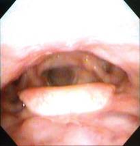
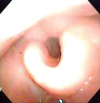
The investigators studied adult patients treated for head and neck cancer at the Vanderbilt-Ingram Cancer Center who were at least 3 months out from the end of their treatment and had no evidence of cancer. External lymphedema was assessed with a clinical exam, using the Foldi scale. Internal lymphedema was assessed with an endoscopic exam, using the Patterson scale for edema of the larynx and pharynx.
The patients were 60 years old, on average. The majority were male (69%) and white (89%). In terms of health behaviors, 66% had a history of smoking and 38% had a history of alcohol use.
In all, 81% of the patients had had locally advanced cancer, and 90% had received at least two treatment modalities. The median time since end of treatment was 20 months.
Study results, reported at the meeting and also recently published, showed that 75% of the patients overall had lymphedema of the head and neck; of those with lymphedema (61 out of 81), 10% had only the external kind, 39% had only the internal kind, and 51% had both (J. Pain Symptom Manage. 2011 July 29 [doi:10.1016/j.jpainsymman.2011.03.019]).
The type and severity of lymphedema were associated with both physical and psychological symptoms, Dr. Deng reported.
As the severity of lymphedema increased, patients were significantly more likely to report difficulty swallowing, issues with mucus or dry mouth, nutritional problems, pain, and voice problems (P = .001 to P = .047, depending on the type of lymphedema and the specific symptom).
Additionally, increasing severity was associated with poorer body image (P = .028 to P = .049). "However, there was no statistically significant relationship between lymphedema and anxiety and depressive symptoms," she noted.
Lymphedema severity was also associated with hearing deficits, limitation of neck range of motion, and impaired quality of life (P = .004 to P = .045).
Analyses identified certain disease and treatment-related factors to be risk factors for the development of lymphedema, according to Dr. Deng.
Namely, patients were more likely to develop lymphedema if they had pharyngeal tumors; were a shorter time out from the end of treatment; received a high total dose of radiation, mirroring what has been seen in breast cancer; received a greater number of treatment modalities, a marker of treatment intensity; or had combined surgery and radiation therapy – specifically, either surgery plus postoperative radiation, or salvage surgery within the irradiated field – as compared with surgery alone.
The higher risk with shorter time since surgery hints that the lymphatics in the area may undergo regeneration over time, she speculated. "This was identified in the murine tail [model of lymphedema]; however, we didn’t know whether or not this phenomenon or similar exists in the head and neck cancer population."
None of the demographic factors or health behaviors assessed were found to be risk factors for the development of lymphedema. But the lack of association between smoking and alcohol consumption and lymphedema may have been related to the fact that patients were asked whether they smoked or drank but not the intensity, or to the cross-sectional nature of the study, Dr. Deng noted. "In the future, longitudinal study is needed to examine whether or not these are risk factors," she said.
Dr. Deng reported that she had no conflicts of interest related to the study. Reed Medical Education and this news organization are owned by Reed Elsevier.
SAN FRANCISCO – Lymphedema is highly common and a source of considerable morbidity among patients who undergo treatment for head and neck cancer, finds a cross-sectional study among 103 survivors.
Fully three-fourths had developed some degree of lymphedema, according to results presented at the annual Oncology Congress presented by Reed Medical Education. The more severe it was, the more likely patients were to have symptoms, functional impairments, and poorer quality of life.
Disease and treatment-related factors such as high radiation dose and combined surgery and radiation therapy were risk factors for the development of lymphedema.
"This is the first study that we are aware of in the United States of this depth to systematically examine lymphedema" in this population, noted lead investigator Jie Deng, Ph.D., R.N., O.C.N., a postdoctoral fellow at the Vanderbilt University, Nashville, Tenn.
"Health care professionals should be aware that lymphedema is a frequent late effect in the head and neck cancer population," she advised. "We need to educate patients about the risk of lymphedema prior to treatment, during treatment, and posttreatment, and we need to conduct external and internal examinations to evaluate related signs and symptoms at each clinic visit."
Patients found to have any signs or symptoms should be referred for lymphedema assessment. Furthermore, "it’s very important we have very detailed documentation so we can follow up on patients’ treatment effect and also identify potential issues in this population," Dr. Deng stressed. "An interdisciplinary approach is needed to best manage lymphedema."
She and her colleagues are now evaluating interventions to treat head and neck lymphedema. Manual lymphatic drainage is one possibility. Elevating the head of the bed at night is another, as anecdotal comments suggest that symptoms worsen in the recumbent position.
"Patients will mention in the morning they feel more tightness, more fluid accumulated in the submental area; around noontime or afternoon, they feel it has drained by itself," she explained.
There are more than half a million survivors of head and neck cancer in the United States, according to Dr. Deng. As a result of their disease and its treatment, these patients can develop both external lymphedema, causing symptoms such as facial puffiness, and internal lymphedema, causing issues such as epiglottal swelling.
The investigators studied adult patients treated for head and neck cancer at the Vanderbilt-Ingram Cancer Center who were at least 3 months out from the end of their treatment and had no evidence of cancer. External lymphedema was assessed with a clinical exam, using the Foldi scale. Internal lymphedema was assessed with an endoscopic exam, using the Patterson scale for edema of the larynx and pharynx.
The patients were 60 years old, on average. The majority were male (69%) and white (89%). In terms of health behaviors, 66% had a history of smoking and 38% had a history of alcohol use.
In all, 81% of the patients had had locally advanced cancer, and 90% had received at least two treatment modalities. The median time since end of treatment was 20 months.
Study results, reported at the meeting and also recently published, showed that 75% of the patients overall had lymphedema of the head and neck; of those with lymphedema (61 out of 81), 10% had only the external kind, 39% had only the internal kind, and 51% had both (J. Pain Symptom Manage. 2011 July 29 [doi:10.1016/j.jpainsymman.2011.03.019]).
The type and severity of lymphedema were associated with both physical and psychological symptoms, Dr. Deng reported.
As the severity of lymphedema increased, patients were significantly more likely to report difficulty swallowing, issues with mucus or dry mouth, nutritional problems, pain, and voice problems (P = .001 to P = .047, depending on the type of lymphedema and the specific symptom).
Additionally, increasing severity was associated with poorer body image (P = .028 to P = .049). "However, there was no statistically significant relationship between lymphedema and anxiety and depressive symptoms," she noted.
Lymphedema severity was also associated with hearing deficits, limitation of neck range of motion, and impaired quality of life (P = .004 to P = .045).
Analyses identified certain disease and treatment-related factors to be risk factors for the development of lymphedema, according to Dr. Deng.
Namely, patients were more likely to develop lymphedema if they had pharyngeal tumors; were a shorter time out from the end of treatment; received a high total dose of radiation, mirroring what has been seen in breast cancer; received a greater number of treatment modalities, a marker of treatment intensity; or had combined surgery and radiation therapy – specifically, either surgery plus postoperative radiation, or salvage surgery within the irradiated field – as compared with surgery alone.
The higher risk with shorter time since surgery hints that the lymphatics in the area may undergo regeneration over time, she speculated. "This was identified in the murine tail [model of lymphedema]; however, we didn’t know whether or not this phenomenon or similar exists in the head and neck cancer population."
None of the demographic factors or health behaviors assessed were found to be risk factors for the development of lymphedema. But the lack of association between smoking and alcohol consumption and lymphedema may have been related to the fact that patients were asked whether they smoked or drank but not the intensity, or to the cross-sectional nature of the study, Dr. Deng noted. "In the future, longitudinal study is needed to examine whether or not these are risk factors," she said.
Dr. Deng reported that she had no conflicts of interest related to the study. Reed Medical Education and this news organization are owned by Reed Elsevier.
FROM THE ANNUAL ONCOLOGY CONGRESS
Major Finding: Fully 75% of patients had lymphedema. The severity of lymphedema was associated with symptoms, functional impairments, and poorer quality of life.
Data Source: A descriptive cross-sectional study among a convenience sample of 103 patients treated for head and neck cancer.
Disclosures: Dr. Deng reported that she had no conflicts of interest related to the study.
Novel Therapies Put Multiple Myeloma 'On the Ropes'
SAN FRANCISCO – A sweep of new agents are poised to deliver what could be a knock-out blow to multiple myeloma, according to the director of the myeloma program at the University of California, San Francisco.
Some are second- or third-generation agents in a mainstay class that appear to have less toxicity than and/or overcome resistance to their predecessors, Dr. Jeffrey L. Wolf said at the annual Oncology Congress here. Others come from classes not previously used in this disease.

"There is a rush to develop new drugs in myeloma," Dr. Wolf told attendees. "We [understand] some mechanisms that the disease seems to favor, so we can interfere with those."
The prospects, in turn, are excellent: "We have made such tremendous headway in myeloma, except for those exceptional cases with 17p deletions and some other adverse prognostic features," he said. "As a disease, it seems to be on the ropes."
A Less-Toxic Proteasome Inhibitor
The first-generation proteasome inhibitor bortezomib (Velcade) improves myeloma outcomes, but maximizing its benefit will require addressing the peripheral neuropathy that limits its use. Three strategies may lessen this toxicity without compromising efficacy, Dr. Wolf suggested: modestly reducing the standard dose, adjusting the schedule from twice to once weekly, and altering the route of administration from intravenous to subcutaneous.
For example, in patients with pretreated myeloma, giving bortezomib subcutaneously instead of intravenously results in an identical overall response rate of 52% (Lancet Oncol. 2011;12:431-40). But there are significant reductions in rates of peripheral neuropathy of any grade (38% vs. 53%) and grade 3 or higher (6% vs. 16%).
"Practically everybody that we see now at UCSF gets subcutaneous [bortezomib]," Dr. Wolf said. It’s a great way to go back and treat patients who maybe otherwise have stopped their therapy because of their neuropathy."
Carfilzomib, an investigational next-generation proteasome inhibitor in phase III trials, is showing good antimyeloma activity and a low rate of peripheral neuropathy. Among patients with pretreated, relapsed, or refractory disease, carfilzomib monotherapy achieved overall response rates of 42% to 53% in a bortezomib-naive group (ASCO 2011 annual meeting. Abstract 8026) and 21% in a bortezomib-exposed group (Haematologica 2010;95:452 Abstract 1099). Median time to progression was at least 8 months for both.
Dr. Wolf said that unpublished data suggest that the response rate was still 17% specifically among patients who had had progression on bortezomib, "so there appears to be some activity in patients who are already refractory to a prior proteasome inhibitor."
Meanwhile, the rates of grade 1/2 and grade 3 peripheral neuropathy were 6% and 1%, respectively. And only a single patient of the 155 had to stop treatment because of this adverse effect.
When carfilzomib is combined with lenalidomide-dexamethasone (the so-called CRd regimen), the overall response rate in patients with pretreated, relapsed, and refractory disease is 78%, and the rate of complete or near complete response is 24% (ASCO 2011 meeting. Abstract 8025).
And, in newly diagnosed myeloma, among 18 patients receiving eight cycles of CRd, the overall response rate was 100%, and the rate of stringent-complete, complete, or near-complete response was 61% (2011 International Myeloma Workshop. Poster P-253). "This is very, very exciting—I don’t think we’ve seen this in any other combination," Dr. Wolf commented. "But these are small numbers of patients; we still need to increase the numbers of patients studied with this combination."
Bortezomib and carfilzomib may soon have company from several investigational proteasome inhibitors available in oral formulations that have shown promise in preclinical testing or have advanced to clinical trials: CEP-18770 (Cephalon), ONX-0912 (Onyx), and MLN-9708 (Millenium).
A Third-Generation IMiD in Trials
Pomalidomide, a third-generation immunomodulatory drug (IMiD), coming after thalidomide (Thalomid), and lenalidomide (Revlimid), is also showing good antimyeloma activity in clinical trials, according to Dr. Wolf.
Among patients with pretreated myeloma, the rate of partial or better response when pomalidomide is combined with dexamethasone has ranged from 25% to 42%, depending on the trial and patient population. Adverse events are primarily hematologic.
And in patients who have previously received lenalidomide, the response rate is similar, at 35% (ASCO 2011 annual meeting. Abstract 8067). "So, as with carfilzomib, where there appear to be responses in patients who have prior resistance to bortezomib, it appears that pomalidomide can give us responses in patients who have already had resistance to lenalidomide," he said.
HDAC Inhibitors Show Activity
The histone deacetylase (HDAC) inhibitor vorinostat (Zolinza) is approved for treatment of lymphoma. But it is being tested in clinical trials for myeloma, in combination therapy, with promising results, according to Dr. Wolf. Overall response rates have ranged from 40% to 94%, depending on the patient population and combination.
Similarly, the HDAC inhibitor romidepsin (Istodax) is approved for lymphoma treatment but is also being studied for antimyeloma activity. And panobinostat, an investigational member of this drug class, is being evaluated as a component of combination therapy in phase II and III myeloma trials.
Monoclonal Antibodies Tested
Elotuzumab is an investigational monoclonal antibody directed against CS1, a glycoprotein that is highly expressed on the surface of plasma cells and implicated in myeloma pathogenesis.
In a phase I trial among patients with relapsed or refractory myeloma, the combination of elotuzumab with lenalidomide and low-dose dexamethasone yielded an overall response rate of 82% (ASCO 2011 meeting. Abstract 8076). The rate was 83% among the subset of patients whose disease was refractory to the most recent therapy and 95% among the subset of lenalidomide-naive patients.
The combination of elotuzumab with bortezomib has also been tested in patients with relapsed or refractory myeloma. But the overall response rate with this combination was less impressive, at 48% (ASH 2010 meeting. Abstract 3023).
Other Agents and Pathways
Several other agents are being eyed for roles in myeloma therapy as well. They include bendamustine (Treanda), an old drug initially revived for lymphoma treatment; aurora kinase inhibitors (for example, MLN-8237); and inhibitors of the mammalian target of rapamycin, or mTOR (for example, INK-128).
Additionally, there is considerable interest in agents that target fibroblast growth factor receptor 3 (FGFR3) for one subgroup. "In patients with the 4;14 translocation, FGFR3 is overexpressed," Dr. Wolf explained. "Finding an inhibitor for that or a direct antibody ... may be quite effective in those patients."
Investigators are also assessing the impact of targeting certain signaling pathways, such as the Jak/Stat pathway and the AKT pathway. For instance, a phase III trial is testing perifosine, an investigational AKT inhibitor, in combination therapy among patients with relapsed or refractory myeloma (NCT01002248).
The Oncology Congress is presented by Reed Medical Education. Reed Medical Education and this news organization are owned by Reed Elsevier Inc.
Dr. Wolf disclosed that he is on the speakers bureaus of Millenium, Celgene, and Ortho-Biotech, and is a consultant to and speaker for Onyx.
SAN FRANCISCO – A sweep of new agents are poised to deliver what could be a knock-out blow to multiple myeloma, according to the director of the myeloma program at the University of California, San Francisco.
Some are second- or third-generation agents in a mainstay class that appear to have less toxicity than and/or overcome resistance to their predecessors, Dr. Jeffrey L. Wolf said at the annual Oncology Congress here. Others come from classes not previously used in this disease.
"There is a rush to develop new drugs in myeloma," Dr. Wolf told attendees. "We [understand] some mechanisms that the disease seems to favor, so we can interfere with those."
The prospects, in turn, are excellent: "We have made such tremendous headway in myeloma, except for those exceptional cases with 17p deletions and some other adverse prognostic features," he said. "As a disease, it seems to be on the ropes."
A Less-Toxic Proteasome Inhibitor
The first-generation proteasome inhibitor bortezomib (Velcade) improves myeloma outcomes, but maximizing its benefit will require addressing the peripheral neuropathy that limits its use. Three strategies may lessen this toxicity without compromising efficacy, Dr. Wolf suggested: modestly reducing the standard dose, adjusting the schedule from twice to once weekly, and altering the route of administration from intravenous to subcutaneous.
For example, in patients with pretreated myeloma, giving bortezomib subcutaneously instead of intravenously results in an identical overall response rate of 52% (Lancet Oncol. 2011;12:431-40). But there are significant reductions in rates of peripheral neuropathy of any grade (38% vs. 53%) and grade 3 or higher (6% vs. 16%).
"Practically everybody that we see now at UCSF gets subcutaneous [bortezomib]," Dr. Wolf said. It’s a great way to go back and treat patients who maybe otherwise have stopped their therapy because of their neuropathy."
Carfilzomib, an investigational next-generation proteasome inhibitor in phase III trials, is showing good antimyeloma activity and a low rate of peripheral neuropathy. Among patients with pretreated, relapsed, or refractory disease, carfilzomib monotherapy achieved overall response rates of 42% to 53% in a bortezomib-naive group (ASCO 2011 annual meeting. Abstract 8026) and 21% in a bortezomib-exposed group (Haematologica 2010;95:452 Abstract 1099). Median time to progression was at least 8 months for both.
Dr. Wolf said that unpublished data suggest that the response rate was still 17% specifically among patients who had had progression on bortezomib, "so there appears to be some activity in patients who are already refractory to a prior proteasome inhibitor."
Meanwhile, the rates of grade 1/2 and grade 3 peripheral neuropathy were 6% and 1%, respectively. And only a single patient of the 155 had to stop treatment because of this adverse effect.
When carfilzomib is combined with lenalidomide-dexamethasone (the so-called CRd regimen), the overall response rate in patients with pretreated, relapsed, and refractory disease is 78%, and the rate of complete or near complete response is 24% (ASCO 2011 meeting. Abstract 8025).
And, in newly diagnosed myeloma, among 18 patients receiving eight cycles of CRd, the overall response rate was 100%, and the rate of stringent-complete, complete, or near-complete response was 61% (2011 International Myeloma Workshop. Poster P-253). "This is very, very exciting—I don’t think we’ve seen this in any other combination," Dr. Wolf commented. "But these are small numbers of patients; we still need to increase the numbers of patients studied with this combination."
Bortezomib and carfilzomib may soon have company from several investigational proteasome inhibitors available in oral formulations that have shown promise in preclinical testing or have advanced to clinical trials: CEP-18770 (Cephalon), ONX-0912 (Onyx), and MLN-9708 (Millenium).
A Third-Generation IMiD in Trials
Pomalidomide, a third-generation immunomodulatory drug (IMiD), coming after thalidomide (Thalomid), and lenalidomide (Revlimid), is also showing good antimyeloma activity in clinical trials, according to Dr. Wolf.
Among patients with pretreated myeloma, the rate of partial or better response when pomalidomide is combined with dexamethasone has ranged from 25% to 42%, depending on the trial and patient population. Adverse events are primarily hematologic.
And in patients who have previously received lenalidomide, the response rate is similar, at 35% (ASCO 2011 annual meeting. Abstract 8067). "So, as with carfilzomib, where there appear to be responses in patients who have prior resistance to bortezomib, it appears that pomalidomide can give us responses in patients who have already had resistance to lenalidomide," he said.
HDAC Inhibitors Show Activity
The histone deacetylase (HDAC) inhibitor vorinostat (Zolinza) is approved for treatment of lymphoma. But it is being tested in clinical trials for myeloma, in combination therapy, with promising results, according to Dr. Wolf. Overall response rates have ranged from 40% to 94%, depending on the patient population and combination.
Similarly, the HDAC inhibitor romidepsin (Istodax) is approved for lymphoma treatment but is also being studied for antimyeloma activity. And panobinostat, an investigational member of this drug class, is being evaluated as a component of combination therapy in phase II and III myeloma trials.
Monoclonal Antibodies Tested
Elotuzumab is an investigational monoclonal antibody directed against CS1, a glycoprotein that is highly expressed on the surface of plasma cells and implicated in myeloma pathogenesis.
In a phase I trial among patients with relapsed or refractory myeloma, the combination of elotuzumab with lenalidomide and low-dose dexamethasone yielded an overall response rate of 82% (ASCO 2011 meeting. Abstract 8076). The rate was 83% among the subset of patients whose disease was refractory to the most recent therapy and 95% among the subset of lenalidomide-naive patients.
The combination of elotuzumab with bortezomib has also been tested in patients with relapsed or refractory myeloma. But the overall response rate with this combination was less impressive, at 48% (ASH 2010 meeting. Abstract 3023).
Other Agents and Pathways
Several other agents are being eyed for roles in myeloma therapy as well. They include bendamustine (Treanda), an old drug initially revived for lymphoma treatment; aurora kinase inhibitors (for example, MLN-8237); and inhibitors of the mammalian target of rapamycin, or mTOR (for example, INK-128).
Additionally, there is considerable interest in agents that target fibroblast growth factor receptor 3 (FGFR3) for one subgroup. "In patients with the 4;14 translocation, FGFR3 is overexpressed," Dr. Wolf explained. "Finding an inhibitor for that or a direct antibody ... may be quite effective in those patients."
Investigators are also assessing the impact of targeting certain signaling pathways, such as the Jak/Stat pathway and the AKT pathway. For instance, a phase III trial is testing perifosine, an investigational AKT inhibitor, in combination therapy among patients with relapsed or refractory myeloma (NCT01002248).
The Oncology Congress is presented by Reed Medical Education. Reed Medical Education and this news organization are owned by Reed Elsevier Inc.
Dr. Wolf disclosed that he is on the speakers bureaus of Millenium, Celgene, and Ortho-Biotech, and is a consultant to and speaker for Onyx.
EXPERT ANALYSIS FROM THE ANNUAL ONCOLOGY CONGRESS
Bioengineered, Sirolimus-Eluting Stent Found ‘Safe,’ and ‘Effective’
SAN FRANCISCO – A new stent that combines antibody bioengineering with drug elution technology works at least as well as a paclitaxel-eluting stent for treating coronary artery lesions, the randomized REMEDEE trial showed.
Investigators led by Dr. Michael Haude found that the 9-month in-stent late lumen loss was statistically noninferior with the combination stent, which elutes sirolimus to inhibit cellular proliferation and has surface antibodies to promote endothelial coverage. The absolute difference between stents was only 0.05 mm, according to data reported at Transcatheter Cardiovascular Therapeutics 2011, which was sponsored by the Cardiovascular Research Foundation.

There were no stent thromboses or cardiac deaths with either stent. The two stents also yielded similar 9-month rates of clinically driven target lesion revascularization, myocardial infarction, and major adverse cardiovascular events.
"In this first-in-man study, the [combination stent] effectively controlled neointimal proliferation," Dr. Haude commented in a press conference. "I think we can conclude that [it] is shown to be effective and safe in this trial."
Given that both stents eluted an antiproliferative agent, patients in the trial received the usual antiplatelet therapy for a year to be on the safe side, said Dr. Haude, professor and director at Lukas Hospital Neuss (Germany).
The long-term goal "is to document that, by having this more secure, more firm, more homogeneous coverage of the stent struts [by endothelium], at the end of the day, you will be able to shorten dual antiplatelet therapy," he said. Indeed, intravascular ultrasound images and optical coherence tomography (OCT) images in a subset of patients showed that the combination stent achieved better coverage than the paclitaxel-eluting stent at 9 months.
"There was a little bit more late [lumen] loss than you would have expected with a Cypher or a Xience or one of the other stents."
Additionally, an ongoing, related trial, REMEDEE-OCT, is comparing the combination stent with the newer everolimus-eluting stents, with special attention to strut coverage at 60 days.
In an interview, Dr. John M. Hodgson, chairman of the department of cardiology at Geisinger Health System in Wilkes-Barre, Pa., commented "We’re clearly in a phase of stent development where we are trying to tailor specific aspects of it, with the feeling that less polymer is better, the feeling that something that may help reendothelialize the stent is better. And I think this trial shows that this stent has some promise for that."
He pointed out that the trial was not powered to evaluate end points other than basic vascular indicators of stent function. On that level, Dr. Hodgson said, "it showed that it’s effective compared to a stent that I think all of us would agree now is pretty obsolete."
Another potential issue was the magnitude of late lumen loss with the combination stent. "There was a little bit more late loss than you would have expected with a Cypher or a Xience or one of the other stents," he said. "So whether that little bit more late loss ends up being a problem in the future, we’ll just have to wait for the big studies."
The combination stent has two features designed to address specific known issues with stenting and current stent technologies, Dr. Haude explained. The first is an abluminal, biodegradable, sirolimus-eluting matrix to reduce cellular proliferation and hence restenosis; the second is an anti-CD34 antibody coating to capture circulating endothelial progenitor cells, promoting endothelial coverage and reducing the risk of late thrombosis.
The combination stent’s drug content is approximately half that of the Cypher stent, but the release profile is similar, he noted.
A total of 183 patients in the trial had single de novo native coronary artery lesions no greater than 20 mm in length. They were randomized 2:1 to receive the combination stent (Combo Dual Therapy Stent, manufactured by OrbusNeich) or a paclitaxel-eluting stent (Taxus Liberté, manufactured by Boston Scientific).
Results of the trial showed that the combination and paclitaxel-eluting stent groups had similar, very high, and statistically indistinguishable rates of device, lesion, and procedure success.
The angiographic 9-month in-stent late lumen loss was 0.39 mm with the combination stent and 0.44 mm with the paclitaxel-eluting stent. Both the absolute difference of 0.05 mm and the upper bound of the 95% confidence interval were well within the predefined margin of 0.20 mm for noninferiority (P for noninferiority = .001; P for superiority = .55).
The combination and paclitaxel-eluting stent groups had statistically indistinguishable rates of angiographic in-stent restenosis (5.5% vs. 9.6%, respectively) and in-segment restenosis (8.3% vs. 13.5%).
No patient in either group experienced stent thrombosis or died from cardiac causes. The two groups had similar rates of clinically driven target lesion revascularization (5.2% vs. 9.5%), myocardial infarction (2.4% vs. 1.7%), and major adverse cardiovascular events (8.7% vs. 11.0%).
Dr. Haude reported that he receives research or grant support from OrbusNeich and Biotronik and consultant fees and honoraria from Medtronic, Abbott Vascular, and Volcano Corp. Dr. Hodgson reported that he receives research or grant support from Boston Scientific, Volcano Corp., and St. Jude Medical, and that he is a consultant to, is a speaker for, or receives honoraria from Volcano Corp. The trial was sponsored by OrbusNeich.
SAN FRANCISCO – A new stent that combines antibody bioengineering with drug elution technology works at least as well as a paclitaxel-eluting stent for treating coronary artery lesions, the randomized REMEDEE trial showed.
Investigators led by Dr. Michael Haude found that the 9-month in-stent late lumen loss was statistically noninferior with the combination stent, which elutes sirolimus to inhibit cellular proliferation and has surface antibodies to promote endothelial coverage. The absolute difference between stents was only 0.05 mm, according to data reported at Transcatheter Cardiovascular Therapeutics 2011, which was sponsored by the Cardiovascular Research Foundation.
There were no stent thromboses or cardiac deaths with either stent. The two stents also yielded similar 9-month rates of clinically driven target lesion revascularization, myocardial infarction, and major adverse cardiovascular events.
"In this first-in-man study, the [combination stent] effectively controlled neointimal proliferation," Dr. Haude commented in a press conference. "I think we can conclude that [it] is shown to be effective and safe in this trial."
Given that both stents eluted an antiproliferative agent, patients in the trial received the usual antiplatelet therapy for a year to be on the safe side, said Dr. Haude, professor and director at Lukas Hospital Neuss (Germany).
The long-term goal "is to document that, by having this more secure, more firm, more homogeneous coverage of the stent struts [by endothelium], at the end of the day, you will be able to shorten dual antiplatelet therapy," he said. Indeed, intravascular ultrasound images and optical coherence tomography (OCT) images in a subset of patients showed that the combination stent achieved better coverage than the paclitaxel-eluting stent at 9 months.
"There was a little bit more late [lumen] loss than you would have expected with a Cypher or a Xience or one of the other stents."
Additionally, an ongoing, related trial, REMEDEE-OCT, is comparing the combination stent with the newer everolimus-eluting stents, with special attention to strut coverage at 60 days.
In an interview, Dr. John M. Hodgson, chairman of the department of cardiology at Geisinger Health System in Wilkes-Barre, Pa., commented "We’re clearly in a phase of stent development where we are trying to tailor specific aspects of it, with the feeling that less polymer is better, the feeling that something that may help reendothelialize the stent is better. And I think this trial shows that this stent has some promise for that."
He pointed out that the trial was not powered to evaluate end points other than basic vascular indicators of stent function. On that level, Dr. Hodgson said, "it showed that it’s effective compared to a stent that I think all of us would agree now is pretty obsolete."
Another potential issue was the magnitude of late lumen loss with the combination stent. "There was a little bit more late loss than you would have expected with a Cypher or a Xience or one of the other stents," he said. "So whether that little bit more late loss ends up being a problem in the future, we’ll just have to wait for the big studies."
The combination stent has two features designed to address specific known issues with stenting and current stent technologies, Dr. Haude explained. The first is an abluminal, biodegradable, sirolimus-eluting matrix to reduce cellular proliferation and hence restenosis; the second is an anti-CD34 antibody coating to capture circulating endothelial progenitor cells, promoting endothelial coverage and reducing the risk of late thrombosis.
The combination stent’s drug content is approximately half that of the Cypher stent, but the release profile is similar, he noted.
A total of 183 patients in the trial had single de novo native coronary artery lesions no greater than 20 mm in length. They were randomized 2:1 to receive the combination stent (Combo Dual Therapy Stent, manufactured by OrbusNeich) or a paclitaxel-eluting stent (Taxus Liberté, manufactured by Boston Scientific).
Results of the trial showed that the combination and paclitaxel-eluting stent groups had similar, very high, and statistically indistinguishable rates of device, lesion, and procedure success.
The angiographic 9-month in-stent late lumen loss was 0.39 mm with the combination stent and 0.44 mm with the paclitaxel-eluting stent. Both the absolute difference of 0.05 mm and the upper bound of the 95% confidence interval were well within the predefined margin of 0.20 mm for noninferiority (P for noninferiority = .001; P for superiority = .55).
The combination and paclitaxel-eluting stent groups had statistically indistinguishable rates of angiographic in-stent restenosis (5.5% vs. 9.6%, respectively) and in-segment restenosis (8.3% vs. 13.5%).
No patient in either group experienced stent thrombosis or died from cardiac causes. The two groups had similar rates of clinically driven target lesion revascularization (5.2% vs. 9.5%), myocardial infarction (2.4% vs. 1.7%), and major adverse cardiovascular events (8.7% vs. 11.0%).
Dr. Haude reported that he receives research or grant support from OrbusNeich and Biotronik and consultant fees and honoraria from Medtronic, Abbott Vascular, and Volcano Corp. Dr. Hodgson reported that he receives research or grant support from Boston Scientific, Volcano Corp., and St. Jude Medical, and that he is a consultant to, is a speaker for, or receives honoraria from Volcano Corp. The trial was sponsored by OrbusNeich.
FROM TRANSCATHETER CARDIOVASCULAR THERAPEUTICS 2011
Major Finding: The 9-month in-stent late lumen loss with the combination stent was not inferior to that with the paclitaxel-eluting stent (0.39 vs. 0.44 mm).
Data Source: A randomized noninferiority trial among 183 patients having single de novo native coronary artery lesions (the REMEDEE trial).
Disclosures: Dr. Haude reported that he receives research or grant support from OrbusNeich and Biotronik and consultant fees and honoraria from Medtronic, Abbott Vascular, and Volcano Corp. Dr. Hodgson reported that he receives research or grant support from Boston Scientific, Volcano Corp., and St. Jude Medical, and that he is a consultant to, is a speaker for, or receives honoraria from Volcano Corp. The trial was sponsored by OrbusNeich.
Rotablation in Complex Calcified Lesions Doesn't Improve Outcomes
SAN FRANCISCO – Although there are compelling reasons to treat complex calcified coronary lesions with rotablation instead of standard balloon dilatation before implanting drug-eluting stents, this strategy does not improve vascular or clinical outcomes in the long run, according to the results of the randomized ROTAXUS trial.
The results showed that rotablation was more effective at improving lumen dimensions early on, but this was offset by a subsequent proliferative response. Late lumen loss at 9 months was 0.44 mm with rotablation (also known as rotational atherectomy) and stenting vs. 0.31 mm with balloon dilatation and stenting, a significant difference. Rates of target lesion revascularization and other cardiovascular events did not differ significantly between groups.
"Superior acute gain ... after rotablation was counterbalanced by increased late loss, resulting in a neutral effect on restenosis," lead investigator Dr. Gert Richardt said at Transcatheter Cardiovascular Therapeutics 2011, sponsored by the Cardiovascular Research Foundation.
"So balloon dilatation with provisional rotablation – that is my suggestion – prior to stenting remains the default strategy for complex calcified lesions even in the drug-eluting stent era," he said.
Dr. Roxana Mehran, professor of medicine at Mount Sinai Medical Center in New York, noted that rotablation remains helpful, at least from a logistic point of view, in bad lesions. "It’s important to note that rotational atherectomy still does have a place in our armamentarium, but it would be in the severely calcified lesions and in complex morphologies where I think it would make our procedure easier," she explained. "But if it is to reduce late loss," this study, "in a randomized fashion, did not show that."
Discussant Dr. Ron Waksman, director of experimental angioplasty and emerging technologies at the Washington Hospital Center in Hyattsville, Md., agreed, and also expressed concern that the trial’s findings might be interpreted as cause to abandon rotablation.
"You don’t do rotablation to [address] the late loss issue, the restenosis. It’s to facilitate stent deployment," he asserted. "In my view, we are underusing rotablation today, and we should use more of this. I’m just afraid that people will interpret this [trial as indicating] that you can do without a rotablator, and that’s not the message. ... We should actually reconsider more usage of rotablation to facilitate stent deployment and [reduce] thromboembolic complications of the procedure."
"Rotablation does not increase the efficacy of drug-eluting stents in calcified lesions."
"We nowadays treat more and more elderly patients with complex and severely calcified lesions," noted Dr. Richardt, who is a professor at the Heart Center, Segeberger Kliniken, in Bad Segeberg, Germany. And there is good rationale for using rotablation before implantation of drug-eluting stents for these lesions, he said.
Calcified lesions not only damage the stent, stripping off its drug-containing polymer coat, but may also impair outward diffusion of the drug. Ablating these lesions facilitates stent deployment and expansion, and possibly also effectiveness.
To be eligible for the trial, patients had to have angina (stable or unstable) and coronary artery disease, plus both of two angiographic criteria (a de novo lesion in a native coronary artery and moderate to severe calcification) and at least one of several others (an ostial location, a bifurcation lesion, or a long lesion).
The 240 patients enrolled in the study were assigned in equal numbers to either rotablation or balloon dilatation, followed by implantation of paclitaxel-eluting stents (Taxus, manufactured by Boston Scientific).
Trial results showed that from the procedural perspective, the rotablation group was more likely to be treated with a 7-French guiding catheter (84% vs. 28%) and had a lower maximal predilatation balloon pressure (13.6 vs. 15.8 atm), Dr. Richardt reported.
In the rotablation group, the maximum burr size was 1.5 mm. "It was mainly the plaque modification concept that we followed, with small burrs, and we usually used just one burr on the study," he commented. The groups were similar with respect to numbers of stents used and the lengths of stented vessels, as well as the use of balloon dilatation after stenting.
Fluoroscopy time was somewhat longer with rotablation, but patients in the two groups received the same amount of contrast dye, according to Dr. Richardt.
The rotablation and balloon dilatation groups had similarly high rates of angiographic success (97% each) and low rates of stent loss (0% and 3%), but the former had a lower rate of crossover (4% vs. 12%).
Despite a greater early gain in lumen dimensions, the rate of in-stent late lumen loss at 9 months – the trial’s primary end point – was higher with rotablation than with balloon dilatation (0.44 vs. 0.31 mm).
The two groups had statistically indistinguishable rates of clinical outcomes such as myocardial infarction, major adverse cardiovascular events, and death. The target lesion revascularization rate – a clinical indicator of restenosis – was about 12% in each group. There was only a single case of definite stent thrombosis, seen in the rotablation group.
"Rotablation and paclitaxel-eluting stent implantation was not superior to balloon dilatation and paclitaxel-eluting stent implantation in reducing the primary end point of late lumen loss at 9 months, indicating that rotablation does not increase the efficacy of drug-eluting stents in calcified lesions," he concluded.
The trial was sponsored by Herz-Kreislauf-Zentrum Segeberger Kliniken GmbH. Dr. Richardt reported that he is a consultant to, is a speaker for, or receives honoraria from Abbott Vascular and Boston Scientific. Dr. Mehran reported that she receives research or grant support from Bristol-Myers Squibb and The Medicines Company; is a consultant to, is a speaker for, or receives honoraria from AstraZeneca and Ortho-McNeil; and receives other, noncategorized financial support from Abbott Vascular. Dr. Waksman reported that he has no relevant conflicts of interest.
FROM TRANSCATHETER CARDIOVASCULAR THERAPEUTICS 2011
Major Finding: In-stent late lumen loss at 9 months was 0.44 mm with rotablation plus paclitaxel-eluting stents vs. 0.31 mm with standard balloon dilatation plus paclitaxel-eluting stents.
Data Source: A randomized trial of 240 patients with angina and complex calcified coronary lesions (the ROTAXUS trial)
Disclosures: The trial was sponsored by Herz-Kreislauf-Zentrum Segeberger Kliniken GmbH. Dr. Richardt reported that he is a consultant to, is a speaker for, or receives honoraria from Abbott Vascular and Boston Scientific. Dr. Mehran reported that she receives research or grant support from Bristol-Myers Squibb and The Medicines Company; is a consultant to, is a speaker for, or receives honoraria from AstraZeneca and Ortho-McNeil; and receives other, noncategorized financial support from Abbott Vascular. Dr. Waksman reported that he has no relevant conflicts of interest.
Zotarolimus-, Everolimus-Eluting Stents Found Equally Safe, Effective
SAN FRANCISCO – Zotarolimus-eluting stents are generally as safe and effective as everolimus-eluting stents for coronary stenting in the real-world setting, according to results from a randomized, noninferiority phase IV trial comparing them head to head.
Among the 1,391 patients studied, all of whom were treated at the tertiary-care Thoraxcentrum Twente in the Netherlands, about 8% experienced events signaling a failure of the target vessel within a year, no matter which of these second-generation drug-eluting stents they received.
The difference between groups was well within the predefined boundary for noninferiority, lead investigator Dr. Clemens von Birgelen reported at Transcatheter Cardiovascular Therapeutics 2011, which is sponsored by the Cardiovascular Research Foundation. Additionally, both groups had very low and statistically indistinguishable rates of definite or probable stent thrombosis.

The bottom line is that the choice between the two stents will come down to a matter of physicians’ personal preference, he said in an interview. "There are emotional arguments" favoring one stent or the other, Dr. von Birgelen noted. He declined to say whether other factors might give one stent the edge.
The trial’s findings confirm those of the similar Resolute All Comers trial (N. Engl. J. Med. 2010;363:136-46), although that trial was larger and multicenter in nature. Collectively, these data show that "both stents are safe and efficacious, and you can use either one or the other," Dr. von Birgelen maintained. "So the physician has to decide at the end of the day which one to use."
The practice of dilatation after stenting – used in most of the procedures in the new trial – may be important to getting the same results, according to Dr. von Birgelen. "Postdilatation of stents is something to consider in the proximal and mid part [of the lesion] to get good stent apposition, and could be part of the reason why we found such nice results in this study," he explained.
Discussant Dr. Ron Waksman, director of Experimental Angioplasty & Emerging Technologies, Cardiovascular Research Institute in Washington, said, "It’s very hard to differentiate between the stents right now. The data is almost identical ... I think it’s really going to be a question of availability of sizes and lengths, pricing, et cetera, but not in terms of performance of the stent."
Dr. John McB. Hodgson, chairman of the department of cardiology at Geisinger Health System in Wilkes-Barre, Pa., agreed and noted that these second-generation drug-eluting stents are a major technological advance. "Both of these platforms are fantastic compared to what we had 5 years ago. We are splitting hairs – these are both great platforms," he commented.

Patients were eligible for the trial, known as the TWENTE trial and jointly supported by Abbott Vascular and Medtronic, if they had stable angina or a non–ST-elevation acute coronary syndrome plus an indication for implantation of a drug-eluting stent.
The enrolled patients, 82% of all those who were eligible, were randomized in balanced fashion to receive either zotarolimus-eluting stents (Resolute, manufactured by Medtronic and currently investigational in the United States) or everolimus-eluting stents (Xience V, manufactured by Abbott). Patients and data analysts, but not treating clinicians, were blinded to group assignment.
There was no limit on the number of lesions or vessels treated, the lesion length, or the vessel size, noted Dr. von Birgelen, who is a professor at the Thoraxcentrum Twente. All types of lesions, including those in bypass grafts and at bifurcations, were permitted.
The study population was 64 years old on average. About one-fifth of patients had diabetes. The slight majority had acute coronary syndromes. Nearly 25% had multiple vessels treated, and 77% had at least one off-label indication for drug-eluting stent implantation, such as stenting of the left main stem, long lesions, or very small vessels.
Roughly two-thirds of treated lesions were complex, and one-quarter were at bifurcations. The stent was implanted directly, without predilatation of the lesion, in 39% of lesions, a rate similar to that seen in other studies. "Postdilatation, however, was performed particularly often in our center, in 82% of the stented lesions," Dr. von Birgelen noted. "It’s a kind of standard strategy in our center for several years with drug-eluting stents, because we think that the postdilatation leads to better apposition of the stent to the vessel wall, with a better drug transition into the vessel wall, and this could avoid stent thrombosis and other events."
Additionally, use of postdilatation increased as the center began stenting longer lesions, given that coronary vessels naturally taper over their length. "We have to choose a stent of a certain size, and then by postdilatation, try to mimic the biological situation by being more aggressive with postdilatation in the proximal part, somewhat less in the mid part, and then distal," he explained.
Study results showed that both treatment groups had nearly 100% rates of lesion success and 96% rates of procedure success.
In intent-to-treat analyses, the 1-year rate of target vessel failure – a composite of cardiac death, target vessel–related myocardial infarction, and clinically driven target vessel revascularization – was 8.2% with zotarolimus-eluting stents and 8.1% with everolimus-eluting stents. The absolute difference of 0.1% and upper limit of the one-sided 95% confidence interval of 2.53% were well within the predefined noninferiority margin of 4.48% (P for noninferiority = .001; P for superiority = .94).
The findings were similar across patient subgroups stratified by disease and lesion characteristics, and similar for each of the three components of the composite end point individually.
There was an interaction between treatment group and diabetes status (P = .045), but more-detailed analysis failed to show a significant benefit of one stent over the other. "This is a trend which is hypothesis generating and interesting," Dr. von Birgelen commented. "It may raise some more pooling of data and analysis."
The trial’s main results were much the same when the investigators used a broader, patient-oriented composite end point of all deaths, any myocardial infarction, and any revascularization (11.2% vs. 10.5%; P for superiority = .69).
The zotarolimus- and everolimus-eluting stent groups also had very low and statistically indistinguishable rates of definite or probable stent thrombosis (0.86% vs. 1.16%). These rates are "relatively low, certainly when you consider the complexity of patients and lesions that were studied in this trial," he noted.
"Zotarolimus-eluting Resolute stents were noninferior to everolimus-eluting Xience V stents in terms of safety and efficacy for treating real-world patients with a vast majority of complex lesions and off-label indications for drug-eluting stents, which were implanted with liberal use of postdilatation," Dr. von Birgelen concluded.
Follow-up continues, he added. "It makes sense, and we must learn from the first-generation drug-eluting stents that we should follow up these patients longer than just 1 year. There is a 2-year follow-up already running at this moment, and we will follow these patients even further," he said.
Dr. von Birgelen reported that he is a consultant to and has received speaker honoraria and/or traveling expenses from Abbott Vascular, Medtronic, and Boston Scientific. Dr. Waksman reported that he had no relevant conflicts of interest. Dr. Hodgson reported that he receives research or grant support from Boston Scientific, Volcano, and St. Jude Medical, and that he is a consultant to, is a speaker for, or receives honoraria from Volcano.
SAN FRANCISCO – Zotarolimus-eluting stents are generally as safe and effective as everolimus-eluting stents for coronary stenting in the real-world setting, according to results from a randomized, noninferiority phase IV trial comparing them head to head.
Among the 1,391 patients studied, all of whom were treated at the tertiary-care Thoraxcentrum Twente in the Netherlands, about 8% experienced events signaling a failure of the target vessel within a year, no matter which of these second-generation drug-eluting stents they received.
The difference between groups was well within the predefined boundary for noninferiority, lead investigator Dr. Clemens von Birgelen reported at Transcatheter Cardiovascular Therapeutics 2011, which is sponsored by the Cardiovascular Research Foundation. Additionally, both groups had very low and statistically indistinguishable rates of definite or probable stent thrombosis.
The bottom line is that the choice between the two stents will come down to a matter of physicians’ personal preference, he said in an interview. "There are emotional arguments" favoring one stent or the other, Dr. von Birgelen noted. He declined to say whether other factors might give one stent the edge.
The trial’s findings confirm those of the similar Resolute All Comers trial (N. Engl. J. Med. 2010;363:136-46), although that trial was larger and multicenter in nature. Collectively, these data show that "both stents are safe and efficacious, and you can use either one or the other," Dr. von Birgelen maintained. "So the physician has to decide at the end of the day which one to use."
The practice of dilatation after stenting – used in most of the procedures in the new trial – may be important to getting the same results, according to Dr. von Birgelen. "Postdilatation of stents is something to consider in the proximal and mid part [of the lesion] to get good stent apposition, and could be part of the reason why we found such nice results in this study," he explained.
Discussant Dr. Ron Waksman, director of Experimental Angioplasty & Emerging Technologies, Cardiovascular Research Institute in Washington, said, "It’s very hard to differentiate between the stents right now. The data is almost identical ... I think it’s really going to be a question of availability of sizes and lengths, pricing, et cetera, but not in terms of performance of the stent."
Dr. John McB. Hodgson, chairman of the department of cardiology at Geisinger Health System in Wilkes-Barre, Pa., agreed and noted that these second-generation drug-eluting stents are a major technological advance. "Both of these platforms are fantastic compared to what we had 5 years ago. We are splitting hairs – these are both great platforms," he commented.
Patients were eligible for the trial, known as the TWENTE trial and jointly supported by Abbott Vascular and Medtronic, if they had stable angina or a non–ST-elevation acute coronary syndrome plus an indication for implantation of a drug-eluting stent.
The enrolled patients, 82% of all those who were eligible, were randomized in balanced fashion to receive either zotarolimus-eluting stents (Resolute, manufactured by Medtronic and currently investigational in the United States) or everolimus-eluting stents (Xience V, manufactured by Abbott). Patients and data analysts, but not treating clinicians, were blinded to group assignment.
There was no limit on the number of lesions or vessels treated, the lesion length, or the vessel size, noted Dr. von Birgelen, who is a professor at the Thoraxcentrum Twente. All types of lesions, including those in bypass grafts and at bifurcations, were permitted.
The study population was 64 years old on average. About one-fifth of patients had diabetes. The slight majority had acute coronary syndromes. Nearly 25% had multiple vessels treated, and 77% had at least one off-label indication for drug-eluting stent implantation, such as stenting of the left main stem, long lesions, or very small vessels.
Roughly two-thirds of treated lesions were complex, and one-quarter were at bifurcations. The stent was implanted directly, without predilatation of the lesion, in 39% of lesions, a rate similar to that seen in other studies. "Postdilatation, however, was performed particularly often in our center, in 82% of the stented lesions," Dr. von Birgelen noted. "It’s a kind of standard strategy in our center for several years with drug-eluting stents, because we think that the postdilatation leads to better apposition of the stent to the vessel wall, with a better drug transition into the vessel wall, and this could avoid stent thrombosis and other events."
Additionally, use of postdilatation increased as the center began stenting longer lesions, given that coronary vessels naturally taper over their length. "We have to choose a stent of a certain size, and then by postdilatation, try to mimic the biological situation by being more aggressive with postdilatation in the proximal part, somewhat less in the mid part, and then distal," he explained.
Study results showed that both treatment groups had nearly 100% rates of lesion success and 96% rates of procedure success.
In intent-to-treat analyses, the 1-year rate of target vessel failure – a composite of cardiac death, target vessel–related myocardial infarction, and clinically driven target vessel revascularization – was 8.2% with zotarolimus-eluting stents and 8.1% with everolimus-eluting stents. The absolute difference of 0.1% and upper limit of the one-sided 95% confidence interval of 2.53% were well within the predefined noninferiority margin of 4.48% (P for noninferiority = .001; P for superiority = .94).
The findings were similar across patient subgroups stratified by disease and lesion characteristics, and similar for each of the three components of the composite end point individually.
There was an interaction between treatment group and diabetes status (P = .045), but more-detailed analysis failed to show a significant benefit of one stent over the other. "This is a trend which is hypothesis generating and interesting," Dr. von Birgelen commented. "It may raise some more pooling of data and analysis."
The trial’s main results were much the same when the investigators used a broader, patient-oriented composite end point of all deaths, any myocardial infarction, and any revascularization (11.2% vs. 10.5%; P for superiority = .69).
The zotarolimus- and everolimus-eluting stent groups also had very low and statistically indistinguishable rates of definite or probable stent thrombosis (0.86% vs. 1.16%). These rates are "relatively low, certainly when you consider the complexity of patients and lesions that were studied in this trial," he noted.
"Zotarolimus-eluting Resolute stents were noninferior to everolimus-eluting Xience V stents in terms of safety and efficacy for treating real-world patients with a vast majority of complex lesions and off-label indications for drug-eluting stents, which were implanted with liberal use of postdilatation," Dr. von Birgelen concluded.
Follow-up continues, he added. "It makes sense, and we must learn from the first-generation drug-eluting stents that we should follow up these patients longer than just 1 year. There is a 2-year follow-up already running at this moment, and we will follow these patients even further," he said.
Dr. von Birgelen reported that he is a consultant to and has received speaker honoraria and/or traveling expenses from Abbott Vascular, Medtronic, and Boston Scientific. Dr. Waksman reported that he had no relevant conflicts of interest. Dr. Hodgson reported that he receives research or grant support from Boston Scientific, Volcano, and St. Jude Medical, and that he is a consultant to, is a speaker for, or receives honoraria from Volcano.
FROM TRANSCATHETER CARDIOVASCULAR THERAPEUTICS 2011
Major Finding: The 1-year rate of target vessel failure was statistically noninferior with zotarolimus-eluting stents compared with everolimus-eluting stents (8.2% vs. 8.1%).
Data Source: A single-center randomized noninferiority trial among 1,391 patients who had an indication for percutaneous coronary intervention with drug-eluting stent implantation (the TWENTE trial)
Disclosures: The study was jointly supported by Abbott Vascular and Medtronic. Dr. von Birgelen reported that he is a consultant to and has received speaker honoraria and/or traveling expenses from Abbott Vascular, Medtronic, and Boston Scientific. Dr. Waksman reported that he had no relevant conflicts of interest. Dr. Hodgson reported that he receives research or grant support from Boston Scientific, Volcano, and St. Jude Medical, and that he is a consultant to, is a speaker for, or receives honoraria from Volcano.
Drug-Eluting Balloon, Bare Metal Stent Combo Disappoints in STEMI
SAN FRANCISCO – Treating a coronary artery with both a drug-eluting balloon and a bare metal stent is feasible in patients with acute ST-elevation myocardial infarction but does not significantly reduce late lumen loss, compared with bare metals stents alone or with drug-eluting stents alone.
The Drug-Eluting Balloon in Acute Myocardial Infarction (DEB-AMI) trial failed to meet its primary end point of a 50% reduction in late lumen loss at 6 months with the combination, Dr. Pieter R. Stella reported at Transcatheter Cardiovascular Therapeutics 2011. There was also no advantage for the combination in terms of clinical outcomes.

The drug-eluting balloon and bare metal stent combination "induces some morphological changes that are seen with [optical coherence tomography] and acetylcholine testing, but this is insufficient to result in superior angiographic results," he said in a press conference. "The drug-eluting stent is better in all comparisons."
However, he added, more detailed analyses suggested that results with the combination may have been influenced by procedural factors and differences across centers and operators.
"The use of the drug-eluting balloon probably needs special dedication by the operator," Dr. Stella maintained. "Further investigation is needed to optimize the results; we think that predilatation is extremely important with a drug-eluting balloon."
The trial took a futuristic approach to STEMI treatment, according to Dr. Juan F. Granada, executive director and chief scientific officer at the Jack H. Skirball Center for Cardiovascular Research, New York.
Much is unknown about the use of drug-eluting balloons in this context, he explained. "We really don’t know what is the drug uptake, especially with high doses delivered in the setting of ruptured plaque and thrombotic burden, and the pharmacokinetic profile in their presence, and the potential for toxicity that exists, especially when you combine drug elution with a bare metal stent."
He said it was "very interesting" to see that results for the combination were between those of a bare metal stent alone and the drug-eluting stent alone.

"So even though I applaud the hypothesis and investigators’ initiative, I think this was a bold approach to do the trial, knowing the pharmacokinetic profiles of drug-eluting balloons," Dr. Granada said.
Explaining the trial’s rationale, Dr. Stella noted that bare metal stents are safe but have high restenosis rates. And drug-eluting stents carry the potential risks of malapposition to the vessel wall and uncovered struts, which could increase the risk of late stent thrombosis.
Thus, hypothetically, there might be advantages to combined use of a drug-eluting balloon and bare metal stents: The combination might permit local drug delivery just to the vulnerable plaque; allow brief exposure of the vessel to an antiproliferative drug, leading to better healing of the endothelium; cause less restenosis than bare metal stents alone; cause less late (acquired) malapposition of the stent, compared with a drug-eluting stent; and preserve endothelial function.
To be eligible for the trial, sponsored by UMC Utrecht, patients had to first undergo successful thrombus aspiration. They were then randomized in balanced fashion to treatment with bare metal stents alone (Magic, manufactured by EuroCor), the combination of a drug-eluting balloon (Dior, manufactured by EuroCor) and bare metal stents (Magic), or drug-eluting stents alone (Taxus Liberté, manufactured by Boston Scientific).
For the sake of comparability, the drug-eluting balloon and drug-eluting stents both delivered paclitaxel, Dr. Stella, an interventional cardiologist at the University Medical Centre Utrecht (the Netherlands), said at the meeting sponsored by the Cardiovascular Research Foundation.
The patients were reevaluated clinically and angiographically with quantitative coronary analysis at 6 months. About one-fourth also underwent optical coherence tomography (OCT) with acetylcholine endothelial testing at that time.
Results for 127 patients showed that the angiographic late lumen loss at 6 months, the trial’s primary end point, averaged 0.78 mm in the group treated with bare metal stents alone, 0.64 mm in the group treated with the drug-eluting balloon and bare metal stent combination (a nonsignificant reduction), and 0.21 mm in the group treated with drug-eluting stents.
Dr. Stella pointed out that although predilatation was mandated in all three groups, it was done in only 60% of cases in the combination group and within that group, the rate of late lumen loss was indeed greater when the vessel was predilated (0.49 vs. 0.85 mm, P = .04).
"We found out that experienced [drug-eluting balloon] operators had better results than nonexperienced operators," he also noted, with a smaller late lumen loss seen for the principal investigator than for the operators overall.
The bare metal stent and balloon plus bare metal stent groups had similar rates of clinical outcomes such as major adverse cardiovascular events (24% and 20%) and target lesion revascularization (18% and 20%). By comparison, the drug-eluting stent group had dramatically lower rates of major adverse cardiovascular events (4%) and target lesion revascularization (2%).
In the subset of patients who underwent OCT, the combination was marginally superior to bare metal stents alone in terms of mean neointimal area (4.14 vs. 2.86 mm2, P = .05). But there was also a trend toward a higher percentage of stent struts showing malapposition (1.74% vs. 0.56%, P = .06). Drug-eluting stents were significantly superior to the combination in terms of maximum neointimal area, mean neointimal area, and the percentage of covered embedded stents.
Acetylcholine testing did not show any significant differences in coronary vasodilation between the combination and the bare metal stents alone. But vasodilation was significantly better for the combination than for drug-eluting stents.
Dr. Stella reported that he is a consultant to, is a speaker for, or receives honoraria from EuroCor, and Sahajanand Medical Technologies (SMT). Dr. Granada reported that he had no relevant conflicts of interest.
DEB-AMI "is a little bit before the time," noted Dr. Samir R. Kapadia.
 |
|
"It’s interesting to note that when they used the drug-eluting balloon and the bare metal stent, there was higher rate of malapposition," Dr. Kapadia said at the meeting. "That tells you that delivering the drug earlier on to the subintima may not be totally harmless. So you still have to worry about the pharmacokinetics and work it out in the stable patients first," before using it in the acute STEMI population.
Dr. Samir R. Kapadia is director of the Sones Catheterization Laboratories at the Cleveland Clinic. Dr. Kapadia reported that he had no relevant conflicts of interest
DEB-AMI "is a little bit before the time," noted Dr. Samir R. Kapadia.
|
|
"It’s interesting to note that when they used the drug-eluting balloon and the bare metal stent, there was higher rate of malapposition," Dr. Kapadia said at the meeting. "That tells you that delivering the drug earlier on to the subintima may not be totally harmless. So you still have to worry about the pharmacokinetics and work it out in the stable patients first," before using it in the acute STEMI population.
Dr. Samir R. Kapadia is director of the Sones Catheterization Laboratories at the Cleveland Clinic. Dr. Kapadia reported that he had no relevant conflicts of interest
SAN FRANCISCO – Treating a coronary artery with both a drug-eluting balloon and a bare metal stent is feasible in patients with acute ST-elevation myocardial infarction but does not significantly reduce late lumen loss, compared with bare metals stents alone or with drug-eluting stents alone.
The Drug-Eluting Balloon in Acute Myocardial Infarction (DEB-AMI) trial failed to meet its primary end point of a 50% reduction in late lumen loss at 6 months with the combination, Dr. Pieter R. Stella reported at Transcatheter Cardiovascular Therapeutics 2011. There was also no advantage for the combination in terms of clinical outcomes.
The drug-eluting balloon and bare metal stent combination "induces some morphological changes that are seen with [optical coherence tomography] and acetylcholine testing, but this is insufficient to result in superior angiographic results," he said in a press conference. "The drug-eluting stent is better in all comparisons."
However, he added, more detailed analyses suggested that results with the combination may have been influenced by procedural factors and differences across centers and operators.
"The use of the drug-eluting balloon probably needs special dedication by the operator," Dr. Stella maintained. "Further investigation is needed to optimize the results; we think that predilatation is extremely important with a drug-eluting balloon."
The trial took a futuristic approach to STEMI treatment, according to Dr. Juan F. Granada, executive director and chief scientific officer at the Jack H. Skirball Center for Cardiovascular Research, New York.
Much is unknown about the use of drug-eluting balloons in this context, he explained. "We really don’t know what is the drug uptake, especially with high doses delivered in the setting of ruptured plaque and thrombotic burden, and the pharmacokinetic profile in their presence, and the potential for toxicity that exists, especially when you combine drug elution with a bare metal stent."
He said it was "very interesting" to see that results for the combination were between those of a bare metal stent alone and the drug-eluting stent alone.
"So even though I applaud the hypothesis and investigators’ initiative, I think this was a bold approach to do the trial, knowing the pharmacokinetic profiles of drug-eluting balloons," Dr. Granada said.
Explaining the trial’s rationale, Dr. Stella noted that bare metal stents are safe but have high restenosis rates. And drug-eluting stents carry the potential risks of malapposition to the vessel wall and uncovered struts, which could increase the risk of late stent thrombosis.
Thus, hypothetically, there might be advantages to combined use of a drug-eluting balloon and bare metal stents: The combination might permit local drug delivery just to the vulnerable plaque; allow brief exposure of the vessel to an antiproliferative drug, leading to better healing of the endothelium; cause less restenosis than bare metal stents alone; cause less late (acquired) malapposition of the stent, compared with a drug-eluting stent; and preserve endothelial function.
To be eligible for the trial, sponsored by UMC Utrecht, patients had to first undergo successful thrombus aspiration. They were then randomized in balanced fashion to treatment with bare metal stents alone (Magic, manufactured by EuroCor), the combination of a drug-eluting balloon (Dior, manufactured by EuroCor) and bare metal stents (Magic), or drug-eluting stents alone (Taxus Liberté, manufactured by Boston Scientific).
For the sake of comparability, the drug-eluting balloon and drug-eluting stents both delivered paclitaxel, Dr. Stella, an interventional cardiologist at the University Medical Centre Utrecht (the Netherlands), said at the meeting sponsored by the Cardiovascular Research Foundation.
The patients were reevaluated clinically and angiographically with quantitative coronary analysis at 6 months. About one-fourth also underwent optical coherence tomography (OCT) with acetylcholine endothelial testing at that time.
Results for 127 patients showed that the angiographic late lumen loss at 6 months, the trial’s primary end point, averaged 0.78 mm in the group treated with bare metal stents alone, 0.64 mm in the group treated with the drug-eluting balloon and bare metal stent combination (a nonsignificant reduction), and 0.21 mm in the group treated with drug-eluting stents.
Dr. Stella pointed out that although predilatation was mandated in all three groups, it was done in only 60% of cases in the combination group and within that group, the rate of late lumen loss was indeed greater when the vessel was predilated (0.49 vs. 0.85 mm, P = .04).
"We found out that experienced [drug-eluting balloon] operators had better results than nonexperienced operators," he also noted, with a smaller late lumen loss seen for the principal investigator than for the operators overall.
The bare metal stent and balloon plus bare metal stent groups had similar rates of clinical outcomes such as major adverse cardiovascular events (24% and 20%) and target lesion revascularization (18% and 20%). By comparison, the drug-eluting stent group had dramatically lower rates of major adverse cardiovascular events (4%) and target lesion revascularization (2%).
In the subset of patients who underwent OCT, the combination was marginally superior to bare metal stents alone in terms of mean neointimal area (4.14 vs. 2.86 mm2, P = .05). But there was also a trend toward a higher percentage of stent struts showing malapposition (1.74% vs. 0.56%, P = .06). Drug-eluting stents were significantly superior to the combination in terms of maximum neointimal area, mean neointimal area, and the percentage of covered embedded stents.
Acetylcholine testing did not show any significant differences in coronary vasodilation between the combination and the bare metal stents alone. But vasodilation was significantly better for the combination than for drug-eluting stents.
Dr. Stella reported that he is a consultant to, is a speaker for, or receives honoraria from EuroCor, and Sahajanand Medical Technologies (SMT). Dr. Granada reported that he had no relevant conflicts of interest.
FROM TRANSCATHETER CARDIOVASCULAR THERAPEUTICS 2011
Major Finding: The combination of a drug-eluting balloon and bare metal stents did not significantly reduce the 6-month angiographic late lumen loss (0.64 mm) when compared with bare metal stents alone (0.78 mm) and with drug-eluting stents alone (0.21 mm).
Data Source: A randomized trial among 127 patients with acute STEMI undergoing primary percutaneous coronary intervention.
Disclosures: Dr. Stella is a consultant to, is a speaker for, or receives honoraria from EuroCor and Sahajanand Medical Technologies (SMT). Dr. Granada and Dr. Kapadia each reported that he had no relevant conflicts of interest. The trial was sponsored by UMC Utrecht.
Transfemoral TAVR Edges Surgery on Quality of Life Factors
SAN FRANCISCO – The relative impact of transcatheter aortic valve replacement on patients’ quality of life depends on which access site is used for the procedure, suggest new data from Cohort A of the randomized PARTNER trial.
The trial compared transcatheter aortic valve replacement (TAVR) with surgical valve replacement among nearly 700 patients at high surgical risk. Lead investigator Dr. David J. Cohen presented its key quality of life findings – notably as perceived by patients themselves – at Transcatheter Cardiovascular Therapeutics 2011.

These findings showed that patients having TAVR by transfemoral access had better summary scores on the Kansas City Cardiomyopathy Questionnaire at 1 month than their surgical counterparts, with a difference between groups of about 10 points on the 100-point scale. The difference disappeared thereafter.
In contrast, patients having TAVR by transapical access had worse scores than their surgical counterparts at 6 months, with a difference between groups of about 8 points, although this difference likewise abated with longer follow-up.
"Taken together with previous data that have been reported, these findings demonstrate that for patients suitable for a transfemoral approach, TAVR provides important benefits compared with surgical aortic valve replacement from the patients’ perspective," Dr. Cohen said in a press conference. "The lack of benefit and suggestion of worse quality of life among patients who were ineligible for the transfemoral approach suggest the transapical approach may not be preferable to surgical aortic valve replacement in such patients."
"Whether further experience and refinements in the transapical approach can overcome these limitations should be the subject of future investigation," he added.
The findings provide reassurance about the transfemoral approach, according to Dr. Michael J. Mack, medical director of cardiovascular surgery at the Baylor Health Care System, Heart Hospital Baylor Plano, in Plano, Tex.
The trial "does validate that transfemoral does improve quality of life over surgery, especially early on. It’s a less invasive procedure, and patients recover quicker. That’s kind of what we always hoped and thought [that] clinically" that would be the case, he commented.
But the findings regarding transapical TAVR are not as worrisome as the data might suggest in light of certain aspects of the trial; in fact, "it’s very dangerous to go down that route to say it" is worse than surgery, he asserted.
Dr. Mack noted that transfemoral TAVR "got a head start," and many centers in the trial were just beginning to use transapical TAVR by the end of the trial. "It is clear there is a learning curve to it, and you had to think of this as a feasibility trial in the United States," he commented. Moreover, transfemoral access was attempted first in patients; thus, "the worst of the worst got left for transapical." A final factor to consider was the better-than-expected performance of surgery in the trial.
"So not to ignore the message and not to say there aren’t some concerns here, but I think that the data everywhere else in the world on transapical, with ... [rare exceptions] all is positive for transapical," he concluded at the meeting, which was sponsored by the Cardiovascular Research Foundation. "So I think, yes, concern. And yes, it should be noted. But I am not as concerned about it as the data may speak."
The trial data that Dr. Cohen reported came out of a prospective, preplanned quality of life substudy within Cohort A of the PARTNER trial, which enrolled 699 patients with severe, symptomatic aortic stenosis who had a high risk for surgical complications.
The patients were randomized 1:1 to undergo TAVR (with an Edwards Sapien valve, Edwards Lifesciences, either transfemoral or transapical) vs. surgical aortic valve replacement (with any other valve).
Previously reported safety and efficacy results for Cohort A indicated that TAVR and surgical aortic valve replacement yielded similar rates of 1-year survival (N. Engl. J. Med. 2011;364:2187-98). The former was associated with a higher rate of major vascular complications, whereas the latter was associated with higher rates of major bleeding and new-onset atrial fibrillation.
Patients’ health-related quality of life was assessed with the Kansas City Cardiomyopathy Questionnaire. Dr. Cohen stressed that this questionnaire captures quality of life data from the patient’s perspective, differentiating it from the New York Heart Association class, which instead reflects the physician’s perspective.
The primary quality of life end point was the summary score on this questionnaire. "This has been shown to predict mortality and cost among patients with heart failure," he noted. "And just to give you some reference, a minimum clinically important difference on this specific scale is 5 points."
Preliminary results showed a significant interaction between treatment effect and access site used for TAVR – transfemoral vs. transapical – and trial outcomes, according to Dr. Cohen, who is director of cardiovascular research at Saint Luke’s Mid-America Heart and Vascular Institute, University of Missouri–Kansas City. Therefore, analyses considered these subgroups separately.
Patients undergoing transfemoral TAVR had better scores on the Kansas City Cardiomyopathy Questionnaire at 1 month than their surgical counterparts (difference, 9.9 points; P less than .001). But there was no longer a significant difference at 6 or 12 months.
Patients undergoing transapical TAVR had worse scores than their surgical counterparts at 6 months (difference, 7.9 points; P = .04). But there was no significant difference at 1 or 12 months.
The findings were much the same for the individual subscales of the Kansas City Cardiomyopathy Questionnaire, and for additional, generic measures of health and quality of life assessed in the trial (the Short Form-12 Health Survey and the EQ-5D questionnaire), according to Dr. Cohen.
Furthermore, the findings held up in sensitivity analyses that were restricted to only patients who actually had attempted valve replacement, that used worst-case values for patients with missing data, and that instead categorized outcomes dichotomously.
The trial was sponsored by Edwards Lifesciences. Dr. Cohen reported that he receives research or grant support from Abbott Vascular, Boston Scientific, Medtronic, AstraZeneca, and Edwards Lifesciences; and is a consultant to, is a speaker for, or receives honoraria from Daiichi-Sankyo/Eli Lilly and Medtronic. Dr. Mack reported that he had no relevant conflicts of interest.
SAN FRANCISCO – The relative impact of transcatheter aortic valve replacement on patients’ quality of life depends on which access site is used for the procedure, suggest new data from Cohort A of the randomized PARTNER trial.
The trial compared transcatheter aortic valve replacement (TAVR) with surgical valve replacement among nearly 700 patients at high surgical risk. Lead investigator Dr. David J. Cohen presented its key quality of life findings – notably as perceived by patients themselves – at Transcatheter Cardiovascular Therapeutics 2011.
These findings showed that patients having TAVR by transfemoral access had better summary scores on the Kansas City Cardiomyopathy Questionnaire at 1 month than their surgical counterparts, with a difference between groups of about 10 points on the 100-point scale. The difference disappeared thereafter.
In contrast, patients having TAVR by transapical access had worse scores than their surgical counterparts at 6 months, with a difference between groups of about 8 points, although this difference likewise abated with longer follow-up.
"Taken together with previous data that have been reported, these findings demonstrate that for patients suitable for a transfemoral approach, TAVR provides important benefits compared with surgical aortic valve replacement from the patients’ perspective," Dr. Cohen said in a press conference. "The lack of benefit and suggestion of worse quality of life among patients who were ineligible for the transfemoral approach suggest the transapical approach may not be preferable to surgical aortic valve replacement in such patients."
"Whether further experience and refinements in the transapical approach can overcome these limitations should be the subject of future investigation," he added.
The findings provide reassurance about the transfemoral approach, according to Dr. Michael J. Mack, medical director of cardiovascular surgery at the Baylor Health Care System, Heart Hospital Baylor Plano, in Plano, Tex.
The trial "does validate that transfemoral does improve quality of life over surgery, especially early on. It’s a less invasive procedure, and patients recover quicker. That’s kind of what we always hoped and thought [that] clinically" that would be the case, he commented.
But the findings regarding transapical TAVR are not as worrisome as the data might suggest in light of certain aspects of the trial; in fact, "it’s very dangerous to go down that route to say it" is worse than surgery, he asserted.
Dr. Mack noted that transfemoral TAVR "got a head start," and many centers in the trial were just beginning to use transapical TAVR by the end of the trial. "It is clear there is a learning curve to it, and you had to think of this as a feasibility trial in the United States," he commented. Moreover, transfemoral access was attempted first in patients; thus, "the worst of the worst got left for transapical." A final factor to consider was the better-than-expected performance of surgery in the trial.
"So not to ignore the message and not to say there aren’t some concerns here, but I think that the data everywhere else in the world on transapical, with ... [rare exceptions] all is positive for transapical," he concluded at the meeting, which was sponsored by the Cardiovascular Research Foundation. "So I think, yes, concern. And yes, it should be noted. But I am not as concerned about it as the data may speak."
The trial data that Dr. Cohen reported came out of a prospective, preplanned quality of life substudy within Cohort A of the PARTNER trial, which enrolled 699 patients with severe, symptomatic aortic stenosis who had a high risk for surgical complications.
The patients were randomized 1:1 to undergo TAVR (with an Edwards Sapien valve, Edwards Lifesciences, either transfemoral or transapical) vs. surgical aortic valve replacement (with any other valve).
Previously reported safety and efficacy results for Cohort A indicated that TAVR and surgical aortic valve replacement yielded similar rates of 1-year survival (N. Engl. J. Med. 2011;364:2187-98). The former was associated with a higher rate of major vascular complications, whereas the latter was associated with higher rates of major bleeding and new-onset atrial fibrillation.
Patients’ health-related quality of life was assessed with the Kansas City Cardiomyopathy Questionnaire. Dr. Cohen stressed that this questionnaire captures quality of life data from the patient’s perspective, differentiating it from the New York Heart Association class, which instead reflects the physician’s perspective.
The primary quality of life end point was the summary score on this questionnaire. "This has been shown to predict mortality and cost among patients with heart failure," he noted. "And just to give you some reference, a minimum clinically important difference on this specific scale is 5 points."
Preliminary results showed a significant interaction between treatment effect and access site used for TAVR – transfemoral vs. transapical – and trial outcomes, according to Dr. Cohen, who is director of cardiovascular research at Saint Luke’s Mid-America Heart and Vascular Institute, University of Missouri–Kansas City. Therefore, analyses considered these subgroups separately.
Patients undergoing transfemoral TAVR had better scores on the Kansas City Cardiomyopathy Questionnaire at 1 month than their surgical counterparts (difference, 9.9 points; P less than .001). But there was no longer a significant difference at 6 or 12 months.
Patients undergoing transapical TAVR had worse scores than their surgical counterparts at 6 months (difference, 7.9 points; P = .04). But there was no significant difference at 1 or 12 months.
The findings were much the same for the individual subscales of the Kansas City Cardiomyopathy Questionnaire, and for additional, generic measures of health and quality of life assessed in the trial (the Short Form-12 Health Survey and the EQ-5D questionnaire), according to Dr. Cohen.
Furthermore, the findings held up in sensitivity analyses that were restricted to only patients who actually had attempted valve replacement, that used worst-case values for patients with missing data, and that instead categorized outcomes dichotomously.
The trial was sponsored by Edwards Lifesciences. Dr. Cohen reported that he receives research or grant support from Abbott Vascular, Boston Scientific, Medtronic, AstraZeneca, and Edwards Lifesciences; and is a consultant to, is a speaker for, or receives honoraria from Daiichi-Sankyo/Eli Lilly and Medtronic. Dr. Mack reported that he had no relevant conflicts of interest.
FROM TRANSCATHETER CARDIOVASCULAR THERAPEUTICS 2011
Major Finding: Compared with surgical aortic valve replacement, transfemoral TAVR yielded better Kansas City Cardiomyopathy Questionnaire summary scores at 1 month (difference, 9.9 points), while transapical TAVR yielded worse scores at 6 months (difference, 7.9 points).
Data Source: A randomized trial among 699 patients comparing transcatheter vs. surgical aortic valve replacement (PARTNER Cohort A trial)
Disclosures: The trial was sponsored by Edwards Lifesciences. Dr. Cohen reported that he receives research or grant support from Abbott Vascular, Boston Scientific, Medtronic, AstraZeneca, and Edwards Lifesciences; and is a consultant to, is a speaker for, or receives honoraria from Daiichi-Sankyo/Eli Lilly and Medtronic. Dr. Mack reported that he had no relevant conflicts of interest.