User login
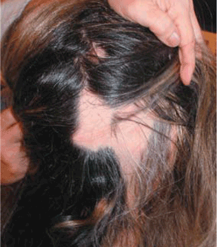
A 30-year-old woman has a 6-month history of episodes of sudden loss of hair in well-demarcated, localized areas of her scalp. She is otherwise well and has had no pruritus or pain. She has had atopic dermatitis since childhood, occasionally treated with topical corticosteroids, and localized facial vitiligo for the past 8 years. She takes no systemic drugs. She has no family history of similar lesions.
Q: What is the most likely diagnosis?
- Telogen effluvium
- Syphilitic alopecia
- Alopecia areata
- Trichotillomania
- Androgenetic alopecia
A: The correct answer is alopecia areata, a disease characterized by recurrent episodes of sudden hair loss and nail changes such as trachyonychia. It can affect any hair-bearing area, but the scalp is the most common site (90% of cases).1 The condition can range from a single patch of hair loss to complete loss of hair on the scalp (alopecia totalis) or the entire body (alopecia universalis).
The characteristic lesion is a round or oval, totally bald, smooth patch, which may have a mild peachy hue. A frequent feature is “exclamation-mark” hairs at the margin of the patch, appearing as broad distal hair shafts, narrow proximally. Active alopecia areata can be confirmed with a positive hair-pull test.
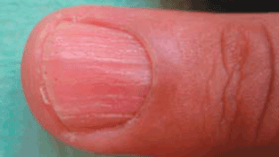
Nail involvement is common in alopecia areata, particularly in severe forms. Pitting is the most common finding, but other abnormalities such as trachyonychia have been reported.1 Nail plate pitting tends to be more shallow and regularly spaced than in psori-atic nail pitting. Trachyonychia, also known as “twenty-nail dystrophy,” is the term used to describe nail plate roughness, pitting, and ridging, affecting 1 to 20 nails. It is associated with a number of skin conditions, including alopecia areata, psoriasis, lichen planus, atopic dermatitis, and ichthyosis vulgaris.4
AN AUTOIMMUNE DISEASE
The precise cause of this common disorder has not been elucidated, but substantial evidence suggests that it is an organ-specific autoimmune disease that targets hair follicles.1–3
The current hypothesis is that the hair follicle is a site of immune privilege. In alopecia areata, increased expression of major histocompatibility complex class I molecules and down-regulation of immunosuppressants allows the immune system to recognize the immune-privileged follicle antigens.2 Several autoimmune diseases, such as thyroid disorders, vitiligo, pernicious anemia, diabetes mellitus, lupus erythematosus, myasthenia gravis, lichen planus, autoimmune polyendocrine syndrome type I, and celiac disease, have been reported in association with alopecia areata1; thyroid disorders and vitiligo have the strongest relationship. Other diseases reported to be associated with alopecia areata at a higher rate than in the general population are atopic dermatitis and Down syndrome.1
NO CURE, BUT TREATABLE
The choice of treatment depends on the patient’s age and the extent of alopecia activity.5 No cure or preventive treatment has been found, so treatments are directed toward halting disease activity.
Corticosteroids are currently the most popular form of treatment, and published reports support their efficacy.1 Topical and intralesional steroids are usually the first-line treatment for localized disease. Systemic steroids are rarely used, except in cases with rapid progression.
Other treatments that have been used with some success include minoxidil (Rogaine), anthralin (Dritho-Scalp), contact sensitizers (dinitrochlorobenzene, diphenylcycloprope-none), PUVA (psoralen plus ultraviolet A light), and cyclosporine (Sandimmune).1 If the alopecia is resistant to therapy, hair pros-theses can be recommended.
Indicators of poor prognosis (ie, hair that will not regrow completely) include atopy, the presence of other immune diseases, young age at onset, family history, nail dystrophy, extensive hair loss, and ophiasis (a continuous band of baldness involving the temporal and occipital margins).
Because current treatments may not bring results for 3 to 6 months, it is essential to reassure and inform the patient about the results that can be expected. Physicians should also inform patients of the chronic relapsing nature of alopecia areata and its unpredictable course. Although the condition does not threaten the patient’s general health and does not cause scarring, its psychological impact is significant. Support groups such as the National Alopecia Areata Foundation (www.naaf.org) can help.
- Wasserman D, Guzman-Sanchez DA, Scott K, McMichael A. Alopecia areata. Int J Dermatol 2007; 46:121–131.
- Paus R, Ito N, Takigawa M, Ito T. The hair follicle and immune privilege. J Investig Dermatol Symp Proc 2003; 8:188–194.
- Alexis AF, Dudda-Subramanya R, Sinha AA. Alopecia areata: autoimmune basis of hair loss. Eur J Dermatol 2004; 14:364–370.
- Blanco FP, Scher RK. Trachyonychia: case report and review of the literature. J Drugs Dermatol 2006; 5:469–472.
- Dombrowski NC, Bergfeld WF. Alopecia areata: what to expect from current treatments. Cleve Clin J Med 2005; 72:758–768.
A 30-year-old woman has a 6-month history of episodes of sudden loss of hair in well-demarcated, localized areas of her scalp. She is otherwise well and has had no pruritus or pain. She has had atopic dermatitis since childhood, occasionally treated with topical corticosteroids, and localized facial vitiligo for the past 8 years. She takes no systemic drugs. She has no family history of similar lesions.
Q: What is the most likely diagnosis?
- Telogen effluvium
- Syphilitic alopecia
- Alopecia areata
- Trichotillomania
- Androgenetic alopecia
A: The correct answer is alopecia areata, a disease characterized by recurrent episodes of sudden hair loss and nail changes such as trachyonychia. It can affect any hair-bearing area, but the scalp is the most common site (90% of cases).1 The condition can range from a single patch of hair loss to complete loss of hair on the scalp (alopecia totalis) or the entire body (alopecia universalis).
The characteristic lesion is a round or oval, totally bald, smooth patch, which may have a mild peachy hue. A frequent feature is “exclamation-mark” hairs at the margin of the patch, appearing as broad distal hair shafts, narrow proximally. Active alopecia areata can be confirmed with a positive hair-pull test.
Nail involvement is common in alopecia areata, particularly in severe forms. Pitting is the most common finding, but other abnormalities such as trachyonychia have been reported.1 Nail plate pitting tends to be more shallow and regularly spaced than in psori-atic nail pitting. Trachyonychia, also known as “twenty-nail dystrophy,” is the term used to describe nail plate roughness, pitting, and ridging, affecting 1 to 20 nails. It is associated with a number of skin conditions, including alopecia areata, psoriasis, lichen planus, atopic dermatitis, and ichthyosis vulgaris.4
AN AUTOIMMUNE DISEASE
The precise cause of this common disorder has not been elucidated, but substantial evidence suggests that it is an organ-specific autoimmune disease that targets hair follicles.1–3
The current hypothesis is that the hair follicle is a site of immune privilege. In alopecia areata, increased expression of major histocompatibility complex class I molecules and down-regulation of immunosuppressants allows the immune system to recognize the immune-privileged follicle antigens.2 Several autoimmune diseases, such as thyroid disorders, vitiligo, pernicious anemia, diabetes mellitus, lupus erythematosus, myasthenia gravis, lichen planus, autoimmune polyendocrine syndrome type I, and celiac disease, have been reported in association with alopecia areata1; thyroid disorders and vitiligo have the strongest relationship. Other diseases reported to be associated with alopecia areata at a higher rate than in the general population are atopic dermatitis and Down syndrome.1
NO CURE, BUT TREATABLE
The choice of treatment depends on the patient’s age and the extent of alopecia activity.5 No cure or preventive treatment has been found, so treatments are directed toward halting disease activity.
Corticosteroids are currently the most popular form of treatment, and published reports support their efficacy.1 Topical and intralesional steroids are usually the first-line treatment for localized disease. Systemic steroids are rarely used, except in cases with rapid progression.
Other treatments that have been used with some success include minoxidil (Rogaine), anthralin (Dritho-Scalp), contact sensitizers (dinitrochlorobenzene, diphenylcycloprope-none), PUVA (psoralen plus ultraviolet A light), and cyclosporine (Sandimmune).1 If the alopecia is resistant to therapy, hair pros-theses can be recommended.
Indicators of poor prognosis (ie, hair that will not regrow completely) include atopy, the presence of other immune diseases, young age at onset, family history, nail dystrophy, extensive hair loss, and ophiasis (a continuous band of baldness involving the temporal and occipital margins).
Because current treatments may not bring results for 3 to 6 months, it is essential to reassure and inform the patient about the results that can be expected. Physicians should also inform patients of the chronic relapsing nature of alopecia areata and its unpredictable course. Although the condition does not threaten the patient’s general health and does not cause scarring, its psychological impact is significant. Support groups such as the National Alopecia Areata Foundation (www.naaf.org) can help.
- Wasserman D, Guzman-Sanchez DA, Scott K, McMichael A. Alopecia areata. Int J Dermatol 2007; 46:121–131.
- Paus R, Ito N, Takigawa M, Ito T. The hair follicle and immune privilege. J Investig Dermatol Symp Proc 2003; 8:188–194.
- Alexis AF, Dudda-Subramanya R, Sinha AA. Alopecia areata: autoimmune basis of hair loss. Eur J Dermatol 2004; 14:364–370.
- Blanco FP, Scher RK. Trachyonychia: case report and review of the literature. J Drugs Dermatol 2006; 5:469–472.
- Dombrowski NC, Bergfeld WF. Alopecia areata: what to expect from current treatments. Cleve Clin J Med 2005; 72:758–768.