User login
A 10-month-old Hispanic female presents to the emergency department with a chief complaint of being limp. The mother states that the first day of the child’s illness began with tactile fever. The patient did not have any diarrhea or vomiting. After presenting to the emergency department she was diagnosed with a left otitis media. At that time, intramuscular ceftriaxone was given and amoxicillin was prescribed. The second day the patient was doing well and there were no new problems. On the third day the patient woke up and was not moving her arms and legs as much as usual, did not want to sit up on her own, and was lying limp. Additionally, she had some decreased oral intake that day and one loose stool.
Upon follow up at the pediatrician’s office there was concern about how limp the child seemed and she was sent back to the emergency department for transfer to the children’s hospital. However, the emergency department did blood work, gave intravenous fluids, and after observing slight improvement in limb movement, discharged the child to home.
At home, the child spiked a temperature. She was brought to the pediatrician’s office the next day where slight improvement in movement and more alert affect were noted; however, concern remained regarding her overall decreased activity. The child was again sent to the emergency department for transport to the pediatric hospital.
Review of systems revealed one vomiting episode and one loose stool, slight runny nose, and decrease in oral intake 1 day prior to admission. They had recently returned form California from a weekend trip. There was no significant past medical history aside from an otitis media 4 months ago. The child was born full-term without any complications. The child does not take any chronic medications, but did take two doses of amoxicillin for one and a half days in addition to acetaminophen for fever. The child does not have any known allergies. Her diet includes breastmilk and table foods. Six month vaccinations are not completed, but prior vaccines had been given on schedule. The child lives with her mother only and family history was not significant. Developmentally the child rolled over at 4 months, sits without support, pulls to stand, and stands with help.
Physical exam showed a temperature of 100.5F, heart rate of 148, respiratory rate of 30, blood pressure 103/70, and a weight of 10.5 kg (90%). Patient is well developed and well nourished without any acute distress. Physical exam was pertinent for supple neck with head lag, generalized weakness with decreased muscular strength in the upper and lower extremities. Upper extremities strength was 4/5 and lower extremities were 3/5. Neck laxity was present, sitting required support, and she refused to stand. Neurologically the patient was alert with reflexes mildly decreased in the lower extremities.
Laboratory evaluation included a complete blood count with a white blood cell (WBC) count of 11.7 and of hemoglobin of 11.4. A comprehensive metabolic panel was within normal limits. Lumbar puncture showed glucose of 50, protein of 38, 2 WBCs, 2 red blood cells, and a negative gram stain. Urinalysis was negative. Erythrocyte sedimentation rate was 17, CPK was 57, and chest radiograph was negative.
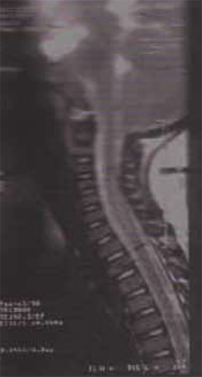
At this time stool was sent for botulism toxin; however, there was no history of eating canned food or honey. While waiting for results the patient showed slight improvement in movement and activity and demonstrated good oral intake. Physical therapy and speech therapy were working with the patient and lower extremity movement was improving, but she still had diffi culty standing. At this time an MRI of the brain and cervical spine was ordered and showed an intramedullary lesion involving the cervical and upper thoracic cord with localized cord enlargement. Enhancement was seen along C5 and C6 to the T1 level without canal stenosis and normal anatomic alignment was noted (Figure 1). In the meantime, stool for botulism toxin returned negative. Treatment was started for the diagnosis of transverse myelitis and included dexamethasone 2 mg/kg once daily with a taper over 5 weeks. She had minimal improvement prior to discharge, but showed slow and steady resolution of symptoms. Follow up with neurology was to be done on an outpatient basis, as well as continuation of physical therapy and occupational therapy.
Discussion
Transverse myelitis or transverse myelopathy (TM) is a syndrome of acute or subacute inflammation involving the spinal cord with partial or complete loss of neurologic function. It is usually limited to a number of spinal segments. The incidence is 1.3 new cases per one million residents annually. It affects all ages and both sexes; however, is uncommon in children less than 10 years of age. There are three theories for the etiology: cell-mediated autoimmune inflammation and/or demyelination of the spinal cord, a direct viral infection of the cord, and a paraneoplastic remote effect of cancer.
Clinical features include abrupt onset of symptoms varying from hours to a few days. One third of patients have a preceding viral illness. An early sign is muscle weakness, especially of the lower limbs. There may be numbness at the midthoracic region, strength loss as extreme as paraplegia, loss of bowel or bladder control, or sensory loss, most commonly involves pain and temperature. Fifty percent may experience a sharp, gnawing back pain at the level of the myelitis. Progression of the disease can lead to spasticity and hyperreflexia.
There is not a confirmation of any pathophysiologic mechanism. Demyelination, neuronal injury, and incomplete or complete necrosis of neural tissue may be associated with inflammatory changes on biopsy.
Differential diagnosis includes idiopathic autoimmune transverse myelitis, acute disseminated encephalomyelitis, multiple sclerosis (MS), Guillain-Barre, viral myelitis, vasculitis, spinal cord infarction, paraneoplastic myelopathy, vascular malformations, and nutritional myelopathy. Additionally, infectious etiologies include HIV myelopathy, spirochetal infection, and poliomyelitis.
Diagnosis is by exclusion. First, a mechanical, compressive lesion must be excluded from the differential. MRI of the spine visualizes the myelin and excludes compression and is used primarily to exclude these possible etiologies. MRI of the brain is useful to diagnose multiple sclerosis. Lumbar puncture results are variable ranging from normal to nonspecifi c inflammatory changes including a mild increase in protein or leukocytosis with an increase in polymorponuclear cells. Glucose is usually normal.
It is important, especially in adolescents, to differentiate TM from MS and Guillain-Barre. In MS symptoms are more likely separated by time, with “attacks” and a relapsing and remitting presentation. MS patients typically present with numbness and weakness in more than one extremity. In Guillain-Barre syndrome the presentation may be similar to TM in that there is weakness in the lower extremity, areflexia, and a possible viral illness precedent. However, a dermatome localization of sensory loss on neurologic exam can differentiate between a spinal cord lesion and a peripheral nerve etiology.
Treatment for TM symptoms is nonspecifi c. If a specific etiology is found then specific treatment should be rendered, otherwise one must rule out infectious or systemic inflammatory disorders. Pharmacologic treatment commonly includes high dose steroids, for example methylprednisone 1 g IV daily for 3–5 days followed with an
oral steroid taper. The evidence in the literature supporting this therapeutic approach includes mostly small studies with inconsistent results. In addition, small, uncontrolled or pilot studies have demonstrated that cyclophosphamide, azathioprine and plasmapheresis alone or in addition to corticosteroids may improve outcomes. Larger scale randomized, blinded, and controlled studies are required to more clearly define the most safe and efficacious treatment for this uncommon pediatric diagnosis.
References
- Knebusch M, Strassburg HM, Reiners K. Acute myelitis in childhood: nine cases and review of the literature. Dev Med Child Neurol. 1998;40:631-9.
- Rolad L. Pathophysiology and clinical feature of Multiple sclerosis. Immune and Infectious Disease.408-11.
- Transverse Myelitis consortium working group. Proposed diagnostic and nosology of acute transverse myelitis. Neurology. 2002;59(4).
- Andronikou S, Albuquerque-Jonathan G, Wilmshurst J, Hewlett R. MRI fi nding in acute idiopathic transverse myelopathy in children. Pediatr Radiol. 2003;33:624-9.
- Corboy J, Price R. Myelitis and toxic, infl ammatory, and infectious disorders. Curr Opin Neurol Neurosurg. 1993;6:564-70.
- Rust R. Multiple sclerosis, acute disseminated encephalomyelitis, and related conditions. Semin Pediatr Neurol. 2000;7:66-90.
- Mewashingh L, Christiaens F, et al. Cervical myelitis from herpes simplex virus type 1. Pediatr Neurol. 2004;30:54-6.
- Dhiwakar M, Buxton N. Acute transverse myelitis mimicking and intramedullary neoplasm. Br J Neurosurg. 2004;18:72-3.
- Fegan, Cheng, Demmeller, Kaplan. Textbook of Pediatric Infectious Disease. 5th Ed. 2004:518-31.
- Samuels, Feske. Offi ce Practice of Neurology. 2nd Ed. 2003
A 10-month-old Hispanic female presents to the emergency department with a chief complaint of being limp. The mother states that the first day of the child’s illness began with tactile fever. The patient did not have any diarrhea or vomiting. After presenting to the emergency department she was diagnosed with a left otitis media. At that time, intramuscular ceftriaxone was given and amoxicillin was prescribed. The second day the patient was doing well and there were no new problems. On the third day the patient woke up and was not moving her arms and legs as much as usual, did not want to sit up on her own, and was lying limp. Additionally, she had some decreased oral intake that day and one loose stool.
Upon follow up at the pediatrician’s office there was concern about how limp the child seemed and she was sent back to the emergency department for transfer to the children’s hospital. However, the emergency department did blood work, gave intravenous fluids, and after observing slight improvement in limb movement, discharged the child to home.
At home, the child spiked a temperature. She was brought to the pediatrician’s office the next day where slight improvement in movement and more alert affect were noted; however, concern remained regarding her overall decreased activity. The child was again sent to the emergency department for transport to the pediatric hospital.
Review of systems revealed one vomiting episode and one loose stool, slight runny nose, and decrease in oral intake 1 day prior to admission. They had recently returned form California from a weekend trip. There was no significant past medical history aside from an otitis media 4 months ago. The child was born full-term without any complications. The child does not take any chronic medications, but did take two doses of amoxicillin for one and a half days in addition to acetaminophen for fever. The child does not have any known allergies. Her diet includes breastmilk and table foods. Six month vaccinations are not completed, but prior vaccines had been given on schedule. The child lives with her mother only and family history was not significant. Developmentally the child rolled over at 4 months, sits without support, pulls to stand, and stands with help.
Physical exam showed a temperature of 100.5F, heart rate of 148, respiratory rate of 30, blood pressure 103/70, and a weight of 10.5 kg (90%). Patient is well developed and well nourished without any acute distress. Physical exam was pertinent for supple neck with head lag, generalized weakness with decreased muscular strength in the upper and lower extremities. Upper extremities strength was 4/5 and lower extremities were 3/5. Neck laxity was present, sitting required support, and she refused to stand. Neurologically the patient was alert with reflexes mildly decreased in the lower extremities.
Laboratory evaluation included a complete blood count with a white blood cell (WBC) count of 11.7 and of hemoglobin of 11.4. A comprehensive metabolic panel was within normal limits. Lumbar puncture showed glucose of 50, protein of 38, 2 WBCs, 2 red blood cells, and a negative gram stain. Urinalysis was negative. Erythrocyte sedimentation rate was 17, CPK was 57, and chest radiograph was negative.
At this time stool was sent for botulism toxin; however, there was no history of eating canned food or honey. While waiting for results the patient showed slight improvement in movement and activity and demonstrated good oral intake. Physical therapy and speech therapy were working with the patient and lower extremity movement was improving, but she still had diffi culty standing. At this time an MRI of the brain and cervical spine was ordered and showed an intramedullary lesion involving the cervical and upper thoracic cord with localized cord enlargement. Enhancement was seen along C5 and C6 to the T1 level without canal stenosis and normal anatomic alignment was noted (Figure 1). In the meantime, stool for botulism toxin returned negative. Treatment was started for the diagnosis of transverse myelitis and included dexamethasone 2 mg/kg once daily with a taper over 5 weeks. She had minimal improvement prior to discharge, but showed slow and steady resolution of symptoms. Follow up with neurology was to be done on an outpatient basis, as well as continuation of physical therapy and occupational therapy.
Discussion
Transverse myelitis or transverse myelopathy (TM) is a syndrome of acute or subacute inflammation involving the spinal cord with partial or complete loss of neurologic function. It is usually limited to a number of spinal segments. The incidence is 1.3 new cases per one million residents annually. It affects all ages and both sexes; however, is uncommon in children less than 10 years of age. There are three theories for the etiology: cell-mediated autoimmune inflammation and/or demyelination of the spinal cord, a direct viral infection of the cord, and a paraneoplastic remote effect of cancer.
Clinical features include abrupt onset of symptoms varying from hours to a few days. One third of patients have a preceding viral illness. An early sign is muscle weakness, especially of the lower limbs. There may be numbness at the midthoracic region, strength loss as extreme as paraplegia, loss of bowel or bladder control, or sensory loss, most commonly involves pain and temperature. Fifty percent may experience a sharp, gnawing back pain at the level of the myelitis. Progression of the disease can lead to spasticity and hyperreflexia.
There is not a confirmation of any pathophysiologic mechanism. Demyelination, neuronal injury, and incomplete or complete necrosis of neural tissue may be associated with inflammatory changes on biopsy.
Differential diagnosis includes idiopathic autoimmune transverse myelitis, acute disseminated encephalomyelitis, multiple sclerosis (MS), Guillain-Barre, viral myelitis, vasculitis, spinal cord infarction, paraneoplastic myelopathy, vascular malformations, and nutritional myelopathy. Additionally, infectious etiologies include HIV myelopathy, spirochetal infection, and poliomyelitis.
Diagnosis is by exclusion. First, a mechanical, compressive lesion must be excluded from the differential. MRI of the spine visualizes the myelin and excludes compression and is used primarily to exclude these possible etiologies. MRI of the brain is useful to diagnose multiple sclerosis. Lumbar puncture results are variable ranging from normal to nonspecifi c inflammatory changes including a mild increase in protein or leukocytosis with an increase in polymorponuclear cells. Glucose is usually normal.
It is important, especially in adolescents, to differentiate TM from MS and Guillain-Barre. In MS symptoms are more likely separated by time, with “attacks” and a relapsing and remitting presentation. MS patients typically present with numbness and weakness in more than one extremity. In Guillain-Barre syndrome the presentation may be similar to TM in that there is weakness in the lower extremity, areflexia, and a possible viral illness precedent. However, a dermatome localization of sensory loss on neurologic exam can differentiate between a spinal cord lesion and a peripheral nerve etiology.
Treatment for TM symptoms is nonspecifi c. If a specific etiology is found then specific treatment should be rendered, otherwise one must rule out infectious or systemic inflammatory disorders. Pharmacologic treatment commonly includes high dose steroids, for example methylprednisone 1 g IV daily for 3–5 days followed with an
oral steroid taper. The evidence in the literature supporting this therapeutic approach includes mostly small studies with inconsistent results. In addition, small, uncontrolled or pilot studies have demonstrated that cyclophosphamide, azathioprine and plasmapheresis alone or in addition to corticosteroids may improve outcomes. Larger scale randomized, blinded, and controlled studies are required to more clearly define the most safe and efficacious treatment for this uncommon pediatric diagnosis.
References
- Knebusch M, Strassburg HM, Reiners K. Acute myelitis in childhood: nine cases and review of the literature. Dev Med Child Neurol. 1998;40:631-9.
- Rolad L. Pathophysiology and clinical feature of Multiple sclerosis. Immune and Infectious Disease.408-11.
- Transverse Myelitis consortium working group. Proposed diagnostic and nosology of acute transverse myelitis. Neurology. 2002;59(4).
- Andronikou S, Albuquerque-Jonathan G, Wilmshurst J, Hewlett R. MRI fi nding in acute idiopathic transverse myelopathy in children. Pediatr Radiol. 2003;33:624-9.
- Corboy J, Price R. Myelitis and toxic, infl ammatory, and infectious disorders. Curr Opin Neurol Neurosurg. 1993;6:564-70.
- Rust R. Multiple sclerosis, acute disseminated encephalomyelitis, and related conditions. Semin Pediatr Neurol. 2000;7:66-90.
- Mewashingh L, Christiaens F, et al. Cervical myelitis from herpes simplex virus type 1. Pediatr Neurol. 2004;30:54-6.
- Dhiwakar M, Buxton N. Acute transverse myelitis mimicking and intramedullary neoplasm. Br J Neurosurg. 2004;18:72-3.
- Fegan, Cheng, Demmeller, Kaplan. Textbook of Pediatric Infectious Disease. 5th Ed. 2004:518-31.
- Samuels, Feske. Offi ce Practice of Neurology. 2nd Ed. 2003