User login
TAVR forges ahead in PARTNER III for low-risk patients
SNOWMASS, COLO. – The Food and Drug Administration has approved the first-ever U.S. randomized clinical trial of transcatheter aortic valve replacement versus open surgical replacement in low–surgical risk patients with symptomatic severe aortic stenosis.
The PARTNER III trial will enroll roughly 1,200 patients age 65 or older, all with a Society of Thoracic Surgeons risk score of less than 4%, at 50 sites beginning this spring, Dr. Vinod H. Thourani said at the Annual Cardiovascular Conference at Snowmass.

This is a noninferiority trial with a primary endpoint comprising a 1-year composite of death, stroke, or rehospitalization. The study is sponsored by Edwards Lifesciences, and patients randomized to transcatheter aortic valve replacement (TAVR) will receive the company’s low-profile Sapien 3 valve.
Coprincipal investigators are Dr. Michael J. Mack of the Baylor Health Care System in Plano, Tex., and Dr. Martin B. Leon of Columbia University, New York. Dr. Thourani is a member of the PARTNER III executive committee.
This is a study that could upend clinical practice, he observed.
“Are we going to have within the next 5 years 80%-90% of all patients who present with severe symptomatic aortic stenosis treated with transcatheter valves? We’re really at a major crossroads here, I believe,” said Dr. Thourani, professor of surgery and medicine and codirector of the structural heart and valve center at Emory University in Atlanta.
He ran down the numbers: Today, roughly 80% of all surgical aortic valve replacements (SAVR) in the United States are performed in low–surgical risk patients. These low-risk patients comprise roughly 65% of the total operable population with severe aortic stenosis. If PARTNER III and other data show that TAVR provides results comparable to SAVR in this group, Dr. Thourani predicted that it’s likely most low–surgical risk patients will opt for the less invasive approach. The appeal is no surgical incision, less pain, a shorter or no ICU stay, and faster return to normal activity.
Right now, U.S. and European guidelines state that TAVR is the preferred or alternative strategy to SAVR only in the relatively small group comprised of inoperable or high–surgical risk patients. In clinical practice, TAVR has already supplanted SAVR in the 10% of operable patients with high surgical risk. And TAVR is poised to do so in the roughly 25% of patients who fall into the intermediate–surgical risk category, according to the cardiothoracic surgeon.
He predicted that the 1-year outcomes of TAVR in more than 1,000 intermediate-risk participants in the PARTNER II trial will create a stir when presented this year, as a late-breaker at the annual meeting of the American College of Cardiology in Chicago. Although he stressed that he doesn’t know the results, the 30-day outcomes presented at last year’s Transcatheter Cardiovascular Therapeutics conference are extremely promising: a 1.1% all-cause mortality rate in patients with an average Society of Thoracic Surgeons risk score of 5.3%, for a stunning observed-to-expected ratio of just 0.21. Plus, a 1.0% rate of disabling stroke in this large multicenter randomized experience.
“That becomes really compelling data for us to think we’re ready now to go to the next step,” Dr. Thourani said. “My belief is at the rate we’re going, we’ll see most intermediate-risk patients going to TAVR.”
To date there has been only one randomized trial of TAVR versus SAVR in low–surgical risk patients: the Nordic Aortic Valve Intervention Trial (NOTION), which included 280 randomized patients with an average Society of Thoracic Surgeons risk score of 3%.
In the 2-year results presented by Dr. Lars Søndergaard of the University of Copenhagen at TCT 2015, all-cause mortality was 2.1% with TAVR and 3.7% with SAVR at 30 days, 4.9% with TAVR and 7.5% with SAVR at 12 months, and 8.0% versus 9.8% at 24 months. The 30-day rates of major bleeding, cardiogenic shock, atrial fibrillation, and acute kidney injury were all substantially lower in the TAVR group. All very impressive. However, Dr. Thourani found the TAVR patients’ pacemaker-requirement rate troubling. At 30 days post TAVR, 34% of patients had a pacemaker, compared with 1.6% of the SAVR group. By 24 months, 41% of the TAVR group had received a pacemaker, compared with just 4% of the SAVR group.
“What’s the acceptable pacemaker rate for someone utilizing TAVR – 5%, 10%, 40%? That’s something we as a community have to look at,” the surgeon observed. He noted that his purchase price for a TAVR valve is roughly $32,500, whereas a SAVR valve costs him $4,500. And at Emory, putting in a pacemaker costs an added $10,000-$15,000 for the device.
“If I’m putting a pacemaker in 40% of my TAVR patients at a cost of $40,000-$45,000 per patient for the valve and pacemaker, that becomes an issue,” Dr. Thourani said.
Other concerns surrounding TAVR, in addition to reimbursement, include the uncertain long-term impact of residual minimal paravalvular leak, which is common.
“We’re not done talking about paravalvular leak rates. As cardiologists you’re not okay with me giving your patient a minimal paravalvular leak post-SAVR. Are we going to change the bar a little bit for TAVR?” he mused.
Another issue is thrombosis of TAVR valve leaflets, Dr. Thourani continued. In a large patient series reported last year, this event occurred in 0.6% of patients, with an average of 181 days from TAVR to confirmatory abnormal imaging (Circ Cardiovasc Interv. 2015 Apr;8[4]. pii: e001779). Two clinical trials are gearing up to examine various anticoagulant strategies to address the problem.
Despite the various concerns, however, Dr. Thourani is extremely optimistic about TAVR’s future. It’s a booming field, with 396 U.S. TAVR centers as of 2015. The indications appear to be on the verge of expansion. Technical progress continues, with half a dozen TAVR valves in development in addition to the two now FDA approved.
“We have just scratched the surface of what we’re going to do in the management of severe aortic stenosis,” the surgeon promised.
The latest results of minimalist TAVR provide another reason for optimism regarding TAVR’s future.
Emory University surgeons and interventional cardiologists have been pacesetters in the minimalist TAVR approach. The key elements of minimalist TAVR are that the procedure is performed in the cardiac catheterization laboratory via transfemoral access, under conscious sedation, with transthoracic echocardiographic guidance, no Swan-Ganz catheter, and no ICU stay for most patients.
Dr. Thourani presented as-yet unpublished data on a recent series of 111 high–surgical risk patients who underwent minimalist TAVR with implantation of a Sapien 3 valve at Emory. Although their Society of Thoracic Surgeons risk score was 8%, there was zero 30-day mortality in this group. One patient had a major stroke, two had major vascular complications, and the 30-day readmission rate was just 3.8%.
“Can we get to these results universally? We think we can. This is the bar we need to start thinking about,” Dr. Thourani said.
Dr. Thourani reported serving as a consultant to Edwards Lifesciences and St. Jude Medical and receiving research grants from Abbott, Boston Scientific, Medtronic, and Sorin.
SNOWMASS, COLO. – The Food and Drug Administration has approved the first-ever U.S. randomized clinical trial of transcatheter aortic valve replacement versus open surgical replacement in low–surgical risk patients with symptomatic severe aortic stenosis.
The PARTNER III trial will enroll roughly 1,200 patients age 65 or older, all with a Society of Thoracic Surgeons risk score of less than 4%, at 50 sites beginning this spring, Dr. Vinod H. Thourani said at the Annual Cardiovascular Conference at Snowmass.
This is a noninferiority trial with a primary endpoint comprising a 1-year composite of death, stroke, or rehospitalization. The study is sponsored by Edwards Lifesciences, and patients randomized to transcatheter aortic valve replacement (TAVR) will receive the company’s low-profile Sapien 3 valve.
Coprincipal investigators are Dr. Michael J. Mack of the Baylor Health Care System in Plano, Tex., and Dr. Martin B. Leon of Columbia University, New York. Dr. Thourani is a member of the PARTNER III executive committee.
This is a study that could upend clinical practice, he observed.
“Are we going to have within the next 5 years 80%-90% of all patients who present with severe symptomatic aortic stenosis treated with transcatheter valves? We’re really at a major crossroads here, I believe,” said Dr. Thourani, professor of surgery and medicine and codirector of the structural heart and valve center at Emory University in Atlanta.
He ran down the numbers: Today, roughly 80% of all surgical aortic valve replacements (SAVR) in the United States are performed in low–surgical risk patients. These low-risk patients comprise roughly 65% of the total operable population with severe aortic stenosis. If PARTNER III and other data show that TAVR provides results comparable to SAVR in this group, Dr. Thourani predicted that it’s likely most low–surgical risk patients will opt for the less invasive approach. The appeal is no surgical incision, less pain, a shorter or no ICU stay, and faster return to normal activity.
Right now, U.S. and European guidelines state that TAVR is the preferred or alternative strategy to SAVR only in the relatively small group comprised of inoperable or high–surgical risk patients. In clinical practice, TAVR has already supplanted SAVR in the 10% of operable patients with high surgical risk. And TAVR is poised to do so in the roughly 25% of patients who fall into the intermediate–surgical risk category, according to the cardiothoracic surgeon.
He predicted that the 1-year outcomes of TAVR in more than 1,000 intermediate-risk participants in the PARTNER II trial will create a stir when presented this year, as a late-breaker at the annual meeting of the American College of Cardiology in Chicago. Although he stressed that he doesn’t know the results, the 30-day outcomes presented at last year’s Transcatheter Cardiovascular Therapeutics conference are extremely promising: a 1.1% all-cause mortality rate in patients with an average Society of Thoracic Surgeons risk score of 5.3%, for a stunning observed-to-expected ratio of just 0.21. Plus, a 1.0% rate of disabling stroke in this large multicenter randomized experience.
“That becomes really compelling data for us to think we’re ready now to go to the next step,” Dr. Thourani said. “My belief is at the rate we’re going, we’ll see most intermediate-risk patients going to TAVR.”
To date there has been only one randomized trial of TAVR versus SAVR in low–surgical risk patients: the Nordic Aortic Valve Intervention Trial (NOTION), which included 280 randomized patients with an average Society of Thoracic Surgeons risk score of 3%.
In the 2-year results presented by Dr. Lars Søndergaard of the University of Copenhagen at TCT 2015, all-cause mortality was 2.1% with TAVR and 3.7% with SAVR at 30 days, 4.9% with TAVR and 7.5% with SAVR at 12 months, and 8.0% versus 9.8% at 24 months. The 30-day rates of major bleeding, cardiogenic shock, atrial fibrillation, and acute kidney injury were all substantially lower in the TAVR group. All very impressive. However, Dr. Thourani found the TAVR patients’ pacemaker-requirement rate troubling. At 30 days post TAVR, 34% of patients had a pacemaker, compared with 1.6% of the SAVR group. By 24 months, 41% of the TAVR group had received a pacemaker, compared with just 4% of the SAVR group.
“What’s the acceptable pacemaker rate for someone utilizing TAVR – 5%, 10%, 40%? That’s something we as a community have to look at,” the surgeon observed. He noted that his purchase price for a TAVR valve is roughly $32,500, whereas a SAVR valve costs him $4,500. And at Emory, putting in a pacemaker costs an added $10,000-$15,000 for the device.
“If I’m putting a pacemaker in 40% of my TAVR patients at a cost of $40,000-$45,000 per patient for the valve and pacemaker, that becomes an issue,” Dr. Thourani said.
Other concerns surrounding TAVR, in addition to reimbursement, include the uncertain long-term impact of residual minimal paravalvular leak, which is common.
“We’re not done talking about paravalvular leak rates. As cardiologists you’re not okay with me giving your patient a minimal paravalvular leak post-SAVR. Are we going to change the bar a little bit for TAVR?” he mused.
Another issue is thrombosis of TAVR valve leaflets, Dr. Thourani continued. In a large patient series reported last year, this event occurred in 0.6% of patients, with an average of 181 days from TAVR to confirmatory abnormal imaging (Circ Cardiovasc Interv. 2015 Apr;8[4]. pii: e001779). Two clinical trials are gearing up to examine various anticoagulant strategies to address the problem.
Despite the various concerns, however, Dr. Thourani is extremely optimistic about TAVR’s future. It’s a booming field, with 396 U.S. TAVR centers as of 2015. The indications appear to be on the verge of expansion. Technical progress continues, with half a dozen TAVR valves in development in addition to the two now FDA approved.
“We have just scratched the surface of what we’re going to do in the management of severe aortic stenosis,” the surgeon promised.
The latest results of minimalist TAVR provide another reason for optimism regarding TAVR’s future.
Emory University surgeons and interventional cardiologists have been pacesetters in the minimalist TAVR approach. The key elements of minimalist TAVR are that the procedure is performed in the cardiac catheterization laboratory via transfemoral access, under conscious sedation, with transthoracic echocardiographic guidance, no Swan-Ganz catheter, and no ICU stay for most patients.
Dr. Thourani presented as-yet unpublished data on a recent series of 111 high–surgical risk patients who underwent minimalist TAVR with implantation of a Sapien 3 valve at Emory. Although their Society of Thoracic Surgeons risk score was 8%, there was zero 30-day mortality in this group. One patient had a major stroke, two had major vascular complications, and the 30-day readmission rate was just 3.8%.
“Can we get to these results universally? We think we can. This is the bar we need to start thinking about,” Dr. Thourani said.
Dr. Thourani reported serving as a consultant to Edwards Lifesciences and St. Jude Medical and receiving research grants from Abbott, Boston Scientific, Medtronic, and Sorin.
EXPERT ANALYSIS FROM THE CARDIOVASCULAR CONFERENCE AT SNOWMASS

Noninvasive Testing of Women with Suspected CAD Simplified
SNOWMASS, COLO. – Current American Heart Association consensus recommendations for noninvasive testing of women with suspected ischemic heart disease utilize a novel and simplified approach to pretest risk stratification.
The writing committee’s thinking was that a new risk assessment method specific to symptomatic women was needed. Conventional risk scores, such as the Framingham and American College of Cardiology/AHA atherosclerotic cardiovascular disease risk calculator, are weighted for risk evaluation in the general population of asymptomatic men and women, committee cochair Dr. Jennifer H. Mieres explained at the Annual Cardiovascular Conference at Snowmass.

The pretest risk assessment strategy for ischemic heart disease (IHD) described in the AHA consensus statement (Circulation. 2014 Jul 22;130[4]:350-79) is applicable only in women who present with chest pain symptoms or an ischemic equivalent such as excessive shortness of breath not attributable to underlying pulmonary disease. It’s designed to avoid overuse of costly noninvasive and invasive diagnostic testing, which has become common in concert with increasing physician awareness that the pattern of symptom presentation is broader in women than men with IHD.
Given the epidemiologic evidence that coronary heart disease tends to occur a decade later in women than men, the female-specific risk assessment method utilizes age as its starting point. In general, women who present with symptoms suggestive of IHD in their 50s are classified as low risk. Those in their 60s are deemed intermediate risk. And symptomatic women in their 70s are categorized as high risk. A patient gets bumped up one risk category if she has multiple cardiac risk factors or functional disability, according to Dr. Mieres, professor of cardiology and population health at Hofstra University in Hempstead, N.Y.
Functional disability is defined as inability to perform activities of daily living or as a limited exercise capacity estimated at less than 5 metabolic equivalents (METs) using the Duke Activities Status Index (DASI), a validated brief 12-item questionnaire suitable for patients to complete in the waiting room.
A symptomatic woman’s baseline fitness level is important for a couple of reasons. If she can’t perform basic activities of daily living or do more than 5 METs of exercise then the exercise treadmill test is not the initial diagnostic test of choice because she won’t be able to achieve maximum stress.
Plus, the landmark Women’s Ischemia Syndrome Evaluation (WISE) study showed that a symptomatic woman’s baseline fitness level is a powerful predictor of her prognosis: Participants who had an estimated exercise capacity of 4.7 METs or less based upon their DASI score had a 3.7-fold greater risk of death or nonfatal MI during follow-up did than those with a DASI score of 10 METs or more. Those with a DASI score of 4.8-7.4 METs were 2.1-fold more likely to experience death or MI, and women with an estimated exercise capacity of 7.5-9.9 METs based upon DASI were at 1.9-fold increased risk of death or MI, compared with those having a DASI score of at least 10 METs (J Am Coll Cardiol. 2006 Feb 7;47[3 Suppl]:S36-43).
In addition, the women’s IHD risk evaluation utilizes several high-risk equivalents which automatically move a symptomatic woman into the high-risk category regardless of her age. These are peripheral artery disease, longstanding poorly controlled diabetes, and a history of cerebrovascular accident, Dr. Mieres continued.
She stressed that most low-risk symptomatic women are not candidates for diagnostic testing. The exercise treadmill test is the first-line diagnostic test for women at intermediate risk for IHD who are functionally capable and have a normal resting ECG.
Stress imaging studies are reserved for intermediate- and high-risk women with resting ST-segment abnormalities, inability to exercise adequately, or an indeterminate exercise treadmill test.
Stress echocardiography gets a class I recommendation in these situations. So does stress myocardial perfusion imaging with SPECT or PET except in premenopausal women, where concerns about radiation exposure dictate that stress echo or stress cardiac magnetic resonance imaging is preferable.
Otherwise, stress cardiac magnetic resonance gets a class IIb – “may be reasonable” – recommendation as an initial imaging test in these situations.
Coronary artery calcium measurement via CT angiography gets a class IIb recommendation in intermediate-risk symptomatic women. This imaging method is unique in that it not only provides accurate information about the obstructive burden of CAD, but it measures the nonobstructive burden as well. That’s particularly important in women because they have a greater burden of nonobstructive CAD, defined as 1%-49% stenosis, than do men. Women with stress test abnormalities and nonobstructive CAD are no longer defined as having a false-positive test, as was traditionally the case; instead, their test is categorized as abnormal because the WISE study has shown they are at elevated IHD risk, Dr. Mieres said.
She reported having no financial conflicts regarding her presentation.
SNOWMASS, COLO. – Current American Heart Association consensus recommendations for noninvasive testing of women with suspected ischemic heart disease utilize a novel and simplified approach to pretest risk stratification.
The writing committee’s thinking was that a new risk assessment method specific to symptomatic women was needed. Conventional risk scores, such as the Framingham and American College of Cardiology/AHA atherosclerotic cardiovascular disease risk calculator, are weighted for risk evaluation in the general population of asymptomatic men and women, committee cochair Dr. Jennifer H. Mieres explained at the Annual Cardiovascular Conference at Snowmass.
The pretest risk assessment strategy for ischemic heart disease (IHD) described in the AHA consensus statement (Circulation. 2014 Jul 22;130[4]:350-79) is applicable only in women who present with chest pain symptoms or an ischemic equivalent such as excessive shortness of breath not attributable to underlying pulmonary disease. It’s designed to avoid overuse of costly noninvasive and invasive diagnostic testing, which has become common in concert with increasing physician awareness that the pattern of symptom presentation is broader in women than men with IHD.
Given the epidemiologic evidence that coronary heart disease tends to occur a decade later in women than men, the female-specific risk assessment method utilizes age as its starting point. In general, women who present with symptoms suggestive of IHD in their 50s are classified as low risk. Those in their 60s are deemed intermediate risk. And symptomatic women in their 70s are categorized as high risk. A patient gets bumped up one risk category if she has multiple cardiac risk factors or functional disability, according to Dr. Mieres, professor of cardiology and population health at Hofstra University in Hempstead, N.Y.
Functional disability is defined as inability to perform activities of daily living or as a limited exercise capacity estimated at less than 5 metabolic equivalents (METs) using the Duke Activities Status Index (DASI), a validated brief 12-item questionnaire suitable for patients to complete in the waiting room.
A symptomatic woman’s baseline fitness level is important for a couple of reasons. If she can’t perform basic activities of daily living or do more than 5 METs of exercise then the exercise treadmill test is not the initial diagnostic test of choice because she won’t be able to achieve maximum stress.
Plus, the landmark Women’s Ischemia Syndrome Evaluation (WISE) study showed that a symptomatic woman’s baseline fitness level is a powerful predictor of her prognosis: Participants who had an estimated exercise capacity of 4.7 METs or less based upon their DASI score had a 3.7-fold greater risk of death or nonfatal MI during follow-up did than those with a DASI score of 10 METs or more. Those with a DASI score of 4.8-7.4 METs were 2.1-fold more likely to experience death or MI, and women with an estimated exercise capacity of 7.5-9.9 METs based upon DASI were at 1.9-fold increased risk of death or MI, compared with those having a DASI score of at least 10 METs (J Am Coll Cardiol. 2006 Feb 7;47[3 Suppl]:S36-43).
In addition, the women’s IHD risk evaluation utilizes several high-risk equivalents which automatically move a symptomatic woman into the high-risk category regardless of her age. These are peripheral artery disease, longstanding poorly controlled diabetes, and a history of cerebrovascular accident, Dr. Mieres continued.
She stressed that most low-risk symptomatic women are not candidates for diagnostic testing. The exercise treadmill test is the first-line diagnostic test for women at intermediate risk for IHD who are functionally capable and have a normal resting ECG.
Stress imaging studies are reserved for intermediate- and high-risk women with resting ST-segment abnormalities, inability to exercise adequately, or an indeterminate exercise treadmill test.
Stress echocardiography gets a class I recommendation in these situations. So does stress myocardial perfusion imaging with SPECT or PET except in premenopausal women, where concerns about radiation exposure dictate that stress echo or stress cardiac magnetic resonance imaging is preferable.
Otherwise, stress cardiac magnetic resonance gets a class IIb – “may be reasonable” – recommendation as an initial imaging test in these situations.
Coronary artery calcium measurement via CT angiography gets a class IIb recommendation in intermediate-risk symptomatic women. This imaging method is unique in that it not only provides accurate information about the obstructive burden of CAD, but it measures the nonobstructive burden as well. That’s particularly important in women because they have a greater burden of nonobstructive CAD, defined as 1%-49% stenosis, than do men. Women with stress test abnormalities and nonobstructive CAD are no longer defined as having a false-positive test, as was traditionally the case; instead, their test is categorized as abnormal because the WISE study has shown they are at elevated IHD risk, Dr. Mieres said.
She reported having no financial conflicts regarding her presentation.
EXPERT ANALYSIS FROM THE CARDIOVASCULAR CONFERENCE AT SNOWMASS

Noninvasive testing of women with suspected CAD simplified
SNOWMASS, COLO. – Current American Heart Association consensus recommendations for noninvasive testing of women with suspected ischemic heart disease utilize a novel and simplified approach to pretest risk stratification.
The writing committee’s thinking was that a new risk assessment method specific to symptomatic women was needed. Conventional risk scores, such as the Framingham and American College of Cardiology/AHA atherosclerotic cardiovascular disease risk calculator, are weighted for risk evaluation in the general population of asymptomatic men and women, committee cochair Dr. Jennifer H. Mieres explained at the Annual Cardiovascular Conference at Snowmass.

The pretest risk assessment strategy for ischemic heart disease (IHD) described in the AHA consensus statement (Circulation. 2014 Jul 22;130[4]:350-79) is applicable only in women who present with chest pain symptoms or an ischemic equivalent such as excessive shortness of breath not attributable to underlying pulmonary disease. It’s designed to avoid overuse of costly noninvasive and invasive diagnostic testing, which has become common in concert with increasing physician awareness that the pattern of symptom presentation is broader in women than men with IHD.
Given the epidemiologic evidence that coronary heart disease tends to occur a decade later in women than men, the female-specific risk assessment method utilizes age as its starting point. In general, women who present with symptoms suggestive of IHD in their 50s are classified as low risk. Those in their 60s are deemed intermediate risk. And symptomatic women in their 70s are categorized as high risk. A patient gets bumped up one risk category if she has multiple cardiac risk factors or functional disability, according to Dr. Mieres, professor of cardiology and population health at Hofstra University in Hempstead, N.Y.
Functional disability is defined as inability to perform activities of daily living or as a limited exercise capacity estimated at less than 5 metabolic equivalents (METs) using the Duke Activities Status Index (DASI), a validated brief 12-item questionnaire suitable for patients to complete in the waiting room.
A symptomatic woman’s baseline fitness level is important for a couple of reasons. If she can’t perform basic activities of daily living or do more than 5 METs of exercise then the exercise treadmill test is not the initial diagnostic test of choice because she won’t be able to achieve maximum stress.
Plus, the landmark Women’s Ischemia Syndrome Evaluation (WISE) study showed that a symptomatic woman’s baseline fitness level is a powerful predictor of her prognosis: Participants who had an estimated exercise capacity of 4.7 METs or less based upon their DASI score had a 3.7-fold greater risk of death or nonfatal MI during follow-up did than those with a DASI score of 10 METs or more. Those with a DASI score of 4.8-7.4 METs were 2.1-fold more likely to experience death or MI, and women with an estimated exercise capacity of 7.5-9.9 METs based upon DASI were at 1.9-fold increased risk of death or MI, compared with those having a DASI score of at least 10 METs (J Am Coll Cardiol. 2006 Feb 7;47[3 Suppl]:S36-43).
In addition, the women’s IHD risk evaluation utilizes several high-risk equivalents which automatically move a symptomatic woman into the high-risk category regardless of her age. These are peripheral artery disease, longstanding poorly controlled diabetes, and a history of cerebrovascular accident, Dr. Mieres continued.
She stressed that most low-risk symptomatic women are not candidates for diagnostic testing. The exercise treadmill test is the first-line diagnostic test for women at intermediate risk for IHD who are functionally capable and have a normal resting ECG.
Stress imaging studies are reserved for intermediate- and high-risk women with resting ST-segment abnormalities, inability to exercise adequately, or an indeterminate exercise treadmill test.
Stress echocardiography gets a class I recommendation in these situations. So does stress myocardial perfusion imaging with SPECT or PET except in premenopausal women, where concerns about radiation exposure dictate that stress echo or stress cardiac magnetic resonance imaging is preferable.
Otherwise, stress cardiac magnetic resonance gets a class IIb – “may be reasonable” – recommendation as an initial imaging test in these situations.
Coronary artery calcium measurement via CT angiography gets a class IIb recommendation in intermediate-risk symptomatic women. This imaging method is unique in that it not only provides accurate information about the obstructive burden of CAD, but it measures the nonobstructive burden as well. That’s particularly important in women because they have a greater burden of nonobstructive CAD, defined as 1%-49% stenosis, than do men. Women with stress test abnormalities and nonobstructive CAD are no longer defined as having a false-positive test, as was traditionally the case; instead, their test is categorized as abnormal because the WISE study has shown they are at elevated IHD risk, Dr. Mieres said.
She reported having no financial conflicts regarding her presentation.
SNOWMASS, COLO. – Current American Heart Association consensus recommendations for noninvasive testing of women with suspected ischemic heart disease utilize a novel and simplified approach to pretest risk stratification.
The writing committee’s thinking was that a new risk assessment method specific to symptomatic women was needed. Conventional risk scores, such as the Framingham and American College of Cardiology/AHA atherosclerotic cardiovascular disease risk calculator, are weighted for risk evaluation in the general population of asymptomatic men and women, committee cochair Dr. Jennifer H. Mieres explained at the Annual Cardiovascular Conference at Snowmass.
The pretest risk assessment strategy for ischemic heart disease (IHD) described in the AHA consensus statement (Circulation. 2014 Jul 22;130[4]:350-79) is applicable only in women who present with chest pain symptoms or an ischemic equivalent such as excessive shortness of breath not attributable to underlying pulmonary disease. It’s designed to avoid overuse of costly noninvasive and invasive diagnostic testing, which has become common in concert with increasing physician awareness that the pattern of symptom presentation is broader in women than men with IHD.
Given the epidemiologic evidence that coronary heart disease tends to occur a decade later in women than men, the female-specific risk assessment method utilizes age as its starting point. In general, women who present with symptoms suggestive of IHD in their 50s are classified as low risk. Those in their 60s are deemed intermediate risk. And symptomatic women in their 70s are categorized as high risk. A patient gets bumped up one risk category if she has multiple cardiac risk factors or functional disability, according to Dr. Mieres, professor of cardiology and population health at Hofstra University in Hempstead, N.Y.
Functional disability is defined as inability to perform activities of daily living or as a limited exercise capacity estimated at less than 5 metabolic equivalents (METs) using the Duke Activities Status Index (DASI), a validated brief 12-item questionnaire suitable for patients to complete in the waiting room.
A symptomatic woman’s baseline fitness level is important for a couple of reasons. If she can’t perform basic activities of daily living or do more than 5 METs of exercise then the exercise treadmill test is not the initial diagnostic test of choice because she won’t be able to achieve maximum stress.
Plus, the landmark Women’s Ischemia Syndrome Evaluation (WISE) study showed that a symptomatic woman’s baseline fitness level is a powerful predictor of her prognosis: Participants who had an estimated exercise capacity of 4.7 METs or less based upon their DASI score had a 3.7-fold greater risk of death or nonfatal MI during follow-up did than those with a DASI score of 10 METs or more. Those with a DASI score of 4.8-7.4 METs were 2.1-fold more likely to experience death or MI, and women with an estimated exercise capacity of 7.5-9.9 METs based upon DASI were at 1.9-fold increased risk of death or MI, compared with those having a DASI score of at least 10 METs (J Am Coll Cardiol. 2006 Feb 7;47[3 Suppl]:S36-43).
In addition, the women’s IHD risk evaluation utilizes several high-risk equivalents which automatically move a symptomatic woman into the high-risk category regardless of her age. These are peripheral artery disease, longstanding poorly controlled diabetes, and a history of cerebrovascular accident, Dr. Mieres continued.
She stressed that most low-risk symptomatic women are not candidates for diagnostic testing. The exercise treadmill test is the first-line diagnostic test for women at intermediate risk for IHD who are functionally capable and have a normal resting ECG.
Stress imaging studies are reserved for intermediate- and high-risk women with resting ST-segment abnormalities, inability to exercise adequately, or an indeterminate exercise treadmill test.
Stress echocardiography gets a class I recommendation in these situations. So does stress myocardial perfusion imaging with SPECT or PET except in premenopausal women, where concerns about radiation exposure dictate that stress echo or stress cardiac magnetic resonance imaging is preferable.
Otherwise, stress cardiac magnetic resonance gets a class IIb – “may be reasonable” – recommendation as an initial imaging test in these situations.
Coronary artery calcium measurement via CT angiography gets a class IIb recommendation in intermediate-risk symptomatic women. This imaging method is unique in that it not only provides accurate information about the obstructive burden of CAD, but it measures the nonobstructive burden as well. That’s particularly important in women because they have a greater burden of nonobstructive CAD, defined as 1%-49% stenosis, than do men. Women with stress test abnormalities and nonobstructive CAD are no longer defined as having a false-positive test, as was traditionally the case; instead, their test is categorized as abnormal because the WISE study has shown they are at elevated IHD risk, Dr. Mieres said.
She reported having no financial conflicts regarding her presentation.
EXPERT ANALYSIS FROM THE CARDIOVASCULAR CONFERENCE AT SNOWMASS

Acute heart failure: What works, what doesn’t
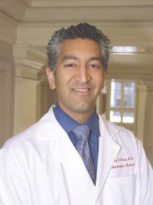
SNOWMASS, COLO. – The primary treatment goal in patients hospitalized for acute decompensated heart failure is aggressive decongestion to get them feeling better and out of the hospital quicker – and the best way to achieve that is with high-dose loop diuretics administered intravenously at a dose equivalent to 2.5 times the previous oral dose, Dr. Akshay S. Desai said at the Annual Cardiovascular Conference at Snowmass.
This was the key lesson of the Diuretic Optimization Strategies Evaluation (DOSE) trial, a prospective double-blind randomized trial that provides physicians with the best available data on how to decongest patients with acute decompensated heart failure (ADHF), according to Dr. Desai, a coinvestigator. The study was conducted by the National Heart, Lung, and Blood Institute Heart Failure Clinical Research Network.
The DOSE trial showed that it really doesn’t matter whether the diuretic is administered intravenously by continuous infusion or a bolus every 12 hours. The important thing is the high-dose strategy. It proved to be safe and was associated with accelerated decongestion as manifest in greater relief of dyspnea, greater weight loss and fluid loss, and a larger reduction in serum brain natriuretic peptide at 72 hours than low-dose therapy equivalent to the patient’s previous oral dose (N Engl J Med. 2011 Mar 3;364[9]:797-805).
“The message for you in practice is that the route of diuretic administration is probably not important as long as you give an adequate dose. I would consider giving the higher dose of diuretic because it’s associated with more effective decongestion and perhaps shorter length of stay,” said Dr. Desai, director of heart failure disease management at Brigham and Women’s Hospital in Boston.
There is a trade-off in ADHF between effective decongestion and worsening renal function as reflected in increased serum creatinine levels. Transient worsening of renal function occurred more frequently with the high-dose strategy in the DOSE trial, but it had no impact on clinical outcomes at 60 days follow-up. This finding is consistent with the results of an important Italian study showing that worsening renal function alone isn’t independently associated with increased risk of death or ADHF readmission; the problems arise in patients with worsening renal function and persistent congestion (Circ Heart Fail. 2012 Jan;5[1]:54-62).
Worsening congestion drives most hospitalizations for heart failure. And patients who are still congested at discharge are at dramatically increased risk for death or readmission in the ensuing 6 months. Yet the limitations of current therapy mean that even in expert hands, a substantial proportion of patients are discharged with clinically significant congestion. For example, in a retrospective analysis of nearly 500 patients enrolled in ADHF studies conducted by physicians in the NHLBI Heart Failure Clinical Research Network, only 52% were discharged free of congestion (Circ Heart Fail. 2015 Jul;8[4]:741-8).
Beyond aggressive treatment with loop diuretics, what else is useful in achieving the goal of hospital discharge with normalized filling pressures? Not much, according to a considerable body of research on the topic.
“The data tell us more about what not to do than what to do,” according to Dr. Desai.
For example, even though aggressive salt and fluid restriction is often forced upon patients hospitalized for ADHF on the rationale that this strategy may make it easier for diuretics to work, it’s not an evidence-based practice. Indeed, in a randomized clinical trial with blinded outcome assessments, an in-hospital diet restricted to a maximum intake of 800 mg of sodium and 800 mL of fluid daily had no effect on weight loss or a clinical congestion score (JAMA Intern Med. 2013 June 24;173[12]:1058-64).
“What it did very effectively was make patients thirsty. There are probably some patients where restriction of sodium and fluid intake is important, but routine use of tight restrictions is probably more harmful than helpful,” he observed.
The list of failed once-promising alternatives to diuretics in the setting of ADHF is impressive. It includes milrinone, tolvaptan, nesiritide, levosimendan, tezosentan, low-dose dopamine, and ultrafiltration. All had a sound mechanistic basis for the belief that they might improve outcomes, but in clinical trials none of them did.
Although routine use of pulmonary artery catheters to guide decongestion therapy in ADHF isn’t warranted because it has not been shown to be better than clinical assessment, there are certain situations where it is extremely helpful. For example, in the patient who isn’t responding to adequate doses of loop diuretics, it becomes important to understand the hemodynamics, which may involve systemic vascular resistance or a cardiac output problem.
Other situations where it’s worthwhile to consider placement of a pulmonary artery catheter include the patient of uncertain fluid status where it’s not possible to confidently estimate cardiac output at the bedside or markedly worsening renal function with empiric therapy.
Dr. Desai reported receiving research funding from Novartis and St. Jude Medical and serving as a paid consultant to Merck, Relypsa, and St. Jude Medical.
EXPERT ANALYSIS FROM THE CARDIOVASCULAR CONFERENCE AT SNOWMASS

What’s next for Watchman stroke prevention device
SNOWMASS, COLO. – The goal is finally in sight following an odyssey to develop the Watchman left atrial appendage closure device as a safe and effective alternative to oral anticoagulation for stroke prevention in patients with nonvalvular atrial fibrillation, Dr. David R. Holmes Jr. said at the Annual Cardiovascular Conference at Snowmass.
It’s all coming together: The Watchman, a small percutaneously delivered parachute-like device, has received FDA marketing approval as the sole authorized left atrial appendage closure device in this country on the strength of two compellingly positive randomized controlled trials. A recent meta-analysis of those trials showed significantly fewer hemorrhagic strokes, fewer cardiovascular or unexplained deaths, and fewer nonprocedural bleeding events in Watchman recipients than in patients randomized to warfarin. And a cost-effectiveness analysis has concluded that after 8 years, the Watchman becomes “the dominant strategy” – meaning more effective and less costly for stroke prevention in patients with atrial fibrillation (AF) having a contraindication to warfarin – compared with the novel oral anticoagulant apixaban (J Am Coll Cardiol. 2015 Dec 22;66[24]:2728-39).

Moreover, the final pieces required for the Watchman to become a mainstream reimbursable therapy are falling into place. The Society for Cardiovascular Angiography and Interventions (SCAI), the American College of Cardiology, and the Heart Rhythm Society (HRS) have jointly issued institutional and operator requirements for left atrial appendage occlusion programs (J Am Coll Cardiol. 2015 Dec 8. pii: S0735-1097[15]07550-6). The ACC’s National Cardiovascular Data Registry has set up a new left atrial occlusion registry. And most important of all from a reimbursement standpoint, the Centers for Medicare and Medicaid Services has released a preliminary National Coverage Determination for left atrial appendage occlusion.
“This will affect your patients and your lives,” noted Dr. Holmes of the Mayo Clinic in Rochester, Minn. He and the Mayo Clinic share a financial interest in the Watchman technology, which has been licensed to Boston Scientific.
The CMS will cover the Watchman only when the catheter procedure is performed by an experienced interventional cardiologist or electrophysiologist in an experienced center as defined by the SCAI/ACC/HRS standards, and only in patients enrolled in the national prospective registry. The registry will monitor operator and device-related complications, stroke and systemic embolism rates, deaths, and major bleeding rates for 5 years post-procedure.
“If you want to be in this field, you will be in that registry because reimbursement will depend on that,” Dr. Holmes explained. “This registry will be incredibly important. It will tell us how we’re doing, what we should be doing, and what we could potentially be doing in the future.”
One hitch is that the preliminary National Coverage Determination states that coverage will be limited to AF patients with high stroke-risk and HAS-BLED scores as well as a contraindication to warfarin, whereas the FDA-approved indication says patients must be deemed by their physician to be suitable for warfarin.
“In this particular case, CMS was not talking with FDA. We don’t know how that will get sorted out,” according to the cardiologist. “But as soon as CMS comes through with their final regulatory coverage determination, I think we will finally be there. We’ll then be able to offer this as a treatment strategy for stroke prevention in selected patients with atrial fibrillation, realizing that with this device there’s a 40% reduction in the composite endpoint of cardiovascular or unexplained death, stroke, and systemic embolism compared to warfarin.”
That figure of a 40% relative risk reduction comes from the 46-month follow-up data in the randomized PROTECT AF trial (JAMA. 2014 Nov 19;312[19]:1988-98).
More recently, Dr. Holmes and his coinvestigators published a patient-level meta-analysis of data from PROTECT AF and PREVAIL, the other randomized trial of the Watchman versus warfarin. They reported that Watchman recipients had a 78% reduction in hemorrhagic strokes, a 52% reduction in cardiovascular or unexplained deaths, and a 49% lower rate of nonprocedural bleeding, compared with patients assigned to warfarin (J Am Coll Cardiol. 2015 Jun 23;65[24]:2614-23).
There have been no randomized trials comparing the Watchman to novel oral anticoagulants.
Dr. Holmes said the worldwide experience to date has been that roughly 95% of AF patients are able to safely go off warfarin or a novel oral anticoagulant 12 months after Watchman placement.
“So instead of taking eight drugs when you’re 75 years old, you can take seven. Most patients think that’s a pretty good deal,” the cardiologist observed.

Session moderator Dr. Samuel J. Asirvatham posed a question: Since recurrence of AF following catheter ablation is common and it’s now thought that up to 20% of AF arises from foci located in the left atrial appendage, what about combining standard catheter ablation of AF via pulmonary vein isolation with placement of the Watchman in a single procedure?
If such a combined procedure can be done efficiently, it should enable recipients of AF catheter ablation to safely go off oral anticoagulation, noted Dr. Asirvatham, an electrophysiologist who is professor of medicine and pediatrics at the Mayo Clinic, Rochester, Minn.
Dr. Holmes said several small studies of patients who have received combined AF ablation and Watchman implantation have been published.
“It’s uncertain whether left atrial appendage closure will affect AF recurrence rates post ablation, but it should reduce stroke risk. It’s a terribly important field of exploration that will be pursued in registries both in Europe and the United States,” he said.
SNOWMASS, COLO. – The goal is finally in sight following an odyssey to develop the Watchman left atrial appendage closure device as a safe and effective alternative to oral anticoagulation for stroke prevention in patients with nonvalvular atrial fibrillation, Dr. David R. Holmes Jr. said at the Annual Cardiovascular Conference at Snowmass.
It’s all coming together: The Watchman, a small percutaneously delivered parachute-like device, has received FDA marketing approval as the sole authorized left atrial appendage closure device in this country on the strength of two compellingly positive randomized controlled trials. A recent meta-analysis of those trials showed significantly fewer hemorrhagic strokes, fewer cardiovascular or unexplained deaths, and fewer nonprocedural bleeding events in Watchman recipients than in patients randomized to warfarin. And a cost-effectiveness analysis has concluded that after 8 years, the Watchman becomes “the dominant strategy” – meaning more effective and less costly for stroke prevention in patients with atrial fibrillation (AF) having a contraindication to warfarin – compared with the novel oral anticoagulant apixaban (J Am Coll Cardiol. 2015 Dec 22;66[24]:2728-39).
Moreover, the final pieces required for the Watchman to become a mainstream reimbursable therapy are falling into place. The Society for Cardiovascular Angiography and Interventions (SCAI), the American College of Cardiology, and the Heart Rhythm Society (HRS) have jointly issued institutional and operator requirements for left atrial appendage occlusion programs (J Am Coll Cardiol. 2015 Dec 8. pii: S0735-1097[15]07550-6). The ACC’s National Cardiovascular Data Registry has set up a new left atrial occlusion registry. And most important of all from a reimbursement standpoint, the Centers for Medicare and Medicaid Services has released a preliminary National Coverage Determination for left atrial appendage occlusion.
“This will affect your patients and your lives,” noted Dr. Holmes of the Mayo Clinic in Rochester, Minn. He and the Mayo Clinic share a financial interest in the Watchman technology, which has been licensed to Boston Scientific.
The CMS will cover the Watchman only when the catheter procedure is performed by an experienced interventional cardiologist or electrophysiologist in an experienced center as defined by the SCAI/ACC/HRS standards, and only in patients enrolled in the national prospective registry. The registry will monitor operator and device-related complications, stroke and systemic embolism rates, deaths, and major bleeding rates for 5 years post-procedure.
“If you want to be in this field, you will be in that registry because reimbursement will depend on that,” Dr. Holmes explained. “This registry will be incredibly important. It will tell us how we’re doing, what we should be doing, and what we could potentially be doing in the future.”
One hitch is that the preliminary National Coverage Determination states that coverage will be limited to AF patients with high stroke-risk and HAS-BLED scores as well as a contraindication to warfarin, whereas the FDA-approved indication says patients must be deemed by their physician to be suitable for warfarin.
“In this particular case, CMS was not talking with FDA. We don’t know how that will get sorted out,” according to the cardiologist. “But as soon as CMS comes through with their final regulatory coverage determination, I think we will finally be there. We’ll then be able to offer this as a treatment strategy for stroke prevention in selected patients with atrial fibrillation, realizing that with this device there’s a 40% reduction in the composite endpoint of cardiovascular or unexplained death, stroke, and systemic embolism compared to warfarin.”
That figure of a 40% relative risk reduction comes from the 46-month follow-up data in the randomized PROTECT AF trial (JAMA. 2014 Nov 19;312[19]:1988-98).
More recently, Dr. Holmes and his coinvestigators published a patient-level meta-analysis of data from PROTECT AF and PREVAIL, the other randomized trial of the Watchman versus warfarin. They reported that Watchman recipients had a 78% reduction in hemorrhagic strokes, a 52% reduction in cardiovascular or unexplained deaths, and a 49% lower rate of nonprocedural bleeding, compared with patients assigned to warfarin (J Am Coll Cardiol. 2015 Jun 23;65[24]:2614-23).
There have been no randomized trials comparing the Watchman to novel oral anticoagulants.
Dr. Holmes said the worldwide experience to date has been that roughly 95% of AF patients are able to safely go off warfarin or a novel oral anticoagulant 12 months after Watchman placement.
“So instead of taking eight drugs when you’re 75 years old, you can take seven. Most patients think that’s a pretty good deal,” the cardiologist observed.
Session moderator Dr. Samuel J. Asirvatham posed a question: Since recurrence of AF following catheter ablation is common and it’s now thought that up to 20% of AF arises from foci located in the left atrial appendage, what about combining standard catheter ablation of AF via pulmonary vein isolation with placement of the Watchman in a single procedure?
If such a combined procedure can be done efficiently, it should enable recipients of AF catheter ablation to safely go off oral anticoagulation, noted Dr. Asirvatham, an electrophysiologist who is professor of medicine and pediatrics at the Mayo Clinic, Rochester, Minn.
Dr. Holmes said several small studies of patients who have received combined AF ablation and Watchman implantation have been published.
“It’s uncertain whether left atrial appendage closure will affect AF recurrence rates post ablation, but it should reduce stroke risk. It’s a terribly important field of exploration that will be pursued in registries both in Europe and the United States,” he said.
EXPERT ANALYSIS FROM THE CARDIOVASCULAR CONFERENCE AT SNOWMASS

New ACC/AHA Guidelines Will Curb DAPT Duration for Some
SNOWMASS, COLO. – Forthcoming guideline updates addressing the duration of dual-antiplatelet therapy will recommend shorter minimum durations following elective coronary stent placement for patients with stable ischemic heart disease in order to provide physicians with more room to exercise clinical judgment and individualize therapy.
“The American College of Cardiology and the American Heart Association have gone through a several month process of revising all recommendations with respect to antiplatelet therapy across six different guidelines. That should be published in the next few months. I suspect it will be similar to what the Europeans have been espousing,” Dr. Patrick T. O’Gara, ACC immediate past president, said at the Annual Cardiovascular Conference at Snowmass.

“If you look at the European Society of Cardiology guidelines, it’s as little as 30 days of DAPT [dual-antiplatelet therapy] following placement of a bare metal stent in a stable ischemic heart disease patient, 3 months for current generation drug-eluting stents, extending that out a little longer for the bioabsorbable stent platforms,” said Dr. O’Gara, professor of medicine at Harvard Medical School and director of clinical cardiology at Brigham and Women’s Hospital, Boston.
The impetus for the updated guidelines on DAPT duration, according to the cardiologist, was a growing appreciation of the trade-offs involved in this potent therapy, which reduces the risk of major adverse cardiovascular events at the price of a significant increase in major bleeding.
The new ACC/AHA recommendation calling for as little as 3 months of DAPT following placement of current generation drug-eluting stents in patients with stable ischemic heart disease (IHD) follows from the observation that the risks of late and very late stent thrombosis are extremely low with these devices, whereas the risk of bleeding remains higher, he continued.
The new, shorter recommended minimum DAPT durations are consistent with the underlying histopathology of stable IHD with intermittent claudication. Plaque rupture is unlikely to occur at that stage of atherosclerotic heart disease, in contrast to the inherent instability of the vulnerable plaque with a thin fibrous cap that’s present in an acute coronary syndrome (ACS). Patients with ACS have increased risks of recurrent ischemia or MI, stent thrombosis, bleeding, and death, compared with those with stable IHD.
“It’s just their biology,” Dr. O’Gara explained. “Think of ACS as a prothrombotic state which has a tail of activity over the next 6-12 months.”
That’s why a minimum of 12 months of DAPT featuring aspirin at 81 mg/day following stent placement in patients who present with ACS will continue to be recommended in the update. The same applies to patients who undergo coronary artery bypass grafting after presenting with ACS, although Dr. O’Gara said many surgeons appear to be unaware of this recommendation.
“You’re obligated to complete 12 months of DAPT in that situation. One of the common mistakes made in clinical practice is for surgeons to simply send those patients home on aspirin without a P2Y12 inhibitor,” according to Dr. O’Gara.
The positive outcomes seen with DAPT lasting for longer than 12 months after stent implantation in patients with ACS in the large DAPT trial and the more recent PEGASUS-TIMI 54 trial have put considerable pressure on guideline-writing committees to give a strong endorsement of extended DAPT. But the steep price paid in terms of major bleeding with this prolonged DAPT strategy, he said, gives one pause.
PEGASUS-TIMI 54 randomized more than 21,000 patients with a history of MI nearly 2 years earlier plus one additional high-risk factor such as diabetes to an average of 33 months of double-blind treatment with ticagrelor (Brilinta) at 60 or 90 mg/day or placebo on top of low-dose aspirin (N Engl J Med. 2015 May 7;372[19]:1791-800).
“For every 1,000 patients treated with prolonged DAPT, there were four fewer major ischemic events but three additional major bleeding events per year. Is this a clear winner? For all the enthusiasm there’s been about PEGASUS, those numbers look relatively small. It gets back to the point that individual judgment trumps everything,” Dr. O’Gara said.
He noted that this theme of a close overall risk/benefit balance for prolonged DAPT received further support at last year’s annual meeting of the European Society of Cardiology, where Dr. Jay A. Udell of the University of Toronto presented a meta-analysis of six randomized trials of prolonged DAPT versus aspirin alone in more than 33,000 high-risk patients with a history of MI. The results, which were recently published, showed a 22% reduction in the relative risk of major adverse cardiovascular events with extended DAPT, but a 73% increase in the risk of major bleeding (Eur Heart J. 2016 Jan 21;37[4]:390-9).
The DAPT trial investigators have developed a risk score in an effort to aid clinicians in deciding whether to extend DAPT for more than 12 months after stent placement for ACS in a given individual. The score, which utilizes eight clinical variables, was unveiled at last November’s AHA scientific sessions. For patients with a DAPT score of 2 or more, the number needed to treat for 18 additional months after the initial 12 months of DAPT in order to prevent one major adverse cardiovascular event was 34, with the number needed to harm in the form of major bleeding being 272. For patients with a DAPT score of less than 2, the number needed to treat was 153, and the number needed to harm was 64.
However, Dr. O’Gara said he doesn’t consider the DAPT score ready for use in daily clinical practice for a couple of reasons: It hasn’t yet been validated externally, and it hasn’t yet been published in a peer-reviewed journal.
“I suspect it’s going through a lot of editorial revisions in terms of its statistical underpinnings,” the cardiologist said.
He reported having no financial conflicts of interest regarding his presentation.
SNOWMASS, COLO. – Forthcoming guideline updates addressing the duration of dual-antiplatelet therapy will recommend shorter minimum durations following elective coronary stent placement for patients with stable ischemic heart disease in order to provide physicians with more room to exercise clinical judgment and individualize therapy.
“The American College of Cardiology and the American Heart Association have gone through a several month process of revising all recommendations with respect to antiplatelet therapy across six different guidelines. That should be published in the next few months. I suspect it will be similar to what the Europeans have been espousing,” Dr. Patrick T. O’Gara, ACC immediate past president, said at the Annual Cardiovascular Conference at Snowmass.
“If you look at the European Society of Cardiology guidelines, it’s as little as 30 days of DAPT [dual-antiplatelet therapy] following placement of a bare metal stent in a stable ischemic heart disease patient, 3 months for current generation drug-eluting stents, extending that out a little longer for the bioabsorbable stent platforms,” said Dr. O’Gara, professor of medicine at Harvard Medical School and director of clinical cardiology at Brigham and Women’s Hospital, Boston.
The impetus for the updated guidelines on DAPT duration, according to the cardiologist, was a growing appreciation of the trade-offs involved in this potent therapy, which reduces the risk of major adverse cardiovascular events at the price of a significant increase in major bleeding.
The new ACC/AHA recommendation calling for as little as 3 months of DAPT following placement of current generation drug-eluting stents in patients with stable ischemic heart disease (IHD) follows from the observation that the risks of late and very late stent thrombosis are extremely low with these devices, whereas the risk of bleeding remains higher, he continued.
The new, shorter recommended minimum DAPT durations are consistent with the underlying histopathology of stable IHD with intermittent claudication. Plaque rupture is unlikely to occur at that stage of atherosclerotic heart disease, in contrast to the inherent instability of the vulnerable plaque with a thin fibrous cap that’s present in an acute coronary syndrome (ACS). Patients with ACS have increased risks of recurrent ischemia or MI, stent thrombosis, bleeding, and death, compared with those with stable IHD.
“It’s just their biology,” Dr. O’Gara explained. “Think of ACS as a prothrombotic state which has a tail of activity over the next 6-12 months.”
That’s why a minimum of 12 months of DAPT featuring aspirin at 81 mg/day following stent placement in patients who present with ACS will continue to be recommended in the update. The same applies to patients who undergo coronary artery bypass grafting after presenting with ACS, although Dr. O’Gara said many surgeons appear to be unaware of this recommendation.
“You’re obligated to complete 12 months of DAPT in that situation. One of the common mistakes made in clinical practice is for surgeons to simply send those patients home on aspirin without a P2Y12 inhibitor,” according to Dr. O’Gara.
The positive outcomes seen with DAPT lasting for longer than 12 months after stent implantation in patients with ACS in the large DAPT trial and the more recent PEGASUS-TIMI 54 trial have put considerable pressure on guideline-writing committees to give a strong endorsement of extended DAPT. But the steep price paid in terms of major bleeding with this prolonged DAPT strategy, he said, gives one pause.
PEGASUS-TIMI 54 randomized more than 21,000 patients with a history of MI nearly 2 years earlier plus one additional high-risk factor such as diabetes to an average of 33 months of double-blind treatment with ticagrelor (Brilinta) at 60 or 90 mg/day or placebo on top of low-dose aspirin (N Engl J Med. 2015 May 7;372[19]:1791-800).
“For every 1,000 patients treated with prolonged DAPT, there were four fewer major ischemic events but three additional major bleeding events per year. Is this a clear winner? For all the enthusiasm there’s been about PEGASUS, those numbers look relatively small. It gets back to the point that individual judgment trumps everything,” Dr. O’Gara said.
He noted that this theme of a close overall risk/benefit balance for prolonged DAPT received further support at last year’s annual meeting of the European Society of Cardiology, where Dr. Jay A. Udell of the University of Toronto presented a meta-analysis of six randomized trials of prolonged DAPT versus aspirin alone in more than 33,000 high-risk patients with a history of MI. The results, which were recently published, showed a 22% reduction in the relative risk of major adverse cardiovascular events with extended DAPT, but a 73% increase in the risk of major bleeding (Eur Heart J. 2016 Jan 21;37[4]:390-9).
The DAPT trial investigators have developed a risk score in an effort to aid clinicians in deciding whether to extend DAPT for more than 12 months after stent placement for ACS in a given individual. The score, which utilizes eight clinical variables, was unveiled at last November’s AHA scientific sessions. For patients with a DAPT score of 2 or more, the number needed to treat for 18 additional months after the initial 12 months of DAPT in order to prevent one major adverse cardiovascular event was 34, with the number needed to harm in the form of major bleeding being 272. For patients with a DAPT score of less than 2, the number needed to treat was 153, and the number needed to harm was 64.
However, Dr. O’Gara said he doesn’t consider the DAPT score ready for use in daily clinical practice for a couple of reasons: It hasn’t yet been validated externally, and it hasn’t yet been published in a peer-reviewed journal.
“I suspect it’s going through a lot of editorial revisions in terms of its statistical underpinnings,” the cardiologist said.
He reported having no financial conflicts of interest regarding his presentation.
EXPERT ANALYSIS FROM THE CARDIOVASCULAR CONFERENCE AT SNOWMASS

New ACC/AHA guidelines will curb DAPT duration for some
SNOWMASS, COLO. – Forthcoming guideline updates addressing the duration of dual-antiplatelet therapy will recommend shorter minimum durations following elective coronary stent placement for patients with stable ischemic heart disease in order to provide physicians with more room to exercise clinical judgment and individualize therapy.
“The American College of Cardiology and the American Heart Association have gone through a several month process of revising all recommendations with respect to antiplatelet therapy across six different guidelines. That should be published in the next few months. I suspect it will be similar to what the Europeans have been espousing,” Dr. Patrick T. O’Gara, ACC immediate past president, said at the Annual Cardiovascular Conference at Snowmass.

“If you look at the European Society of Cardiology guidelines, it’s as little as 30 days of DAPT [dual-antiplatelet therapy] following placement of a bare metal stent in a stable ischemic heart disease patient, 3 months for current generation drug-eluting stents, extending that out a little longer for the bioabsorbable stent platforms,” said Dr. O’Gara, professor of medicine at Harvard Medical School and director of clinical cardiology at Brigham and Women’s Hospital, Boston.
The impetus for the updated guidelines on DAPT duration, according to the cardiologist, was a growing appreciation of the trade-offs involved in this potent therapy, which reduces the risk of major adverse cardiovascular events at the price of a significant increase in major bleeding.
The new ACC/AHA recommendation calling for as little as 3 months of DAPT following placement of current generation drug-eluting stents in patients with stable ischemic heart disease (IHD) follows from the observation that the risks of late and very late stent thrombosis are extremely low with these devices, whereas the risk of bleeding remains higher, he continued.
The new, shorter recommended minimum DAPT durations are consistent with the underlying histopathology of stable IHD with intermittent claudication. Plaque rupture is unlikely to occur at that stage of atherosclerotic heart disease, in contrast to the inherent instability of the vulnerable plaque with a thin fibrous cap that’s present in an acute coronary syndrome (ACS). Patients with ACS have increased risks of recurrent ischemia or MI, stent thrombosis, bleeding, and death, compared with those with stable IHD.
“It’s just their biology,” Dr. O’Gara explained. “Think of ACS as a prothrombotic state which has a tail of activity over the next 6-12 months.”
That’s why a minimum of 12 months of DAPT featuring aspirin at 81 mg/day following stent placement in patients who present with ACS will continue to be recommended in the update. The same applies to patients who undergo coronary artery bypass grafting after presenting with ACS, although Dr. O’Gara said many surgeons appear to be unaware of this recommendation.
“You’re obligated to complete 12 months of DAPT in that situation. One of the common mistakes made in clinical practice is for surgeons to simply send those patients home on aspirin without a P2Y12 inhibitor,” according to Dr. O’Gara.
The positive outcomes seen with DAPT lasting for longer than 12 months after stent implantation in patients with ACS in the large DAPT trial and the more recent PEGASUS-TIMI 54 trial have put considerable pressure on guideline-writing committees to give a strong endorsement of extended DAPT. But the steep price paid in terms of major bleeding with this prolonged DAPT strategy, he said, gives one pause.
PEGASUS-TIMI 54 randomized more than 21,000 patients with a history of MI nearly 2 years earlier plus one additional high-risk factor such as diabetes to an average of 33 months of double-blind treatment with ticagrelor (Brilinta) at 60 or 90 mg/day or placebo on top of low-dose aspirin (N Engl J Med. 2015 May 7;372[19]:1791-800).
“For every 1,000 patients treated with prolonged DAPT, there were four fewer major ischemic events but three additional major bleeding events per year. Is this a clear winner? For all the enthusiasm there’s been about PEGASUS, those numbers look relatively small. It gets back to the point that individual judgment trumps everything,” Dr. O’Gara said.
He noted that this theme of a close overall risk/benefit balance for prolonged DAPT received further support at last year’s annual meeting of the European Society of Cardiology, where Dr. Jay A. Udell of the University of Toronto presented a meta-analysis of six randomized trials of prolonged DAPT versus aspirin alone in more than 33,000 high-risk patients with a history of MI. The results, which were recently published, showed a 22% reduction in the relative risk of major adverse cardiovascular events with extended DAPT, but a 73% increase in the risk of major bleeding (Eur Heart J. 2016 Jan 21;37[4]:390-9).
The DAPT trial investigators have developed a risk score in an effort to aid clinicians in deciding whether to extend DAPT for more than 12 months after stent placement for ACS in a given individual. The score, which utilizes eight clinical variables, was unveiled at last November’s AHA scientific sessions. For patients with a DAPT score of 2 or more, the number needed to treat for 18 additional months after the initial 12 months of DAPT in order to prevent one major adverse cardiovascular event was 34, with the number needed to harm in the form of major bleeding being 272. For patients with a DAPT score of less than 2, the number needed to treat was 153, and the number needed to harm was 64.
However, Dr. O’Gara said he doesn’t consider the DAPT score ready for use in daily clinical practice for a couple of reasons: It hasn’t yet been validated externally, and it hasn’t yet been published in a peer-reviewed journal.
“I suspect it’s going through a lot of editorial revisions in terms of its statistical underpinnings,” the cardiologist said.
He reported having no financial conflicts of interest regarding his presentation.
SNOWMASS, COLO. – Forthcoming guideline updates addressing the duration of dual-antiplatelet therapy will recommend shorter minimum durations following elective coronary stent placement for patients with stable ischemic heart disease in order to provide physicians with more room to exercise clinical judgment and individualize therapy.
“The American College of Cardiology and the American Heart Association have gone through a several month process of revising all recommendations with respect to antiplatelet therapy across six different guidelines. That should be published in the next few months. I suspect it will be similar to what the Europeans have been espousing,” Dr. Patrick T. O’Gara, ACC immediate past president, said at the Annual Cardiovascular Conference at Snowmass.
“If you look at the European Society of Cardiology guidelines, it’s as little as 30 days of DAPT [dual-antiplatelet therapy] following placement of a bare metal stent in a stable ischemic heart disease patient, 3 months for current generation drug-eluting stents, extending that out a little longer for the bioabsorbable stent platforms,” said Dr. O’Gara, professor of medicine at Harvard Medical School and director of clinical cardiology at Brigham and Women’s Hospital, Boston.
The impetus for the updated guidelines on DAPT duration, according to the cardiologist, was a growing appreciation of the trade-offs involved in this potent therapy, which reduces the risk of major adverse cardiovascular events at the price of a significant increase in major bleeding.
The new ACC/AHA recommendation calling for as little as 3 months of DAPT following placement of current generation drug-eluting stents in patients with stable ischemic heart disease (IHD) follows from the observation that the risks of late and very late stent thrombosis are extremely low with these devices, whereas the risk of bleeding remains higher, he continued.
The new, shorter recommended minimum DAPT durations are consistent with the underlying histopathology of stable IHD with intermittent claudication. Plaque rupture is unlikely to occur at that stage of atherosclerotic heart disease, in contrast to the inherent instability of the vulnerable plaque with a thin fibrous cap that’s present in an acute coronary syndrome (ACS). Patients with ACS have increased risks of recurrent ischemia or MI, stent thrombosis, bleeding, and death, compared with those with stable IHD.
“It’s just their biology,” Dr. O’Gara explained. “Think of ACS as a prothrombotic state which has a tail of activity over the next 6-12 months.”
That’s why a minimum of 12 months of DAPT featuring aspirin at 81 mg/day following stent placement in patients who present with ACS will continue to be recommended in the update. The same applies to patients who undergo coronary artery bypass grafting after presenting with ACS, although Dr. O’Gara said many surgeons appear to be unaware of this recommendation.
“You’re obligated to complete 12 months of DAPT in that situation. One of the common mistakes made in clinical practice is for surgeons to simply send those patients home on aspirin without a P2Y12 inhibitor,” according to Dr. O’Gara.
The positive outcomes seen with DAPT lasting for longer than 12 months after stent implantation in patients with ACS in the large DAPT trial and the more recent PEGASUS-TIMI 54 trial have put considerable pressure on guideline-writing committees to give a strong endorsement of extended DAPT. But the steep price paid in terms of major bleeding with this prolonged DAPT strategy, he said, gives one pause.
PEGASUS-TIMI 54 randomized more than 21,000 patients with a history of MI nearly 2 years earlier plus one additional high-risk factor such as diabetes to an average of 33 months of double-blind treatment with ticagrelor (Brilinta) at 60 or 90 mg/day or placebo on top of low-dose aspirin (N Engl J Med. 2015 May 7;372[19]:1791-800).
“For every 1,000 patients treated with prolonged DAPT, there were four fewer major ischemic events but three additional major bleeding events per year. Is this a clear winner? For all the enthusiasm there’s been about PEGASUS, those numbers look relatively small. It gets back to the point that individual judgment trumps everything,” Dr. O’Gara said.
He noted that this theme of a close overall risk/benefit balance for prolonged DAPT received further support at last year’s annual meeting of the European Society of Cardiology, where Dr. Jay A. Udell of the University of Toronto presented a meta-analysis of six randomized trials of prolonged DAPT versus aspirin alone in more than 33,000 high-risk patients with a history of MI. The results, which were recently published, showed a 22% reduction in the relative risk of major adverse cardiovascular events with extended DAPT, but a 73% increase in the risk of major bleeding (Eur Heart J. 2016 Jan 21;37[4]:390-9).
The DAPT trial investigators have developed a risk score in an effort to aid clinicians in deciding whether to extend DAPT for more than 12 months after stent placement for ACS in a given individual. The score, which utilizes eight clinical variables, was unveiled at last November’s AHA scientific sessions. For patients with a DAPT score of 2 or more, the number needed to treat for 18 additional months after the initial 12 months of DAPT in order to prevent one major adverse cardiovascular event was 34, with the number needed to harm in the form of major bleeding being 272. For patients with a DAPT score of less than 2, the number needed to treat was 153, and the number needed to harm was 64.
However, Dr. O’Gara said he doesn’t consider the DAPT score ready for use in daily clinical practice for a couple of reasons: It hasn’t yet been validated externally, and it hasn’t yet been published in a peer-reviewed journal.
“I suspect it’s going through a lot of editorial revisions in terms of its statistical underpinnings,” the cardiologist said.
He reported having no financial conflicts of interest regarding his presentation.
EXPERT ANALYSIS FROM THE CARDIOVASCULAR CONFERENCE AT SNOWMASS

Compelling case for NOACs in VTE
SNOWMASS, COLO. – All four Food and Drug Administration–approved novel oral anticoagulants offer impressive safety advantages over the traditional strategy of low-molecular-weight heparin bridging to warfarin for treatment of acute venous thromboembolism, Dr. Patrick T. O’Gara observed at the Annual Cardiovascular Conference at Snowmass.
He highlighted a European analysis of six phase III clinical trials totaling more than 27,000 patients with venous thromboembolism (VTE) in which dabigatran (Pradaxa), rivaroxaban(Xarelto), apixaban (Eliquis), or edoxaban (Savaysa) was compared to the traditional strategy of unfractionated or low-molecular-weight heparin (LMWH) bridging to warfarin or another vitamin K antagonist. All four NOACs proved statistically noninferior to the traditional strategy in terms of efficacy as defined by prevention of recurrent VTE. Efficacy of NOACs and warfarin was similar regardless of body weight, chronic kidney disease, age, cancer, and pulmonary embolism versus deep venous thrombosis.

In terms of safety, it was no contest: The NOACs were collectively associated with a 39% lower risk of major bleeding, a 64% lower risk of fatal bleeding, and a 63% reduction in intracranial bleeding compared to LMWH/warfarin (Blood 2014 Sep 18;124[12]:1968-75).
“This is a big-ticket winner for the novel oral anticoagulants in the longer-term management of patients who have venous thromboembolic disease – not inferior to a strategy of low-molecular-weight heparin bridging to warfarin and much better with respect to serious consequences of a safety nature,” said Dr. O’Gara, professor of medicine at Harvard Medical School and director of clinical cardiology at Brigham and Women’s Hospital, Boston.
The pivotal trials for NOACs in VTE were generally structured statistically as noninferiority trials, with one exception: edoxaban has been shown to be superior to warfarin in a prespecified subgroup with submassive pulmonary embolism.
Further strengthening the case for routine use of NOACs in treating acute VTE is the emergence of fast-acting antidotes to the drugs in the event a patient develops a bleeding complication. Idarucizumab (Praxbind) received FDA approval last October as a reversal agent for dabigatran. Many experts think andexanet alpha will likely receive regulatory approval later this year as a universal antidote to all the factor Xa inhibitors, he noted.
It’s estimated that 70% of patients with pulmonary embolism can be classified as low risk and thus eligible for consideration for early hospital discharge and home treatment, provided their social situation is suitable. Pulmonary embolism patients are categorized as low risk if they are hemodynamically stable, don’t require supplemental oxygen, don’t show right ventricular dilatation on CT imaging in the emergency department, and lack serum biomarker evidence of right ventricular strain or injury.
In making decisions about outpatient therapy for VTE, a point worth considering is that two NOACs, rivaroxaban and apixaban, possess the practical advantage of being single-agent therapy. That is, they don’t require a heparin bridge prior to their introduction, as established in the EINSTEIN trial for rivaroxaban and in the AMPLIFY study for apixaban. However, a loading dose is necessary. Rivaroxaban is given at 15 mg b.i.d. for 3 weeks before dropping down to 20 mg once daily. Apixaban has a loading dose of 10 mg b.i.d. for the first 7 days followed by 5 mg b.i.d. thereafter.
“You’ll note that you give a higher loading dose for these particular agents for events that occur on the venous side of the circulation compared with the management of patients who have nonvalvular atrial fibrillation,” Dr. O’Gara said.
In contrast, both dabigatran and edoxaban require either unfractionated or LMWH as bridge before switching to oral therapy.
Dr. O’Gara reported having no financial conflicts of interest regarding his presentation.
SNOWMASS, COLO. – All four Food and Drug Administration–approved novel oral anticoagulants offer impressive safety advantages over the traditional strategy of low-molecular-weight heparin bridging to warfarin for treatment of acute venous thromboembolism, Dr. Patrick T. O’Gara observed at the Annual Cardiovascular Conference at Snowmass.
He highlighted a European analysis of six phase III clinical trials totaling more than 27,000 patients with venous thromboembolism (VTE) in which dabigatran (Pradaxa), rivaroxaban(Xarelto), apixaban (Eliquis), or edoxaban (Savaysa) was compared to the traditional strategy of unfractionated or low-molecular-weight heparin (LMWH) bridging to warfarin or another vitamin K antagonist. All four NOACs proved statistically noninferior to the traditional strategy in terms of efficacy as defined by prevention of recurrent VTE. Efficacy of NOACs and warfarin was similar regardless of body weight, chronic kidney disease, age, cancer, and pulmonary embolism versus deep venous thrombosis.
In terms of safety, it was no contest: The NOACs were collectively associated with a 39% lower risk of major bleeding, a 64% lower risk of fatal bleeding, and a 63% reduction in intracranial bleeding compared to LMWH/warfarin (Blood 2014 Sep 18;124[12]:1968-75).
“This is a big-ticket winner for the novel oral anticoagulants in the longer-term management of patients who have venous thromboembolic disease – not inferior to a strategy of low-molecular-weight heparin bridging to warfarin and much better with respect to serious consequences of a safety nature,” said Dr. O’Gara, professor of medicine at Harvard Medical School and director of clinical cardiology at Brigham and Women’s Hospital, Boston.
The pivotal trials for NOACs in VTE were generally structured statistically as noninferiority trials, with one exception: edoxaban has been shown to be superior to warfarin in a prespecified subgroup with submassive pulmonary embolism.
Further strengthening the case for routine use of NOACs in treating acute VTE is the emergence of fast-acting antidotes to the drugs in the event a patient develops a bleeding complication. Idarucizumab (Praxbind) received FDA approval last October as a reversal agent for dabigatran. Many experts think andexanet alpha will likely receive regulatory approval later this year as a universal antidote to all the factor Xa inhibitors, he noted.
It’s estimated that 70% of patients with pulmonary embolism can be classified as low risk and thus eligible for consideration for early hospital discharge and home treatment, provided their social situation is suitable. Pulmonary embolism patients are categorized as low risk if they are hemodynamically stable, don’t require supplemental oxygen, don’t show right ventricular dilatation on CT imaging in the emergency department, and lack serum biomarker evidence of right ventricular strain or injury.
In making decisions about outpatient therapy for VTE, a point worth considering is that two NOACs, rivaroxaban and apixaban, possess the practical advantage of being single-agent therapy. That is, they don’t require a heparin bridge prior to their introduction, as established in the EINSTEIN trial for rivaroxaban and in the AMPLIFY study for apixaban. However, a loading dose is necessary. Rivaroxaban is given at 15 mg b.i.d. for 3 weeks before dropping down to 20 mg once daily. Apixaban has a loading dose of 10 mg b.i.d. for the first 7 days followed by 5 mg b.i.d. thereafter.
“You’ll note that you give a higher loading dose for these particular agents for events that occur on the venous side of the circulation compared with the management of patients who have nonvalvular atrial fibrillation,” Dr. O’Gara said.
In contrast, both dabigatran and edoxaban require either unfractionated or LMWH as bridge before switching to oral therapy.
Dr. O’Gara reported having no financial conflicts of interest regarding his presentation.
EXPERT ANALYSIS FROM THE CARDIOVASCULAR CONFERENCE AT SNOWMASS

In refractory AF, think weight loss before ablation
SNOWMASS, COLO. – Don’t be in a rush to refer a patient with drug-refractory, symptomatic atrial fibrillation (AF) for catheter ablation of the arrhythmia, a prominent electrophysiologist advised at the Annual Cardiovascular Conference at Snowmass.
“AF ablation is not salvation – and that’s coming from somebody who does these procedures. One really needs to be very selective in referring patients for this,” said Dr. N.A. Mark Estes III, professor of medicine and director of cardiac arrhythmia services at Tufts University, Boston.

Misconceptions about AF catheter ablation outcomes abound among nonelectrophysiologists. Results have often been overstated, Dr. Estes continued. And there’s a far more attractive alternative treatment option for those AF patients who are overweight or obese: weight loss.
In the Australian LEGACY study, which he considers practice changing, patients with AF who had a body mass index of at least 27 kg/m2 who participated in a simple structured weight management program and achieved a sustained loss of at least 10% of their body weight had a 65% reduction in their AF burden as objectively documented by repeated 7-day ambulatory monitoring over 5 years of follow-up. Moreover, 46% of patients who maintained that degree of weight reduction were totally free of AF without use of drugs or ablation procedures (J Am Coll Cardiol. 2015 May 26;65[20]:2159-69).
In a related study, the Australian investigators, led by Dr. Rajeev K. Pathak of the University of Adelaide, showed in the same study population that participation in a tailored exercise program paid added dividends on top of the weight loss. Patients who achieved at least a 2-MET increase in cardiorespiratory fitness had a significantly greater rate of freedom from AF than those who didn’t reach that fitness threshold (J Am Coll Cardiol. Sep 1;66[9]:985-96).
“The data are compelling for improved outcomes, including reduced AF burden, with lifestyle modification in obese patients with AF. This is first-line therapy. You can bet it will be in the guidelines soon. It should be in your practice now,” Dr. Estes declared.
“The starting point is weight reduction, even before sending patients to an electrophysiologist for ablation,” he continued. “And if you’ve got patients on drugs who’ve had ablation in whom there continues to be AF, weight reduction – particularly reaching that 10% threshold – results in a dramatic decline in the burden of AF,” he said.
One of the common misconceptions about catheter ablation for AF is that if the pulmonary vein isolation procedure is successful in eliminating the arrhythmia, then the patient can discontinue oral anticoagulant therapy.
“That rationale, while logical, doesn’t really hold up. In many of the ablation trials, including the AFFIRM trial, if you discontinue anticoagulation in patients in sinus rhythm the stroke rate goes back to the same as in patients with AF,” according to the cardiologist.
In a meta-analysis of prospective studies published through 2007, the single-procedure success rate for radiofrequency ablation in achieving sinus rhythm without the use of antiarrhythmic drugs was 57%, climbing to 71% with multiple ablation procedures. In contrast, antiarrhythmic drugs were substantially less successful, with about a 50% success rate as compared with a 25% placebo response (Circ Arrhythm Electrophysiol. 2009 Aug;2[4]:349-61).
“It’s notable that antiarrhythmic drug development has almost been stopped because the drugs don’t work, with the possible exception of amiodarone, which requires an individualized risk/benefit assessment,” according to Dr. Estes.
There is a major caveat regarding the ablation studies: They’ve mainly enrolled patients who are in their 50s, when AF is far less common than in later decades.
“Whether these results are going to hold up long-term in elderly patients who are hypertensive, diabetic, and may have sleep apnea really remains an unanswered question,” Dr. Estes observed.
Also, significant periprocedural complications occur in roughly 1 in 20 patients undergoing radiofrequency catheter ablation, although the safety data for cryoablation look somewhat better, he continued.
Dr. Estes predicted that the future of catheter ablation of AF hangs on three major ongoing rigorous randomized clinical trials comparing it to drug therapy with hard endpoints including all-cause mortality and cardiovascular hospitalizations. These are CASTLE-AF, with 420 patients; CABANA, with 2,200; and the German EAST study, with roughly 3,000 patients. Results are expected in 2018-2019.
Dr. Estes reported serving as a consultant to Boston Scientific, Medtronic, and St. Jude Medical.
SNOWMASS, COLO. – Don’t be in a rush to refer a patient with drug-refractory, symptomatic atrial fibrillation (AF) for catheter ablation of the arrhythmia, a prominent electrophysiologist advised at the Annual Cardiovascular Conference at Snowmass.
“AF ablation is not salvation – and that’s coming from somebody who does these procedures. One really needs to be very selective in referring patients for this,” said Dr. N.A. Mark Estes III, professor of medicine and director of cardiac arrhythmia services at Tufts University, Boston.
Misconceptions about AF catheter ablation outcomes abound among nonelectrophysiologists. Results have often been overstated, Dr. Estes continued. And there’s a far more attractive alternative treatment option for those AF patients who are overweight or obese: weight loss.
In the Australian LEGACY study, which he considers practice changing, patients with AF who had a body mass index of at least 27 kg/m2 who participated in a simple structured weight management program and achieved a sustained loss of at least 10% of their body weight had a 65% reduction in their AF burden as objectively documented by repeated 7-day ambulatory monitoring over 5 years of follow-up. Moreover, 46% of patients who maintained that degree of weight reduction were totally free of AF without use of drugs or ablation procedures (J Am Coll Cardiol. 2015 May 26;65[20]:2159-69).
In a related study, the Australian investigators, led by Dr. Rajeev K. Pathak of the University of Adelaide, showed in the same study population that participation in a tailored exercise program paid added dividends on top of the weight loss. Patients who achieved at least a 2-MET increase in cardiorespiratory fitness had a significantly greater rate of freedom from AF than those who didn’t reach that fitness threshold (J Am Coll Cardiol. Sep 1;66[9]:985-96).
“The data are compelling for improved outcomes, including reduced AF burden, with lifestyle modification in obese patients with AF. This is first-line therapy. You can bet it will be in the guidelines soon. It should be in your practice now,” Dr. Estes declared.
“The starting point is weight reduction, even before sending patients to an electrophysiologist for ablation,” he continued. “And if you’ve got patients on drugs who’ve had ablation in whom there continues to be AF, weight reduction – particularly reaching that 10% threshold – results in a dramatic decline in the burden of AF,” he said.
One of the common misconceptions about catheter ablation for AF is that if the pulmonary vein isolation procedure is successful in eliminating the arrhythmia, then the patient can discontinue oral anticoagulant therapy.
“That rationale, while logical, doesn’t really hold up. In many of the ablation trials, including the AFFIRM trial, if you discontinue anticoagulation in patients in sinus rhythm the stroke rate goes back to the same as in patients with AF,” according to the cardiologist.
In a meta-analysis of prospective studies published through 2007, the single-procedure success rate for radiofrequency ablation in achieving sinus rhythm without the use of antiarrhythmic drugs was 57%, climbing to 71% with multiple ablation procedures. In contrast, antiarrhythmic drugs were substantially less successful, with about a 50% success rate as compared with a 25% placebo response (Circ Arrhythm Electrophysiol. 2009 Aug;2[4]:349-61).
“It’s notable that antiarrhythmic drug development has almost been stopped because the drugs don’t work, with the possible exception of amiodarone, which requires an individualized risk/benefit assessment,” according to Dr. Estes.
There is a major caveat regarding the ablation studies: They’ve mainly enrolled patients who are in their 50s, when AF is far less common than in later decades.
“Whether these results are going to hold up long-term in elderly patients who are hypertensive, diabetic, and may have sleep apnea really remains an unanswered question,” Dr. Estes observed.
Also, significant periprocedural complications occur in roughly 1 in 20 patients undergoing radiofrequency catheter ablation, although the safety data for cryoablation look somewhat better, he continued.
Dr. Estes predicted that the future of catheter ablation of AF hangs on three major ongoing rigorous randomized clinical trials comparing it to drug therapy with hard endpoints including all-cause mortality and cardiovascular hospitalizations. These are CASTLE-AF, with 420 patients; CABANA, with 2,200; and the German EAST study, with roughly 3,000 patients. Results are expected in 2018-2019.
Dr. Estes reported serving as a consultant to Boston Scientific, Medtronic, and St. Jude Medical.
EXPERT ANALYSIS FROM THE CARDIOVASCULAR CONFERENCE AT SNOWMASS

Therapeutic hypothermia called biggest recent advance in cardiac arrest
SNOWMASS, COLO. – By far the most-important contributor to improved outcomes following out-of-hospital cardiac arrest during the past decade has been therapeutic hypothermia, Dr. N.A. Mark Estes III said at the Annual Cardiovascular Conference at Snowmass.
The No. 1 cause of in-hospital death in patients who arrive at the hospital with a perfusable rhythm following resuscitation from out-of-hospital cardiac arrest isn’t sepsis, hepatic or renal failure, or cardiogenic shock. It’s neurologic death caused by anoxic brain injury, which begins several hours after cardiac arrest and continues for about 48 hours. This is where therapeutic hypothermia has made a huge difference, said Dr. Estes, professor of medicine and director of cardiac arrhythmia services at Tufts University, Boston.

“One-half of out-of-hospital cardiac arrest survivors experience secondary anoxic brain damage of varying degrees. Until recently, there was no treatment with documented efficacy in preventing this damage. Despite multiple agents being looked at for neuroprevention, none really has worked. But therapeutic hypothermia has drastically improved outcomes. More than half of patients who arrive at the hospital with a perfusable rhythm and receive therapeutic hypothermia are discharged relatively neurologically intact. That’s a huge difference from what we used to see,” the electrophysiologist observed.
Indeed, the proportion of U.S. patients who experience out-of-hospital cardiac arrest and survive to hospital discharge neurologically intact is “dismal” at about 10%, he noted.
Virtually all specialized cardiac arrest centers now provide therapeutic hypothermia using various protocols. The demonstrated effectiveness of this postresuscitation therapy was an impetus for the American Heart Association policy statement calling for creation of regional cardiac resuscitation systems of care (Circulation. 2010 Feb 9;121[5]:709-29). To date, however, such organized systems exist in only a handful of states or portions of states.
Nonetheless, when an out-of-hospital cardiac arrest patient arrives at a community hospital that can’t provide emergency coronary angiography and therapeutic hypothermia, it’s appropriate to stabilize that patient in the emergency department and then transfer to a hospital that can, according to Dr. Estes.
The mechanism by which therapeutic hypothermia works has been well elucidated. The treatment curbs the process by which ischemia as a second blow triggers formation of oxygen free radicals, glutamate release, calcium shifts, and mitochondrial dysfunction, with resultant destruction of brain tissue.
Roughly 250,000 sudden cardiac deaths (SCDs) occur annually in this country. In addition to more widespread availability of therapeutic hypothermia and other forms of specialized postresuscitation care through creation of regional systems of care for out-of-hospital cardiac arrest, there are other opportunities for improving outcomes. These include earlier activation of the chain of survival that begins with a bystander dialing 911 as well as greater availability of public access defibrillation.
Dr. Estes emphasized that while these measures will further improve outcomes of cardiac arrest, they won’t actually reduce its frequency. By far the greatest opportunity in that realm lies in primordial prevention of coronary artery disease; that is, prevention of the risk factors for CAD. After all, he noted, 80% of all SCDs are associated with underlying ischemic heart disease. In 30% of SCDs, the fatal event is the first manifestation of previously unrecognized CAD. Another one-third of SCDs occur in patients with known CAD, but who weren’t considered at high risk for SCD because of their preserved left ventricular ejection fraction.
“There are a number of luminaries in the field who feel that if we’re really going to make an impact on sudden cardiac death, it’s going to be through primordial prevention of CAD,” the cardiologist said.

For this reason, he was thrilled to hear Dr. Robert A. Vogel elsewhere at the conference describe research by investigators at Affiris AG in Vienna who’ve created a peptide-based vaccine that inhibits PCSK9. Moreover, they showed it to be effective in sharply lowering LDL in mice (PLoS One. 2014 Dec 4;9[12]:e114469).
“I believe that in my lifetime, we will have an antiatherosclerotic vaccine that will lower LDL to an extent where this disease will not disappear but may get to a manageable extent, perhaps a 10% lifetime risk instead of the 55% lifetime risk of MI or stroke that we as Americans currently have,” predicted Dr. Vogel of the University of Colorado, Denver.
Dr. Vogel reported serving as a consultant to the National Football League and the Pritikin Longevity Center as well as acting as the national coordinator for the Sanofi-sponsored ODYSSEY Outcomes trial studying the PCSK9 inhibitor alirocumab (Praluent).
Dr. Estes reported serving as a consultant to Boston Scientific, Medtronic, and St. Jude Medical.
SNOWMASS, COLO. – By far the most-important contributor to improved outcomes following out-of-hospital cardiac arrest during the past decade has been therapeutic hypothermia, Dr. N.A. Mark Estes III said at the Annual Cardiovascular Conference at Snowmass.
The No. 1 cause of in-hospital death in patients who arrive at the hospital with a perfusable rhythm following resuscitation from out-of-hospital cardiac arrest isn’t sepsis, hepatic or renal failure, or cardiogenic shock. It’s neurologic death caused by anoxic brain injury, which begins several hours after cardiac arrest and continues for about 48 hours. This is where therapeutic hypothermia has made a huge difference, said Dr. Estes, professor of medicine and director of cardiac arrhythmia services at Tufts University, Boston.
“One-half of out-of-hospital cardiac arrest survivors experience secondary anoxic brain damage of varying degrees. Until recently, there was no treatment with documented efficacy in preventing this damage. Despite multiple agents being looked at for neuroprevention, none really has worked. But therapeutic hypothermia has drastically improved outcomes. More than half of patients who arrive at the hospital with a perfusable rhythm and receive therapeutic hypothermia are discharged relatively neurologically intact. That’s a huge difference from what we used to see,” the electrophysiologist observed.
Indeed, the proportion of U.S. patients who experience out-of-hospital cardiac arrest and survive to hospital discharge neurologically intact is “dismal” at about 10%, he noted.
Virtually all specialized cardiac arrest centers now provide therapeutic hypothermia using various protocols. The demonstrated effectiveness of this postresuscitation therapy was an impetus for the American Heart Association policy statement calling for creation of regional cardiac resuscitation systems of care (Circulation. 2010 Feb 9;121[5]:709-29). To date, however, such organized systems exist in only a handful of states or portions of states.
Nonetheless, when an out-of-hospital cardiac arrest patient arrives at a community hospital that can’t provide emergency coronary angiography and therapeutic hypothermia, it’s appropriate to stabilize that patient in the emergency department and then transfer to a hospital that can, according to Dr. Estes.
The mechanism by which therapeutic hypothermia works has been well elucidated. The treatment curbs the process by which ischemia as a second blow triggers formation of oxygen free radicals, glutamate release, calcium shifts, and mitochondrial dysfunction, with resultant destruction of brain tissue.
Roughly 250,000 sudden cardiac deaths (SCDs) occur annually in this country. In addition to more widespread availability of therapeutic hypothermia and other forms of specialized postresuscitation care through creation of regional systems of care for out-of-hospital cardiac arrest, there are other opportunities for improving outcomes. These include earlier activation of the chain of survival that begins with a bystander dialing 911 as well as greater availability of public access defibrillation.
Dr. Estes emphasized that while these measures will further improve outcomes of cardiac arrest, they won’t actually reduce its frequency. By far the greatest opportunity in that realm lies in primordial prevention of coronary artery disease; that is, prevention of the risk factors for CAD. After all, he noted, 80% of all SCDs are associated with underlying ischemic heart disease. In 30% of SCDs, the fatal event is the first manifestation of previously unrecognized CAD. Another one-third of SCDs occur in patients with known CAD, but who weren’t considered at high risk for SCD because of their preserved left ventricular ejection fraction.
“There are a number of luminaries in the field who feel that if we’re really going to make an impact on sudden cardiac death, it’s going to be through primordial prevention of CAD,” the cardiologist said.
For this reason, he was thrilled to hear Dr. Robert A. Vogel elsewhere at the conference describe research by investigators at Affiris AG in Vienna who’ve created a peptide-based vaccine that inhibits PCSK9. Moreover, they showed it to be effective in sharply lowering LDL in mice (PLoS One. 2014 Dec 4;9[12]:e114469).
“I believe that in my lifetime, we will have an antiatherosclerotic vaccine that will lower LDL to an extent where this disease will not disappear but may get to a manageable extent, perhaps a 10% lifetime risk instead of the 55% lifetime risk of MI or stroke that we as Americans currently have,” predicted Dr. Vogel of the University of Colorado, Denver.
Dr. Vogel reported serving as a consultant to the National Football League and the Pritikin Longevity Center as well as acting as the national coordinator for the Sanofi-sponsored ODYSSEY Outcomes trial studying the PCSK9 inhibitor alirocumab (Praluent).
Dr. Estes reported serving as a consultant to Boston Scientific, Medtronic, and St. Jude Medical.
EXPERT ANALYSIS FROM THE CARDIOVASCULAR CONFERENCE AT SNOWMASS