User login
American Society for Laser Medicine and Surgery (ASLMS): Laser 2015 ASLMS Annual Conference
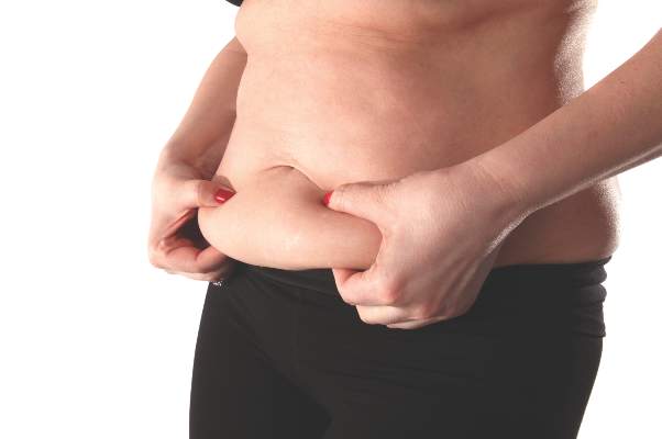
Radiofrequency Device Significantly Cut Abdominal Fat
KISSIMMEE, FLA. – Eight treatments with a noninvasive radiofrequency device reduced unwanted abdominal fat by an average of 428 cc in a prospective study.
“Can we consider this permanent fat loss? Yes, if a healthy lifestyle is maintained,” said Dr. Diane Duncan of Plastic Surgical Associates in Fort Collins, Colo. “I think the best candidates for these treatments are thin to modestly overweight patients, because while we did see significant fat reductions in obese patients, they want all their fat gone, and this won’t do that,” she said at the annual conference of the American Society for Laser Medicine and Surgery.

The study assessed the effects of the BodyFX device (Invasix, Yokneam, Israel) on 19 women aged 20-65 years who had localized abdominal adiposity, with body mass indices ranging from 18 to 30 kg/m2. Dr. Duncan used an energy setting of 42 watts until the target area reached 41° Celsius, which typically took 3-5 minutes, she said. Then she administered high-voltage pulses of oscillating radiofrequency waves in order to create holes in the cell walls of adipocytes, which triggered gradual cell death.
After adjusting for age and height, eight treatments with the device led to a 53% decrease in abdominal fat thickness at a 3-month follow-up visit, Dr. Duncan reported. “Visual results were similar to liposuction,” she added. “But generally, not much change was noted before the fourth treatment.”
The analysis included sequences of three-dimensional Vectra ultrasound measurements, which were taken at four sites around the abdomen at the moment when patients reached their maximum exhale, Dr. Duncan said. She excluded two patients from final analyses because they had gained or lost more than 5 pounds, compared with baseline.
“I think the mechanism of action of this device is what makes it so special,” said Dr. Duncan. Scanning electron microscope photographs showed that adipocytes treated with the radiofrequency device underwent pyroptosis, which combines programmed cell death with the proinflammatory features of necrosis. That process stimulates formation of new collagen, resulting in localized skin tightening, she noted. “These new SEM data show a different and more beneficial process is taking place,” she explained. “Apoptosis, by definition, would not stimulate neocollagenesis, so no tissue tightening would occur.”
Dr. Duncan reported no funding sources or relevant financial disclosures.
KISSIMMEE, FLA. – Eight treatments with a noninvasive radiofrequency device reduced unwanted abdominal fat by an average of 428 cc in a prospective study.
“Can we consider this permanent fat loss? Yes, if a healthy lifestyle is maintained,” said Dr. Diane Duncan of Plastic Surgical Associates in Fort Collins, Colo. “I think the best candidates for these treatments are thin to modestly overweight patients, because while we did see significant fat reductions in obese patients, they want all their fat gone, and this won’t do that,” she said at the annual conference of the American Society for Laser Medicine and Surgery.
The study assessed the effects of the BodyFX device (Invasix, Yokneam, Israel) on 19 women aged 20-65 years who had localized abdominal adiposity, with body mass indices ranging from 18 to 30 kg/m2. Dr. Duncan used an energy setting of 42 watts until the target area reached 41° Celsius, which typically took 3-5 minutes, she said. Then she administered high-voltage pulses of oscillating radiofrequency waves in order to create holes in the cell walls of adipocytes, which triggered gradual cell death.
After adjusting for age and height, eight treatments with the device led to a 53% decrease in abdominal fat thickness at a 3-month follow-up visit, Dr. Duncan reported. “Visual results were similar to liposuction,” she added. “But generally, not much change was noted before the fourth treatment.”
The analysis included sequences of three-dimensional Vectra ultrasound measurements, which were taken at four sites around the abdomen at the moment when patients reached their maximum exhale, Dr. Duncan said. She excluded two patients from final analyses because they had gained or lost more than 5 pounds, compared with baseline.
“I think the mechanism of action of this device is what makes it so special,” said Dr. Duncan. Scanning electron microscope photographs showed that adipocytes treated with the radiofrequency device underwent pyroptosis, which combines programmed cell death with the proinflammatory features of necrosis. That process stimulates formation of new collagen, resulting in localized skin tightening, she noted. “These new SEM data show a different and more beneficial process is taking place,” she explained. “Apoptosis, by definition, would not stimulate neocollagenesis, so no tissue tightening would occur.”
Dr. Duncan reported no funding sources or relevant financial disclosures.
AT LASER 2015
Radiofrequency device significantly cut abdominal fat
KISSIMMEE, FLA. – Eight treatments with a noninvasive radiofrequency device reduced unwanted abdominal fat by an average of 428 cc in a prospective study.
“Can we consider this permanent fat loss? Yes, if a healthy lifestyle is maintained,” said Dr. Diane Duncan of Plastic Surgical Associates in Fort Collins, Colo. “I think the best candidates for these treatments are thin to modestly overweight patients, because while we did see significant fat reductions in obese patients, they want all their fat gone, and this won’t do that,” she said at the annual conference of the American Society for Laser Medicine and Surgery.

The study assessed the effects of the BodyFX device (Invasix, Yokneam, Israel) on 19 women aged 20-65 years who had localized abdominal adiposity, with body mass indices ranging from 18 to 30 kg/m2. Dr. Duncan used an energy setting of 42 watts until the target area reached 41° Celsius, which typically took 3-5 minutes, she said. Then she administered high-voltage pulses of oscillating radiofrequency waves in order to create holes in the cell walls of adipocytes, which triggered gradual cell death.
After adjusting for age and height, eight treatments with the device led to a 53% decrease in abdominal fat thickness at a 3-month follow-up visit, Dr. Duncan reported. “Visual results were similar to liposuction,” she added. “But generally, not much change was noted before the fourth treatment.”
The analysis included sequences of three-dimensional Vectra ultrasound measurements, which were taken at four sites around the abdomen at the moment when patients reached their maximum exhale, Dr. Duncan said. She excluded two patients from final analyses because they had gained or lost more than 5 pounds, compared with baseline.
“I think the mechanism of action of this device is what makes it so special,” said Dr. Duncan. Scanning electron microscope photographs showed that adipocytes treated with the radiofrequency device underwent pyroptosis, which combines programmed cell death with the proinflammatory features of necrosis. That process stimulates formation of new collagen, resulting in localized skin tightening, she noted. “These new SEM data show a different and more beneficial process is taking place,” she explained. “Apoptosis, by definition, would not stimulate neocollagenesis, so no tissue tightening would occur.”
Dr. Duncan reported no funding sources or relevant financial disclosures.
KISSIMMEE, FLA. – Eight treatments with a noninvasive radiofrequency device reduced unwanted abdominal fat by an average of 428 cc in a prospective study.
“Can we consider this permanent fat loss? Yes, if a healthy lifestyle is maintained,” said Dr. Diane Duncan of Plastic Surgical Associates in Fort Collins, Colo. “I think the best candidates for these treatments are thin to modestly overweight patients, because while we did see significant fat reductions in obese patients, they want all their fat gone, and this won’t do that,” she said at the annual conference of the American Society for Laser Medicine and Surgery.
The study assessed the effects of the BodyFX device (Invasix, Yokneam, Israel) on 19 women aged 20-65 years who had localized abdominal adiposity, with body mass indices ranging from 18 to 30 kg/m2. Dr. Duncan used an energy setting of 42 watts until the target area reached 41° Celsius, which typically took 3-5 minutes, she said. Then she administered high-voltage pulses of oscillating radiofrequency waves in order to create holes in the cell walls of adipocytes, which triggered gradual cell death.
After adjusting for age and height, eight treatments with the device led to a 53% decrease in abdominal fat thickness at a 3-month follow-up visit, Dr. Duncan reported. “Visual results were similar to liposuction,” she added. “But generally, not much change was noted before the fourth treatment.”
The analysis included sequences of three-dimensional Vectra ultrasound measurements, which were taken at four sites around the abdomen at the moment when patients reached their maximum exhale, Dr. Duncan said. She excluded two patients from final analyses because they had gained or lost more than 5 pounds, compared with baseline.
“I think the mechanism of action of this device is what makes it so special,” said Dr. Duncan. Scanning electron microscope photographs showed that adipocytes treated with the radiofrequency device underwent pyroptosis, which combines programmed cell death with the proinflammatory features of necrosis. That process stimulates formation of new collagen, resulting in localized skin tightening, she noted. “These new SEM data show a different and more beneficial process is taking place,” she explained. “Apoptosis, by definition, would not stimulate neocollagenesis, so no tissue tightening would occur.”
Dr. Duncan reported no funding sources or relevant financial disclosures.
AT LASER 2015
Key clinical point: A noninvasive radiofrequency device significantly reduced abdominal fat in adults.
Major finding: Eight treatments with the device reduced abdominal fat by an average of 428 cc.
Data source: Prospective, 5-month study of 17 patients with localized abdominal adiposity.
Disclosures: Dr. Duncan reported no funding sources or relevant financial disclosures.

Early combination laser treatment boosts scar healing
KISSIMMEE, FLA. – Early treatment with fractional carbon dioxide ablative laser and light-emitting diode phototherapy sped healing and improved texture, discoloration, and range of motion in hypertrophic burn scars in a controlled, blinded study of 20 patients.
“I think as long as we have intact epithelium, we’re going to have improvements if we can laser these patients as early as possible,” Dr. Jill Waibel said at the annual conference of the American Society for Laser Medicine and Surgery.

Approximately 100 million new scars occur annually in developed countries, almost a third of which result from trauma or burns, noted Dr. Waibel, a dermatologist specializing in treating burn scars at the Miami Dermatology and Laser Institute. Scarred areas begin to hypertrophy about 3-7 months after injury, when they undergo increased collagen production and slowed collagen breakdown, said Dr. Waibel. “Traditionally, reconstructive efforts have been delayed until a year after injury,” she emphasized. “But by this time, many patients have formed hypertrophic scars and have significant decreases in range of motion.”
To determine how early treatment with a combination of laser/LED therapy might interrupt hypertrophy and improve outcomes, Dr. Waibel and coinvestigator Ashley Rudnick treated 20 patients aged 21-55 years with moderate to severe burns on at least 5% of the body surface area that had occurred in the past 1-3 months. For each patient, they divided similarly scarred areas into four 5- by 5-cm squares, one of which served as an untreated control. They treated the other three areas with 120-micron spot fractional ablative carbon dioxide laser only, continuous wave LED phototherapy only, or fractional ablative laser followed by LED. They spaced the three laser sessions 2 months apart, and used eight 20-minute LED treatments per laser session. The fractional laser wavelength was 10,600 nm, energy was 80-120 mJ per pixel, and the frequency was up to 200 Hz, Dr. Waibel said. The LED wavelength was 830 nm, and energy intensity was 40-100 mW per cm2.
All treated areas improved more than the untreated control areas of scarring, but combining the modalities was associated with the fastest healing, according to blinded photographic reviews by physicians based on the visual analog Manchester scar scale, Dr. Waibel reported.
Phototherapy with LED can penetrate up to an inch of tissue and promotes selective ATP production and DNA and RNA activity, she added. “The positive effects occur only in injured cells,” she said. “LED has wound healing and anti-inflammatory effects that might benefit acutely wounded skin.”
Based on her experience, patients usually need from three to five sessions of fractional laser/LED sessions to see clear results, Dr. Waibel said in response to a question from an audience member. “We know that every time we treat a hypertrophic scar, it will get better,” she added. “When patients stop coming for treatment, you know they’re happy. If you can combine modalities softly and safely, you’ll need fewer treatments.”
Dr. Waibel disclosed financial and advisory relationships with ALMA, Cutera, DUSA, Harvest Technologies, L’Oreal/SkinCeuticals, Lumenis, Lutronics, Sebacia, Syneron-Candela, Valeant, and Zeltiq.
KISSIMMEE, FLA. – Early treatment with fractional carbon dioxide ablative laser and light-emitting diode phototherapy sped healing and improved texture, discoloration, and range of motion in hypertrophic burn scars in a controlled, blinded study of 20 patients.
“I think as long as we have intact epithelium, we’re going to have improvements if we can laser these patients as early as possible,” Dr. Jill Waibel said at the annual conference of the American Society for Laser Medicine and Surgery.
Approximately 100 million new scars occur annually in developed countries, almost a third of which result from trauma or burns, noted Dr. Waibel, a dermatologist specializing in treating burn scars at the Miami Dermatology and Laser Institute. Scarred areas begin to hypertrophy about 3-7 months after injury, when they undergo increased collagen production and slowed collagen breakdown, said Dr. Waibel. “Traditionally, reconstructive efforts have been delayed until a year after injury,” she emphasized. “But by this time, many patients have formed hypertrophic scars and have significant decreases in range of motion.”
To determine how early treatment with a combination of laser/LED therapy might interrupt hypertrophy and improve outcomes, Dr. Waibel and coinvestigator Ashley Rudnick treated 20 patients aged 21-55 years with moderate to severe burns on at least 5% of the body surface area that had occurred in the past 1-3 months. For each patient, they divided similarly scarred areas into four 5- by 5-cm squares, one of which served as an untreated control. They treated the other three areas with 120-micron spot fractional ablative carbon dioxide laser only, continuous wave LED phototherapy only, or fractional ablative laser followed by LED. They spaced the three laser sessions 2 months apart, and used eight 20-minute LED treatments per laser session. The fractional laser wavelength was 10,600 nm, energy was 80-120 mJ per pixel, and the frequency was up to 200 Hz, Dr. Waibel said. The LED wavelength was 830 nm, and energy intensity was 40-100 mW per cm2.
All treated areas improved more than the untreated control areas of scarring, but combining the modalities was associated with the fastest healing, according to blinded photographic reviews by physicians based on the visual analog Manchester scar scale, Dr. Waibel reported.
Phototherapy with LED can penetrate up to an inch of tissue and promotes selective ATP production and DNA and RNA activity, she added. “The positive effects occur only in injured cells,” she said. “LED has wound healing and anti-inflammatory effects that might benefit acutely wounded skin.”
Based on her experience, patients usually need from three to five sessions of fractional laser/LED sessions to see clear results, Dr. Waibel said in response to a question from an audience member. “We know that every time we treat a hypertrophic scar, it will get better,” she added. “When patients stop coming for treatment, you know they’re happy. If you can combine modalities softly and safely, you’ll need fewer treatments.”
Dr. Waibel disclosed financial and advisory relationships with ALMA, Cutera, DUSA, Harvest Technologies, L’Oreal/SkinCeuticals, Lumenis, Lutronics, Sebacia, Syneron-Candela, Valeant, and Zeltiq.
AT LASER 2015
Key clinical point: Fractional carbon dioxide ablative laser with light-emitting diode (LED) phototherapy sped healing and improved the appearance and function of burn scars.
Major finding: Both laser and LED alone improved scar healing, compared with untreated control scars, but combining the modalities led to the best outcomes. The fractional laser wavelength was 10,600 nm, and the LED wavelength was 830 nm.
Data source: Prospective, blinded, controlled study of 20 adults with acute moderate to severe burns.
Disclosures: Dr. Waibel disclosed financial and advisory relationships with ALMA, Cutera, DUSA, Harvest Technologies, L’Oreal/Skinceuticals, Lumenis, Lutronic, Sebacia, Syneron-Candela, Valeant, and Zeltiq.

Microwave system cuts axillary hair by 70%
KISSIMMEE, FLA. – A noninvasive microwave system reduced light and dark axillary hair by approximately 70%, with durable results 1 year after treatment, according to data from a prospective multicenter study of 44 patients.
The device is Food and Drug Administration–approved for thermal treatment of axillary sweat glands, and has been used for about 35,000 such procedures, Dr. Jeremy Brauer said at the annual meeting of the American Society for Laser Medicine and Surgery.
The system targets microwave energy at the interface between the dermis and the hypodermis, “and we hypothesized that heat at this level in the skin may also have an important effect on the hair bulb,” he said.
Hair reduction is the third most common cosmetic procedure that U.S. dermatologists perform, said Dr. Brauer, a dermatologist with the Laser & Skin Surgery Center of New York. Laser is the primary modality, but is not always effective and can cause serious tissue damage if melanin concentrations in the hair and skin are too similar, he said.
For the study, Dr. Bauer and his associates used the microwave device to treat the axillae of 44 patients during either one or two treatment sessions. Average age of the patients was 33 years (range, 18-61 years), 80% were female, and 62% had dark axillary hair. Most had Fitzpatrick skin type II (33%), III (27%), or I (23%), but about 18% had type IV or V. During follow-up visits at 3, 6, 9, and 12 months, three blinded assessors counted hairs within a 2 cm x 2 cm area in the center of each axilla, and also qualitatively compared follow-up and baseline photos.
The blinded assessors correctly chose the baseline photos for all the photographic series, Dr. Brauer said. At 12 months, mean hair reduction was 75% for all patients, 72% for light-haired patients, and 76% for dark-haired patients, he reported. Ranges of response within these subgroups were relatively narrow, but patients who were treated at higher energy levels (settings of 3 or 4) achieved significantly more hair reduction (P < .0001) at 12 months than did patients who were treated at a setting of 1, he said. He recommended that future studies examine dose response and optimal number of treatment sessions.
Dr. Brauer reported consulting fees from Miramar, maker of the device.
AT LASER 2015
Key clinical point: A noninvasive microwave device reduced axillary hair by about 70%, with stable results 1 year later.
Major finding: Mean reduction in both light and dark axillary hair was about 70%, 12 months after one to two treatment sessions.
Data source: Prospective multicenter study of 44 patients with blinded assessments.
Disclosures: Dr. Brauer reported consulting fees from Miramar, maker of the device.
Up the anesthesia when treating axillary hyperhidrosis
KISSIMMEE, FLA. – Patients who received more local anesthesia before undergoing microwave-based treatment for axillary hyperhidrosis were happier with results and achieved the same objective improvements as patients who received less anesthesia, a follow-up study found.
“Patient satisfaction with the procedure was extremely high,” said Dr. Carolyn Jacob, a dermatologist and founding director of Chicago Cosmetic Surgery and Dermatology. “You can’t regenerate sweat glands – once they’re gone, they’re gone, and these patients have sustained results.”
The microwave system has been used commercially for more than 30,000 treatment sessions over 3 years and achieved high rates of efficacy and patient satisfaction in two pivotal trials, Dr. Jacob noted (Dermatol. Surg. 2012;38:185-91; Dermatol. Surg. 2012;38:728-35).
But clinicians often have kept to the relatively low volumes of local anesthesia used in the trials because they were concerned that higher volumes would interact with microwave energy and diminish efficacy, she said at the annual meeting of the American Society for Laser Medicine and Surgery.
To investigate that concern and assess overall longer-term treatment outcomes, Dr. Jacob and her associates compared 110 patients who received 10-25 cc of local lidocaine/epinephrine solution per axilla (the amount used in the clinical trials) with 30 patients who received 60-275 cc per axilla before undergoing microwave-based treatment of hyperhidrosis. At baseline, the two groups scored similarly on the 4-point measure of hyperhidrosis severity and on 10-point measures of sweat and odor severity (average scores of 3.4, 8.5, and 5.2, respectively, for the group that received less anesthesia and 3.3, 8.7, and 5.8, respectively, for the group that received more anesthesia), said Dr. Jacob. Patients were treated at energy levels of 3-5, and most were female. The researchers followed the lower anesthesia group and the higher anesthesia group for an average of 6 and 9 months, respectively.
At follow-up, 88% of patients in the lower-volume anesthesia group scored 1 or 2 on the 4-point measure of hyperhidrosis, as did 93% of patients in the higher-volume group, said Dr. Jacob. The groups also had very similar efficacy results on the 10-point measures of sweat and odor severity, she said (respective average scores, 3.0 and 3.2 on the sweat measure, and 1.9 and 2.0 on the odor measure). In all, 85% of patients who received less anesthesia said they were satisfied with results, as did 93% of patients in the higher-volume group.
Patients usually needed two treatment sessions that lasted 60-75 minutes each, depending on the size of the treatment area, Dr. Jacob noted. A few needed only one session. “There’s a high efficacy that’s consistent with the previous publications we have,” she concluded. “The high volumes of anesthesia seem to work just as well as the lower volumes.”
AT LASER 2015
Key clinical point: Higher volumes of local anesthesia did not adversely affect the efficacy of microwave-based treatment of axillary hyperhidrosis.
Major finding: At follow-up, 88% of the lower-volume group and 93% of the higher-volume group<b/>scored 1 or 2 on the 4-point measure of hyperhidrosis.
Data source: Multicenter follow-up study of 110 patients who received the standard low amount of local anesthesia and 30 who received higher volumes.
Disclosures: Dr. Jacob reported financial relationships with Miramar, Abbvie, Allergan, Galderma, and Medicis/Valeant.
LASER: How to succeed in perioperative ablative laser resurfacing
KISSIMMEE, FLA. – Ablative laser resurfacing may be the most effective cosmetic treatment for the periorbital area, excluding blepharoplasty, according to Dr. Suzanne L. Kilmer, a dermatologist specializing in laser and skin surgery in Sacramento, Calif.
“It gives you definite tightening and has the most predictable results,” Dr. Kilmer said at the annual meeting of the American Society for Laser Medicine and Surgery. “It also has the worst down time and side effects, but not for the upper lid – I’ve never had any scarring on the upper eyelid.”

In addition to its cosmetic potential, ablative laser resurfacing can remove epidermal lesions that might otherwise become actinic keratoses or basal cell carcinomas, and can smooth out syringomas, nevi, and seborrheic keratoses, Dr. Kilmer said. She shared some of her top tips for successful use of this technique.
Technique, safety, and analgesia
When treating patients with perioperative ablative laser resurfacing, Dr. Kilmer said she performs the first pass with a carbon dioxide laser at computerized pattern-generator (CPG) settings of pattern 3, size 9, density 6. She follows that with erbium laser resurfacing at a depth of 75-100 microns. “The first pass removes the epidermis,” she said. “Be sure to feather peripherally, and wipe everywhere you will do a second pass.”
The skin usually tightens visibly with the second pass, which can include the upper eyelids if patients have lax skin in this area, Dr. Kilmer noted. “You can be aggressive, like 3-9-5 [CPG settings],” she added. She then performs a “very light” second pass to the lower lids, with lower CPG settings of 3-7-3 or 3-6-2. She again follows with erbium ablation, including to the areas of the central face and midcheek.
The third pass includes the glabella and perioral area, as well as the upper eyelids if they are especially lax, Dr. Kilmer said. She ends with a sculpting pass with erbium laser.
Dr. Kilmer explained that she always treats the upper eyelids with ablative laser therapy in combination with fractional carbon dioxide laser treatment. “I also will include any area I think looks safe to do ablative on an individual patient,” she added. She noted that she sometimes combines ablative and fractional therapies with botulinum toxin treatment.
Patients should always wear eye shields during laser treatment sessions, Dr. Kilmer emphasized. Acceptable options include David-Baker lid clamps, Jaeger lid plates, and individual steel eye shields, but plastic corneal protectors should be avoided because they can sustain thermal damage with laser exposure, she said.
Options for analgesia during treatment include codeine, hydrocodone, hydromorphone, or nonsteroidal anti-inflammatory agents such as Toradol (ketorolac), which Dr. Kilmer gives intramuscularly at a dose of 30-60 mg. Furthermore, EMLA (lidocaine/prilocaine) cream enhances comfort, even if patients are also receiving conscious sedation or blocks, she said. The cream helps prevent several adverse effects, including superficial coagulative thermal damage, prolonged erythema, and delayed hypopigmentation, and does not seem to decrease efficacy, she added.
Before treatment
Clinicians should consider how a patient’s unique characteristics might affect treatment outcomes, Dr. Kilmer said. For example, ablation is more likely to cause hypopigmentation in patients with lighter skin types, and is unlikely to sufficiently resolve wrinkles that involve the facial muscles. “Always discuss Botox [botulinum toxin],” added Dr Kilmer. “If you’re tightening up the skin, and the area underneath is moving a lot, you’re not going to get as much tightening.” Clinicians also should discuss a face lift as an option for patients with substantial excess facial skin, she said.
The overall treatment plan should address existing skin lesions, too. Larger syringomas, trichoepitheliomas, and hyperplastic sebaceous glands should be “drilled out” with erbium ablation, but small tumors usually need only multiple or fractional laser treatment, said Dr. Kilmer.
For antimicrobial prophylaxis, patients should receive acyclovir, valcyclovir, or famvir starting 1 day before the procedure and continuing for 10-14 days, Dr. Kilmer said. “If I’m just treating around the eyes, I don’t worry as much about antivirals. I think about them more if I’m doing the full face,” she noted. Likewise, preoperative antibiotics such as doxycycline, cephalexin, azithromycin, or ciprofloxacin are especially important for full face or perioral ablative resurfacing, she said. To help prevent yeast infections, she gives patients a dose of fluconazole on day 3 after the procedure. “If they have a history of multiple yeast infections, I give it four times a day,” she said.
After the procedure
Postoperative diligence is key to good outcomes, Dr. Kilmer emphasized. During the first 10-14 days after the procedure, patients and clinicians should watch for signs of complications such as pain, purulence, itching, and worsening redness. If patients develop pain accompanied by purulence and redness, clinicians should perform bacterial cultures and switch patients to a quinolone, Dr. Kilmer said. Patients who develop pain consistent with herpes simplex virus infection should receive valcyclovir, she added. “If they’re itchy and red, think about yeast [infection] or contact dermatitis, and treat for both if you’re unsure,” she said.
Dr. Kilmer disclosed advisory relationships with Candela-Syneron, Living Proof, Lumeis, Miramar, Ulthera, and Zeltiq. She reported having received research funding from the same companies, and also from Allergan, CoolTouch, Cutera, Cynosure, Palomar, Sciton, and Solta.
KISSIMMEE, FLA. – Ablative laser resurfacing may be the most effective cosmetic treatment for the periorbital area, excluding blepharoplasty, according to Dr. Suzanne L. Kilmer, a dermatologist specializing in laser and skin surgery in Sacramento, Calif.
“It gives you definite tightening and has the most predictable results,” Dr. Kilmer said at the annual meeting of the American Society for Laser Medicine and Surgery. “It also has the worst down time and side effects, but not for the upper lid – I’ve never had any scarring on the upper eyelid.”
In addition to its cosmetic potential, ablative laser resurfacing can remove epidermal lesions that might otherwise become actinic keratoses or basal cell carcinomas, and can smooth out syringomas, nevi, and seborrheic keratoses, Dr. Kilmer said. She shared some of her top tips for successful use of this technique.
Technique, safety, and analgesia
When treating patients with perioperative ablative laser resurfacing, Dr. Kilmer said she performs the first pass with a carbon dioxide laser at computerized pattern-generator (CPG) settings of pattern 3, size 9, density 6. She follows that with erbium laser resurfacing at a depth of 75-100 microns. “The first pass removes the epidermis,” she said. “Be sure to feather peripherally, and wipe everywhere you will do a second pass.”
The skin usually tightens visibly with the second pass, which can include the upper eyelids if patients have lax skin in this area, Dr. Kilmer noted. “You can be aggressive, like 3-9-5 [CPG settings],” she added. She then performs a “very light” second pass to the lower lids, with lower CPG settings of 3-7-3 or 3-6-2. She again follows with erbium ablation, including to the areas of the central face and midcheek.
The third pass includes the glabella and perioral area, as well as the upper eyelids if they are especially lax, Dr. Kilmer said. She ends with a sculpting pass with erbium laser.
Dr. Kilmer explained that she always treats the upper eyelids with ablative laser therapy in combination with fractional carbon dioxide laser treatment. “I also will include any area I think looks safe to do ablative on an individual patient,” she added. She noted that she sometimes combines ablative and fractional therapies with botulinum toxin treatment.
Patients should always wear eye shields during laser treatment sessions, Dr. Kilmer emphasized. Acceptable options include David-Baker lid clamps, Jaeger lid plates, and individual steel eye shields, but plastic corneal protectors should be avoided because they can sustain thermal damage with laser exposure, she said.
Options for analgesia during treatment include codeine, hydrocodone, hydromorphone, or nonsteroidal anti-inflammatory agents such as Toradol (ketorolac), which Dr. Kilmer gives intramuscularly at a dose of 30-60 mg. Furthermore, EMLA (lidocaine/prilocaine) cream enhances comfort, even if patients are also receiving conscious sedation or blocks, she said. The cream helps prevent several adverse effects, including superficial coagulative thermal damage, prolonged erythema, and delayed hypopigmentation, and does not seem to decrease efficacy, she added.
Before treatment
Clinicians should consider how a patient’s unique characteristics might affect treatment outcomes, Dr. Kilmer said. For example, ablation is more likely to cause hypopigmentation in patients with lighter skin types, and is unlikely to sufficiently resolve wrinkles that involve the facial muscles. “Always discuss Botox [botulinum toxin],” added Dr Kilmer. “If you’re tightening up the skin, and the area underneath is moving a lot, you’re not going to get as much tightening.” Clinicians also should discuss a face lift as an option for patients with substantial excess facial skin, she said.
The overall treatment plan should address existing skin lesions, too. Larger syringomas, trichoepitheliomas, and hyperplastic sebaceous glands should be “drilled out” with erbium ablation, but small tumors usually need only multiple or fractional laser treatment, said Dr. Kilmer.
For antimicrobial prophylaxis, patients should receive acyclovir, valcyclovir, or famvir starting 1 day before the procedure and continuing for 10-14 days, Dr. Kilmer said. “If I’m just treating around the eyes, I don’t worry as much about antivirals. I think about them more if I’m doing the full face,” she noted. Likewise, preoperative antibiotics such as doxycycline, cephalexin, azithromycin, or ciprofloxacin are especially important for full face or perioral ablative resurfacing, she said. To help prevent yeast infections, she gives patients a dose of fluconazole on day 3 after the procedure. “If they have a history of multiple yeast infections, I give it four times a day,” she said.
After the procedure
Postoperative diligence is key to good outcomes, Dr. Kilmer emphasized. During the first 10-14 days after the procedure, patients and clinicians should watch for signs of complications such as pain, purulence, itching, and worsening redness. If patients develop pain accompanied by purulence and redness, clinicians should perform bacterial cultures and switch patients to a quinolone, Dr. Kilmer said. Patients who develop pain consistent with herpes simplex virus infection should receive valcyclovir, she added. “If they’re itchy and red, think about yeast [infection] or contact dermatitis, and treat for both if you’re unsure,” she said.
Dr. Kilmer disclosed advisory relationships with Candela-Syneron, Living Proof, Lumeis, Miramar, Ulthera, and Zeltiq. She reported having received research funding from the same companies, and also from Allergan, CoolTouch, Cutera, Cynosure, Palomar, Sciton, and Solta.
EXPERT ANALYSIS FROM LASER 2015

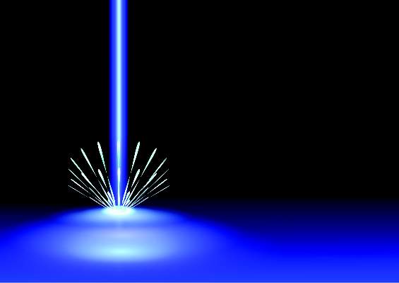
Laser treatment cut the fat on flank and abs
KISSIMMEE, FLA. – Single noninvasive treatments with a prototype 1,060-nm diode laser led to statistically significant, visually discernible reductions of adipose tissue covering the abdomen and flanks in two prospective multicenter studies.
“The treatment is well tolerated, with only mild and transient side effects,” said Dr. Bruce Katz of Mount Sinai School of Medicine in New York, who presented the results of the flank study at the annual meeting of the American Society for Laser Medicine and Surgery.
Most patients were “satisfied” or “very satisfied” with the results, added Dr. Sean Doherty, a plastic surgeon in group practice in Boston, who presented the results of the abdominal study.

Noninvasive fat reduction procedures rose 43% in the past year, said Dr. Doherty. The 1,060-nm diode laser damages adipocytes in targeted areas, eventually resulting in apoptosis, he added. Sessions in both studies lasted 25 minutes, with irradiance at 0.9-1.4 W/cm2 and treatment areas of 48-144 cm2 for the flanks and 192-288 cm2 for the abdomen, the investigators said.
The abdominal study included 34 patients, and the flank study included 42 patients. Investigators treated only one side for each patient in the flank study, reserving the other as a “sham” or control, Dr. Katz said. A blinded review of photographs taken before and 12 weeks after the procedures served as the primary endpoint in both studies. Assessors correctly identified an average of 95% of “before” photographs of the abdomen and 90.3% of those of the flank, Dr. Doherty and Dr. Katz reported.
The secondary endpoint was change in thickness of the adipose layer at 12 weeks post procedure, as measured by ultrasound. The average reduction was 3.1 mm for the abdomen (standard deviation, 1.7 mm) and 2.6 mm for the flank (standard deviation not reported; P < .001 for both), the researchers said. In the abdominal study, 85% of patients said they were “satisfied” or “extremely satisfied” with the results, while 6% were “slightly satisfied,” Dr. Doherty said. In the flank study, 86% of patients said they were “satisfied” or “extremely satisfied,” while 10% were “slightly satisfied.”
Mean pain scores were 3.7 out of 10 for the abdominal procedure and 4.0 out of 10 for the flank procedure. The most common side effect was mild to moderate tenderness that usually subsided within about 2 weeks of treatment. Other side effects included localized firmness, edema, and ecchymosis. No patients had serious adverse effects, the researchers said.
Most of the patients were approximately 45-49 years of age. Those in the abdominal study had an average body mass index of 25.5 kg/m2, while those in the flank study averaged 26.4 kg/m2, the researchers reported. Most of the patients in both studies were women (94% of abdominal patients and 86% of flank patients).
The average weight gain at 12 weeks for the abdominal study was 0.1 pounds (range, –5.2 to 5.6 pounds), while patients in the flank study gained an average of 0.8 pounds (range, –0.7 to 13 pounds), the researchers noted. Six patients in the flank study gained more than 5 pounds, and one gained more than 37 pounds and was therefore excluded from the efficacy analysis, Dr. Katz noted.
The researchers did not report funding sources for the studies. They disclosed advisory and financial relationships with Allergan, Merz Pharmaceuticals, Cynosure, Valeant, Syneron, and several other pharmaceutical companies.
KISSIMMEE, FLA. – Single noninvasive treatments with a prototype 1,060-nm diode laser led to statistically significant, visually discernible reductions of adipose tissue covering the abdomen and flanks in two prospective multicenter studies.
“The treatment is well tolerated, with only mild and transient side effects,” said Dr. Bruce Katz of Mount Sinai School of Medicine in New York, who presented the results of the flank study at the annual meeting of the American Society for Laser Medicine and Surgery.
Most patients were “satisfied” or “very satisfied” with the results, added Dr. Sean Doherty, a plastic surgeon in group practice in Boston, who presented the results of the abdominal study.
Noninvasive fat reduction procedures rose 43% in the past year, said Dr. Doherty. The 1,060-nm diode laser damages adipocytes in targeted areas, eventually resulting in apoptosis, he added. Sessions in both studies lasted 25 minutes, with irradiance at 0.9-1.4 W/cm2 and treatment areas of 48-144 cm2 for the flanks and 192-288 cm2 for the abdomen, the investigators said.
The abdominal study included 34 patients, and the flank study included 42 patients. Investigators treated only one side for each patient in the flank study, reserving the other as a “sham” or control, Dr. Katz said. A blinded review of photographs taken before and 12 weeks after the procedures served as the primary endpoint in both studies. Assessors correctly identified an average of 95% of “before” photographs of the abdomen and 90.3% of those of the flank, Dr. Doherty and Dr. Katz reported.
The secondary endpoint was change in thickness of the adipose layer at 12 weeks post procedure, as measured by ultrasound. The average reduction was 3.1 mm for the abdomen (standard deviation, 1.7 mm) and 2.6 mm for the flank (standard deviation not reported; P < .001 for both), the researchers said. In the abdominal study, 85% of patients said they were “satisfied” or “extremely satisfied” with the results, while 6% were “slightly satisfied,” Dr. Doherty said. In the flank study, 86% of patients said they were “satisfied” or “extremely satisfied,” while 10% were “slightly satisfied.”
Mean pain scores were 3.7 out of 10 for the abdominal procedure and 4.0 out of 10 for the flank procedure. The most common side effect was mild to moderate tenderness that usually subsided within about 2 weeks of treatment. Other side effects included localized firmness, edema, and ecchymosis. No patients had serious adverse effects, the researchers said.
Most of the patients were approximately 45-49 years of age. Those in the abdominal study had an average body mass index of 25.5 kg/m2, while those in the flank study averaged 26.4 kg/m2, the researchers reported. Most of the patients in both studies were women (94% of abdominal patients and 86% of flank patients).
The average weight gain at 12 weeks for the abdominal study was 0.1 pounds (range, –5.2 to 5.6 pounds), while patients in the flank study gained an average of 0.8 pounds (range, –0.7 to 13 pounds), the researchers noted. Six patients in the flank study gained more than 5 pounds, and one gained more than 37 pounds and was therefore excluded from the efficacy analysis, Dr. Katz noted.
The researchers did not report funding sources for the studies. They disclosed advisory and financial relationships with Allergan, Merz Pharmaceuticals, Cynosure, Valeant, Syneron, and several other pharmaceutical companies.
AT LASER 2015
Key clinical point: One 25-minute noninvasive treatment with a 1,060-nm laser significantly reduced abdominal and flank fat.
Major finding: At 12 weeks, blinded assessors correctly chose 95% and 90.3% of “before” (compared with “after”) photographs of the abdomen and flank, respectively.
Data source: Two prospective multicenter studies of 34 patients who underwent abdominal treatment and 42 patients who underwent flank treatment.
Disclosures: Dr. Katz and Dr. Doherty did not report funding sources for the studies. They disclosed advisory and financial relationships with Allergan, Merz Pharmaceuticals, Cynosure, Valeant, Syneron, and several other pharmaceutical companies.
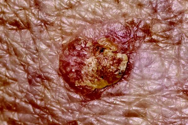
Imaging guides BCC laser ablation
KISSIMMEE, FLA. – Reflective confocal microscopic imaging successfully guided carbon dioxide laser ablation of basal cell carcinomas, and imaging results fully matched those from Mohs histology, a small study found.
“Our results suggest that reflective confocal microscopy can accurately guide carbon dioxide laser ablation of superficial and early nodular basal cell carcinomas,” said Dr. Brian Hibler of Memorial Sloan Kettering Cancer Center in New York. The technique provides a real-time, noninvasive way to delineate the tumor area before ablation and to check for residual tumor between passes with the laser, he said at the annual meeting of the American Society for Laser Medicine and surgery.
While conventional and Mohs microscopic surgeries remain the gold standard for removing basal carcinomas (BCC), surgery is not an option for some patients because of tumor location, comorbidities, or personal preferences, Dr. Hibler noted. Past studies have reported good clinical and cosmetic outcomes with laser ablation of BCCs, but use of the modality has been limited because there was no way to assess response without excision or biopsies. Reflective confocal microscopy (RCM) uses a low-powered laser system that provides cellular-level imaging and can distinguish BCCs, he said.
Dr. Hibler and his colleagues performed baseline RCM of eight BCCs (three on the trunk, three on the extremities, and two on the head and neck) from seven patients aged 29-83 years. Two patients were men and five were women. The patients then underwent carbon dioxide laser ablation with a wavelength of 10,600 nm, pulse duration of 750 microseconds, and fluence of 7.5 J/cm2, using a square pattern and density of 30%. If RCM revealed residual BCC, the researchers repeated the process up to two more times, for a maximum of three passes. They then removed the entire lesion using Mohs micrographic surgery, performing vertical histologic sectioning of the tissue.
Reflective confocal microscopy generated reliable cellular-level images in real time on the tumor surface and up to 150 mcm deep, Dr. Hibler reported. Tissue from BCCs appears as dense nodular areas with adjacent spaces and red blood cell trafficking, he noted. Microscopy results were consistent with Mohs histology findings in all eight cases, including six in which the tumor was completely removed and two with residual tumor. One of these two cases was the only infiltrative BCC in the series, while the other might have been tissue artifact, Dr. Hibler said.
Patients experienced no adverse effects from the interventions, Dr. Hibler reported. “Future studies are planned are planned without Mohs, so we can use reflective confocal microscopy to longitudinally monitor for recurrence,” he added.
The study won an award at the meeting.
Dr. Hibler reported no funding sources and made no disclosures.
Key clinical point: Reflective confocal microscopy offers noninvasive, real-time imaging to guide laser ablation of basal cell carcinomas.
Major finding: Results from RCM matched those from Mohs histology in all patients.
Data source: Prospective study of eight BCCs in seven patients.
Disclosures: Dr. Hibler reported no funding sources and made no disclosures.
Radiofrequency improves lower face laxity
KISSIMMEE, FLA. – Three treatments with a noninvasive radiofrequency device significantly improved mild to moderate laxity of the lower face and neck, a prospective study of 30 patients showed.
Downtime after treatment ranged from 2 to 4 days, and adverse effects included mild stippling and purpura at the treatment sites, Dr. Girish Munavalli reported at the annual meeting of the American Society for Laser Medicine and Surgery.

Aging causes progressive loosening of the tissues of the lower face and neck, which manifests as narrowing of the cervicomental angle (between the neck and the lowest point under the chin) and the gnathion angle (between the lowest point under the chin and the most anterior or prominent point on the front of the chin). Historically, patients resorted to invasive surgical lifting procedures to restore these angles, said Dr. Munavalli of Wake Forest University, Charlotte, N.C.
To explore noninvasive alternatives, he and his associates conducted a pilot study of a bipolar fractionated microneedle radiofrequency device that consisted of a handpiece and a 1 cm2 square disposable microneedle tip with 49 proximally insulated 34-G microneedle electrodes. The study comprised 7 men and 23 women aged 37-71 years. One physician treated all patients, and each patient underwent three treatment sessions lasting about 45 minutes and spaced a month apart.
Each session included three passes across the lower face and submental area at a maximum tissue depth of 1-3 mm, Dr. Munavalli said. Thicker skin requires deeper needle penetration, which in turn requires more treatment energy and longer pulse duration, he noted. Therefore, the physician performed the first pass at a depth of about 2.5 mm with an energy setting of 9-11 and a pulse duration of 280-320 msec, the second pass at a depth of 1.5 mm with a setting of 8-9 and a pulse duration of 230-250 msec, and the third pass at a depth of 1 mm with a setting of 6 and a pulse duration of 160 msec.
Six months after the end of treatment, patients underwent computerized measurements of the cervicomental and gnathion angles, and a panel of blinded investigators compared pre- and posttreatment photographs, Dr. Munavalli said. On average, the cervicomental angle increased by 27 degrees (range, 18 to 36 degrees; P < .01), and the gnathion angle increased by 16 degrees (range, 12 to 20 degrees; P < .01). The blinded assessors correctly chose the posttreatment photographs 90% of the time. Taken together, the results suggest that the device is a safe and effective option for improving laxity of the lower face and neck, Dr. Munavalli concluded.
Lutronic Corp is the maker of the Infini device, which the FDA approved for dermatologic use in July 2013. Dr. Munavalli reported no funding sources or financial disclosures.
KISSIMMEE, FLA. – Three treatments with a noninvasive radiofrequency device significantly improved mild to moderate laxity of the lower face and neck, a prospective study of 30 patients showed.
Downtime after treatment ranged from 2 to 4 days, and adverse effects included mild stippling and purpura at the treatment sites, Dr. Girish Munavalli reported at the annual meeting of the American Society for Laser Medicine and Surgery.
Aging causes progressive loosening of the tissues of the lower face and neck, which manifests as narrowing of the cervicomental angle (between the neck and the lowest point under the chin) and the gnathion angle (between the lowest point under the chin and the most anterior or prominent point on the front of the chin). Historically, patients resorted to invasive surgical lifting procedures to restore these angles, said Dr. Munavalli of Wake Forest University, Charlotte, N.C.
To explore noninvasive alternatives, he and his associates conducted a pilot study of a bipolar fractionated microneedle radiofrequency device that consisted of a handpiece and a 1 cm2 square disposable microneedle tip with 49 proximally insulated 34-G microneedle electrodes. The study comprised 7 men and 23 women aged 37-71 years. One physician treated all patients, and each patient underwent three treatment sessions lasting about 45 minutes and spaced a month apart.
Each session included three passes across the lower face and submental area at a maximum tissue depth of 1-3 mm, Dr. Munavalli said. Thicker skin requires deeper needle penetration, which in turn requires more treatment energy and longer pulse duration, he noted. Therefore, the physician performed the first pass at a depth of about 2.5 mm with an energy setting of 9-11 and a pulse duration of 280-320 msec, the second pass at a depth of 1.5 mm with a setting of 8-9 and a pulse duration of 230-250 msec, and the third pass at a depth of 1 mm with a setting of 6 and a pulse duration of 160 msec.
Six months after the end of treatment, patients underwent computerized measurements of the cervicomental and gnathion angles, and a panel of blinded investigators compared pre- and posttreatment photographs, Dr. Munavalli said. On average, the cervicomental angle increased by 27 degrees (range, 18 to 36 degrees; P < .01), and the gnathion angle increased by 16 degrees (range, 12 to 20 degrees; P < .01). The blinded assessors correctly chose the posttreatment photographs 90% of the time. Taken together, the results suggest that the device is a safe and effective option for improving laxity of the lower face and neck, Dr. Munavalli concluded.
Lutronic Corp is the maker of the Infini device, which the FDA approved for dermatologic use in July 2013. Dr. Munavalli reported no funding sources or financial disclosures.
Key clinical point: A noninvasive bipolar fractionated microneedle radiofrequency device significantly improved laxity of the lower face and neck.
Major finding: After three treatment sessions, the cervicomental and gnathion angles increased by an average of 27 degrees and 16 degrees, respectively.
Data source: Prospective study of 30 patients with mild to moderate laxity of the lower face and neck.
Disclosures: Dr. Munavalli reported no funding sources or financial disclosures.
Fractional laser resurfacing plus ALA-PDT upped AK clearance
KISSIMMEE, FLA. – Fractional carbon dioxide laser resurfacing followed by 30 minutes of aminolevulinic acid plus blue light photodynamic therapy cleared 94% of actinic keratoses, significantly more than ALA-PDT alone, according to the findings of a randomized, single-blinded, split-face study of 20 patients.
Laser resurfacing was associated with worse short-term erythema, but erythema resolved in about 7 days and was not associated with other adverse events, Dr. Macrene Alexiades-Armenakas said at the annual meeting of the American Society for Laser Medicine and Surgery.
Historically, two sessions of 20% topical ALA and blue light PDT have yielded actinic keratosis cure rates of 78% to 89%, but only with ALA incubation times of 14-18 hours, said Dr. Alexiades-Armenakas, associate clinical professor at Yale University, New Haven, Conn.

“Increasing drug penetration may serve to enhance PDT efficacy and shorten incubation time,” she said.
To test that hypothesis, she compared the safety and efficacy of 15- and 30-minute incubations of ALA and blue light PDT, with or without CO2 laser resurfacing. After cleaning patients’ faces with acetone wipes and applying a topical anesthetic for 1 hour, she randomly selected one half of each patient’s face for pretreatment with fractional CO2 laser, using settings of 15-28 W, 500 mcm dot spacing, and 600-800 microsecond dwell time.
Next, she applied 5-ALA to the entire face, then performed blue light illumination for 1,000 seconds. Half of the 20 patients were randomly assigned ALA incubation times of 15 minutes, while the other half underwent 30-minute incubations. She rechecked patients at 1 week, 4 weeks, and 8 weeks, and took digital photographs at baseline and at each recheck using identical lighting conditions. A blinded evaluator scored each side of each face, defining clearance as complete regression of actinic keratosis.
At 8 weeks, the rate of complete clearance for the 10 patients who underwent 15-minute ALA incubations was 88% for laser resurfacing followed by ALA-PDT, compared with 74% for ALA-PDT alone (P < .05), Dr. Alexiades-Armenakas reported. Clearance rates for the 30-minute incubation group were 94% for laser followed by ALA-PDT and 82% for ALA-PDT alone (P < .05).
Skin treated only with ALA-PDT developed minimal to moderate erythema that resolved within 5-7 days for all patients, but the laser-resurfaced skin developed “moderate to significant” erythema that resolved within 5-7 days with home care, she said.
Taken together, the results indicate that fractional CO2 laser treatment yields safe and effective clearance of actinic keratoses with “ultra-short” incubation times, Dr. Alexiades-Armenakas said.
Deka manufactures the fractional CO2 laser tested in the study, and DUSA Pharmaceuticals manufactures the blue light PDT device and the ALA product. Dr. Alexiades-Armenakas reported receiving clinical research grants from Deka, DUSA Pharmaceuticals, Alma, and Syneron.
KISSIMMEE, FLA. – Fractional carbon dioxide laser resurfacing followed by 30 minutes of aminolevulinic acid plus blue light photodynamic therapy cleared 94% of actinic keratoses, significantly more than ALA-PDT alone, according to the findings of a randomized, single-blinded, split-face study of 20 patients.
Laser resurfacing was associated with worse short-term erythema, but erythema resolved in about 7 days and was not associated with other adverse events, Dr. Macrene Alexiades-Armenakas said at the annual meeting of the American Society for Laser Medicine and Surgery.
Historically, two sessions of 20% topical ALA and blue light PDT have yielded actinic keratosis cure rates of 78% to 89%, but only with ALA incubation times of 14-18 hours, said Dr. Alexiades-Armenakas, associate clinical professor at Yale University, New Haven, Conn.
“Increasing drug penetration may serve to enhance PDT efficacy and shorten incubation time,” she said.
To test that hypothesis, she compared the safety and efficacy of 15- and 30-minute incubations of ALA and blue light PDT, with or without CO2 laser resurfacing. After cleaning patients’ faces with acetone wipes and applying a topical anesthetic for 1 hour, she randomly selected one half of each patient’s face for pretreatment with fractional CO2 laser, using settings of 15-28 W, 500 mcm dot spacing, and 600-800 microsecond dwell time.
Next, she applied 5-ALA to the entire face, then performed blue light illumination for 1,000 seconds. Half of the 20 patients were randomly assigned ALA incubation times of 15 minutes, while the other half underwent 30-minute incubations. She rechecked patients at 1 week, 4 weeks, and 8 weeks, and took digital photographs at baseline and at each recheck using identical lighting conditions. A blinded evaluator scored each side of each face, defining clearance as complete regression of actinic keratosis.
At 8 weeks, the rate of complete clearance for the 10 patients who underwent 15-minute ALA incubations was 88% for laser resurfacing followed by ALA-PDT, compared with 74% for ALA-PDT alone (P < .05), Dr. Alexiades-Armenakas reported. Clearance rates for the 30-minute incubation group were 94% for laser followed by ALA-PDT and 82% for ALA-PDT alone (P < .05).
Skin treated only with ALA-PDT developed minimal to moderate erythema that resolved within 5-7 days for all patients, but the laser-resurfaced skin developed “moderate to significant” erythema that resolved within 5-7 days with home care, she said.
Taken together, the results indicate that fractional CO2 laser treatment yields safe and effective clearance of actinic keratoses with “ultra-short” incubation times, Dr. Alexiades-Armenakas said.
Deka manufactures the fractional CO2 laser tested in the study, and DUSA Pharmaceuticals manufactures the blue light PDT device and the ALA product. Dr. Alexiades-Armenakas reported receiving clinical research grants from Deka, DUSA Pharmaceuticals, Alma, and Syneron.
AT LASER 2015
Key clinical point: Performing fractional CO2 laser resurfacing before aminolevulinic acid blue light photodynamic therapy cleared significantly more actinic keratoses than ALA-PDT alone.
Major finding: For the 30-minute incubation group, laser plus ALA-PDT cleared 94% of actinic keratoses, compared with 82% for ALA-PDT alone.
Data source: A randomized, single-blinded, split-face study of 20 patients.
Disclosures: Deka made the fractional CO2 laser tested in the study and DUSA Pharmaceuticals made the blue light PDT device and the ALA product. Dr. Alexiades-Armenakas reported receiving clinical research grants from Deka, DUSA Pharmaceuticals, Alma, and Syneron.