User login
OsteoArthritis Research Society International (OARSI): World Congress on Osteoarthritis
Four factors predict likelihood of future knee replacement
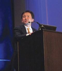
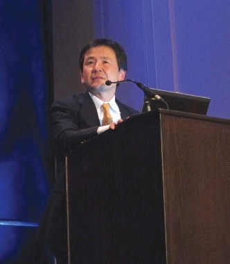
PHILADELPHIA – Knowledge of a patient’s age, gender, quality of life score, and radiographic knee osteoarthritis severity helps predict the odds of a patient undergoing total knee arthroplasty in the next 5 years.
Few studies have looked at the predictors of total knee arthroplasty (TKA) in community-based cohorts, despite the heavy burden of TKA in the United States, said Dr. Marc C. Hochberg, head of rheumatology and clinical immunology, University of Maryland, Baltimore. An estimated 4 million Americans already live with TKA, and more than half of adults diagnosed with knee osteoarthritis (OA) will undergo TKA (J. Bone Joint Surg. Am. 2013;95:385-92).
For the current analysis, Dr. Hochberg and his associates obtained 48 months of annual clinical follow-up data on 6,406 patients in the Osteoarthritis Initiative who had symptomatic, radiographic knee OA or who were at risk for the condition. Follow-up at 60 months involved only questionnaires. Consensus readings of knee radiographs were used for the analysis, along with knee-specific multiple variable regression models. The best models were selected based on Chi-square values and area under the receiver operating characteristic curve (AUC). Radiographic knee OA Kellgren-Lawrence (KL) severity grade 0, 1, 2, 3, and 4 was present in the right knee of 36%, 18%, 28%, 14%, and 3.5% of patients and in the left knee of 38%, 18%, 26%, 14.4%, and 3.3% of patients.

A little more than half of participants were women (58.4%), mean body mass index was 28.7 kg/m2, mean Western Ontario and McMaster Universities Osteoarthritis Index (WOMAC) pain score was 2.47 (0-20 scale), mean WOMAC function score was 8.33 (0-68 scale), and mean Knee injury and Osteoarthritis Outcome Score (KOOS) (quality of life) was 66.10 (0-100 scale). Their average age was 62.7, he said at the World Congress on Osteoarthritis, sponsored by the Osteoarthritis Research Society International.
During the 60 months of follow-up, there were 91 right and 102 left TKAs. The AUC for age, sex, and BMI was 0.64 for the right knee and 0.67 for the left knee.
The best TKA prediction models included age, sex, BMI, and KOOS quality-of-life score, improving the AUC to 0.78 for the right knee and 0.80 for the left knee, Dr. Hochberg said. Addition of the WOMAC pain score was associated with a marginal, but significant improvement in the AUC (0.79 and 0.80, respectively). When KL grade was added to these models, the AUC reached 0.89 and 0.91 for the right and left knees, respectively, he said. Sensitivity analyses failed to demonstrate additional effects of other baseline variables when added.
Limitations of the study included the relatively few TKAs, that most patients had KL grade 2 at baseline, proportional hazards analyses were not used, and the study did not consider a correlation between knees or the change in independent variables over time, he said.
In a prospective Canadian study, willingness to undergo surgery was the strongest predictor of the time to first hip or knee total joint replacement (TJR) (Arthritis Rheum. 2006;54:3212-20), while an international OARSI Task Force reported that pain and disability was higher in patients recommended for hip or knee TJR (Osteoarthritis Cartilage 2011;19:147-54). The Task Force, which included Dr. Hochberg, could not, however, identify cut points for pain and disability that would discriminate between those who did or did not get the nod for surgery.
The Osteoarthritis Initiative is sponsored by the National Institutes of Health. Dr. Hochberg reported serving as a consultant for Allergan, Bioiberica, Iroko, Merck, and Pfizer, and as a scientific/medical advisory board member for Eli Lilly and Merck.
PHILADELPHIA – Knowledge of a patient’s age, gender, quality of life score, and radiographic knee osteoarthritis severity helps predict the odds of a patient undergoing total knee arthroplasty in the next 5 years.
Few studies have looked at the predictors of total knee arthroplasty (TKA) in community-based cohorts, despite the heavy burden of TKA in the United States, said Dr. Marc C. Hochberg, head of rheumatology and clinical immunology, University of Maryland, Baltimore. An estimated 4 million Americans already live with TKA, and more than half of adults diagnosed with knee osteoarthritis (OA) will undergo TKA (J. Bone Joint Surg. Am. 2013;95:385-92).
For the current analysis, Dr. Hochberg and his associates obtained 48 months of annual clinical follow-up data on 6,406 patients in the Osteoarthritis Initiative who had symptomatic, radiographic knee OA or who were at risk for the condition. Follow-up at 60 months involved only questionnaires. Consensus readings of knee radiographs were used for the analysis, along with knee-specific multiple variable regression models. The best models were selected based on Chi-square values and area under the receiver operating characteristic curve (AUC). Radiographic knee OA Kellgren-Lawrence (KL) severity grade 0, 1, 2, 3, and 4 was present in the right knee of 36%, 18%, 28%, 14%, and 3.5% of patients and in the left knee of 38%, 18%, 26%, 14.4%, and 3.3% of patients.
A little more than half of participants were women (58.4%), mean body mass index was 28.7 kg/m2, mean Western Ontario and McMaster Universities Osteoarthritis Index (WOMAC) pain score was 2.47 (0-20 scale), mean WOMAC function score was 8.33 (0-68 scale), and mean Knee injury and Osteoarthritis Outcome Score (KOOS) (quality of life) was 66.10 (0-100 scale). Their average age was 62.7, he said at the World Congress on Osteoarthritis, sponsored by the Osteoarthritis Research Society International.
During the 60 months of follow-up, there were 91 right and 102 left TKAs. The AUC for age, sex, and BMI was 0.64 for the right knee and 0.67 for the left knee.
The best TKA prediction models included age, sex, BMI, and KOOS quality-of-life score, improving the AUC to 0.78 for the right knee and 0.80 for the left knee, Dr. Hochberg said. Addition of the WOMAC pain score was associated with a marginal, but significant improvement in the AUC (0.79 and 0.80, respectively). When KL grade was added to these models, the AUC reached 0.89 and 0.91 for the right and left knees, respectively, he said. Sensitivity analyses failed to demonstrate additional effects of other baseline variables when added.
Limitations of the study included the relatively few TKAs, that most patients had KL grade 2 at baseline, proportional hazards analyses were not used, and the study did not consider a correlation between knees or the change in independent variables over time, he said.
In a prospective Canadian study, willingness to undergo surgery was the strongest predictor of the time to first hip or knee total joint replacement (TJR) (Arthritis Rheum. 2006;54:3212-20), while an international OARSI Task Force reported that pain and disability was higher in patients recommended for hip or knee TJR (Osteoarthritis Cartilage 2011;19:147-54). The Task Force, which included Dr. Hochberg, could not, however, identify cut points for pain and disability that would discriminate between those who did or did not get the nod for surgery.
The Osteoarthritis Initiative is sponsored by the National Institutes of Health. Dr. Hochberg reported serving as a consultant for Allergan, Bioiberica, Iroko, Merck, and Pfizer, and as a scientific/medical advisory board member for Eli Lilly and Merck.
AT OARSI
Major finding: The best TKA prediction models included age, sex, BMI, and KOOS quality-of-life score, improving the AUC to 0.78 for the right knee and 0.80 for the left knee.
Data source: Retrospective analysis of 91 right and 102 left TKAs among 6,406 patients with knee osteoarthritis in the multicenter, observational Osteoarthritis Initiative study.
Disclosures: The Osteoarthritis Initiative is sponsored by the National Institutes of Health. Dr. Hochberg reported serving as a consultant for Allergan, Bioiberica, Iroko, Merck, and Pfizer, and as a scientific/medical advisory board member for Eli Lilly and Merck.

DVDs buoy knee osteoarthritis exercise, until the novelty fades
PHILADELPHIA – Home exercise DVDs do not enhance long-term adherence to prescribed exercise in patients with knee osteoarthritis, despite producing substantial improvements in pain and physical function at 2 years in a randomized controlled trial.
As with most home exercise DVDs, things started off great.
Patients who took home a 30-minute DVD after watching it alongside a physiotherapist reported exercising 5.5 and 5.1 times per week at 3 months and 6 months, compared with just 4.3 and 3.9 times per week among controls given detailed verbal and hands-on instruction in a quadriceps exercise protocol, but no video (P = .0358; P =.0369).
At 12 months and 24 months, however, there was no significant difference in exercise adherence between the two groups (P = .169; P = .324), Dr. Harukazu Tohyama said at the World Congress on Osteoarthritis.
The investigators also hypothesized that use of a home exercise DVD could prevent radiographic progression of knee osteoarthritis (OA), but this was not the case among the study’s 107 patients.
The DVD group showed a significant increase in femorotibial angle at 12 and 24 months over baseline values (both P less than .05), but the difference between groups was not significant at either time point (P = .334; P = .293), said Dr. Tohyama, who is on the faculty of the department of sports medicine at Hokkaido University in Sapporo, Japan.
Joint space area, minimum joint space width, and osteophyte area were also similar between groups, as assessed using a fully automated knee OA computer-aided diagnosis (KOA-CAD) measuring system (Osteoarthritis Cartilage. 2008;16:1300-6).
The DVD-based program encompassed muscle stretching, active range-of-motion exercises, and five forms of muscle strengthening. Both the DVD and control group had their intervention reinforced at a clinic visit 4 weeks after randomization.
Members of the audience suggested that a single visit at 4 weeks without additional boosters is not enough to promote adherence. They also questioned whether a 24-month follow-up is long enough to detect whether OA progression is occurring, and cautioned that the study numbers may be too small to draw such a conclusion.
Dr. Tohyama responded that a 24-month follow-up is sufficient for joint OA, but that "for joint [space] narrowing, 24 months is not enough. We need longer follow-up."
Of the 107 patients randomized, 48 were available for analysis in the DVD group and 23 in the control group. The remainder was lost to follow-up or refused the allocation.
The patients were more than 50 years old and had Kellgren-Lawrence grade 2, 3, or 4 knee OA and knee pain. Patients requiring regular or intermittent use of steroids or nonsteroidal anti-inflammatory drugs were excluded.
Scores on the WOMAC (Western Ontario and McMaster Universities Arthritis Index) for all categories were significantly greater in the DVD group than in the control group at 3, 6, 12, and 24 months, Dr. Tohyama said at the meeting, which was sponsored by the Osteoarthritis Research Society International.
WOMAC scores at 6 months in the DVD and control groups were 2.7 vs. 0.2 for pain (P = .0001), 0.60 vs. –0.20 for stiffness (P = .0020), and 8.3 vs. 0.7 for function (P = .0001).
At 24 months, the corresponding scores were 2.2 vs. 0.6 (P = .023), 0.60 vs. –0.07 (P = .011), and 7.3 vs. 2.3 (P = .014).
Improvement in the physical component of the Short Form-8 Health Survey was also significantly greater in the DVD group than in the control group at 3, 6, and 12 months (12 months: 8.3 vs. 2.3; P = .002), but not at 24 months (7.1 vs. 4.1; P = .07).
No significant between-group differences were observed on the SF-8 mental component at any of the time points, he said.
Body mass decreased significantly early on in the DVD group, at 3 and 6 months, compared with controls, but the decline waned along with adherence, and was no longer significant at 12 or 24 months.
Although exercise DVDs may be popular and engaging, "I think what is most important for adherence improvement is to form an exercise habit," Dr. Tohyama said in an interview.
The Japanese Orthopaedic Association sponsored the study. Dr. Tohyama reported having no financial disclosures.
AT OARSI
Major finding: At 12 and 24 months, there was no significant difference in exercise adherence between those with home exercise videos and those without (P = .169; P = .324).
Data source: Randomized, placebo-controlled trial in 107 consecutive patients with knee osteoarthritis.
Disclosures: The Japanese Orthopaedic Association sponsored the study. Dr. Tohyama reported having no financial disclosures.
Data mixed on role of exercise in older, obese, osteoarthritic patients
PHILADELPHIA – There are few certainties in medicine, but most would agree that exercise is good for you.
In older, largely postmenopausal women, a recent systematic review showed that exercise does indeed prevent weight gain, preserves lean body mass when combined with weight loss, and is a key determinant for the positive effects of weight loss later in life (Maturitas. 2012;72:13-22).
What’s not clear, however, is whether exercise plays the same positive role in weight loss in older, obese patients with knee osteoarthritis, Stephen P. Messier, Ph.D., said at the World Congress on Osteoarthritis.
"Once you take that leap – make that decision to lose weight – the question is: ‘Does exercise enhance, impede, or have no effect on the process and the potential outcomes?,’ " said Dr. Messier, professor and director of the J.B. Snow Biomechanics Laboratory at Wake Forest University, Winston-Salem, N.C.

The question is highly relevant, as the risk of developing knee OA is up to 12 times higher in obese men and up to 17 times higher in obese women (Arthritis Res. Ther. 2010;12:R88).
Dr. Messier reviewed data from two long-running randomized intervention trials at Wake Forest, suggesting that exercise does not impede weight loss. Compliance was 61% for diet only and 63% for diet plus exercise in ADAPT (Arthritis, Diet and Activity Promotion).
On the other hand, adding exercise to diet doesn’t dramatically enhance weight loss, acknowledged Dr. Messier, principal investigator of ADAPT and of the IDEA (Intensive Diet and Exercise for Arthritis) trial, which had a weight loss goal of at least 10% of body weight, or twice the goal of ADAPT.
Patients who adhered to a diet with or without exercise lost more weight than did those who did neither in ADAPT, but just 2 kilograms of weight loss separated the diet-plus-exercise group from the diet-only group in IDEA. Fat loss was similar.
What was concerning was that the addition of exercise to diet did not prevent the loss of lean mass, Dr. Messier said. The mean change in lean mass was –4.2 with intensive dietary restriction vs. –4.7 with the same diet plus 15 minutes of walking and 20 minutes of weight training thrice-weekly in IDEA.
The silver lining
Knee OA patients and their physicians can take heart, as virtually every clinical outcome favored diet plus exercise over diet alone, Dr. Messier observed.
When exercise was combined with diet for 18 months, patients experienced a 30% decrease in pain and 24% improvement in function in ADAPT, with even higher results of 51% and 47% in IDEA.
"The big take-home message was that the diet and exercise group had a 50% reduction in pain, and you don’t find that with any pharmacologic intervention," he said in an interview.
Dr. Messier speculated that the loss of lean body mass with diet plus exercise could be offset by adding strength training – a tactic that is currently being studied in 370 patients with knee OA in START (Strength Training for Arthritis Trial).
"A more intensive strength training regimen may build muscle and attenuate the loss of lean body mass," he said. "If it works, we could get the best of both worlds."
Diet driving mechanistic changes
Interestingly, the data suggest that diet alone may be slightly better than diet plus exercise in improving mechanistic outcomes, Dr. Messier said.
Among the 142 sedentary overweight/obese older adults with knee OA in ADAPT, every 1 pound of weight loss resulted in a fourfold reduction in compressive forces exerted on the knee per step (Arthritis Rheum. 2005;52:2026-32).
The average change in knee compressive forces from baseline in IDEA was 11% with diet alone, 9% with diet plus exercise, and 5% with exercise alone. Reductions in interleukin-6, a marker of physical disability in older adults, were also significantly better with diet than with diet plus exercise, and the decrease was independent of gender, body mass index and other baseline values.
"Once again, it is the weight loss that counts," he said.
That said, should clinicians push their knee OA patients to exercise and diet?
Diet plus exercise should be the standard of care in our older adults with osteoarthritis of the knee, Dr. Messier concluded.
"OA and other obesity-related diseases place an enormous physical and financial burden on our health care system," he said. "Intensive weight loss, when combined with exercise, can safely achieve a mean long-term weight loss of 11.4% for compliant patients and they can expect significant improvement in symptoms relative to either exercise or diet alone. Wider adoption of intensive weight loss combined with exercise can reduce the burden of disability related to OA and obesity in our aging population."
The congress was sponsored by Osteoarthritis Research Society International. Dr. Messier reported no relevant conflicts of interest.
PHILADELPHIA – There are few certainties in medicine, but most would agree that exercise is good for you.
In older, largely postmenopausal women, a recent systematic review showed that exercise does indeed prevent weight gain, preserves lean body mass when combined with weight loss, and is a key determinant for the positive effects of weight loss later in life (Maturitas. 2012;72:13-22).
What’s not clear, however, is whether exercise plays the same positive role in weight loss in older, obese patients with knee osteoarthritis, Stephen P. Messier, Ph.D., said at the World Congress on Osteoarthritis.
"Once you take that leap – make that decision to lose weight – the question is: ‘Does exercise enhance, impede, or have no effect on the process and the potential outcomes?,’ " said Dr. Messier, professor and director of the J.B. Snow Biomechanics Laboratory at Wake Forest University, Winston-Salem, N.C.
The question is highly relevant, as the risk of developing knee OA is up to 12 times higher in obese men and up to 17 times higher in obese women (Arthritis Res. Ther. 2010;12:R88).
Dr. Messier reviewed data from two long-running randomized intervention trials at Wake Forest, suggesting that exercise does not impede weight loss. Compliance was 61% for diet only and 63% for diet plus exercise in ADAPT (Arthritis, Diet and Activity Promotion).
On the other hand, adding exercise to diet doesn’t dramatically enhance weight loss, acknowledged Dr. Messier, principal investigator of ADAPT and of the IDEA (Intensive Diet and Exercise for Arthritis) trial, which had a weight loss goal of at least 10% of body weight, or twice the goal of ADAPT.
Patients who adhered to a diet with or without exercise lost more weight than did those who did neither in ADAPT, but just 2 kilograms of weight loss separated the diet-plus-exercise group from the diet-only group in IDEA. Fat loss was similar.
What was concerning was that the addition of exercise to diet did not prevent the loss of lean mass, Dr. Messier said. The mean change in lean mass was –4.2 with intensive dietary restriction vs. –4.7 with the same diet plus 15 minutes of walking and 20 minutes of weight training thrice-weekly in IDEA.
The silver lining
Knee OA patients and their physicians can take heart, as virtually every clinical outcome favored diet plus exercise over diet alone, Dr. Messier observed.
When exercise was combined with diet for 18 months, patients experienced a 30% decrease in pain and 24% improvement in function in ADAPT, with even higher results of 51% and 47% in IDEA.
"The big take-home message was that the diet and exercise group had a 50% reduction in pain, and you don’t find that with any pharmacologic intervention," he said in an interview.
Dr. Messier speculated that the loss of lean body mass with diet plus exercise could be offset by adding strength training – a tactic that is currently being studied in 370 patients with knee OA in START (Strength Training for Arthritis Trial).
"A more intensive strength training regimen may build muscle and attenuate the loss of lean body mass," he said. "If it works, we could get the best of both worlds."
Diet driving mechanistic changes
Interestingly, the data suggest that diet alone may be slightly better than diet plus exercise in improving mechanistic outcomes, Dr. Messier said.
Among the 142 sedentary overweight/obese older adults with knee OA in ADAPT, every 1 pound of weight loss resulted in a fourfold reduction in compressive forces exerted on the knee per step (Arthritis Rheum. 2005;52:2026-32).
The average change in knee compressive forces from baseline in IDEA was 11% with diet alone, 9% with diet plus exercise, and 5% with exercise alone. Reductions in interleukin-6, a marker of physical disability in older adults, were also significantly better with diet than with diet plus exercise, and the decrease was independent of gender, body mass index and other baseline values.
"Once again, it is the weight loss that counts," he said.
That said, should clinicians push their knee OA patients to exercise and diet?
Diet plus exercise should be the standard of care in our older adults with osteoarthritis of the knee, Dr. Messier concluded.
"OA and other obesity-related diseases place an enormous physical and financial burden on our health care system," he said. "Intensive weight loss, when combined with exercise, can safely achieve a mean long-term weight loss of 11.4% for compliant patients and they can expect significant improvement in symptoms relative to either exercise or diet alone. Wider adoption of intensive weight loss combined with exercise can reduce the burden of disability related to OA and obesity in our aging population."
The congress was sponsored by Osteoarthritis Research Society International. Dr. Messier reported no relevant conflicts of interest.
EXPERT ANALYSIS FROM OARSI

Prearthroplasty exercise benefits short lived
PHILADELPHIA – Preoperative neuromuscular exercise does not significantly improve function or pain 3 months after total hip or knee replacement, according to a randomized, controlled trial in 165 patients.
The study’s primary endpoint of self-reported activities of daily living (ADLs) at 3 months was similar in patients who exercised prior to surgery and those who did not (mean between-group difference 4.4 points; P = .096). The change from baseline in the group who exercised prior to surgery was 29 points vs. 25 points in the surgery-only group, on a 100-point, worst-to-best scale.

Joint-related quality of life (mean 4.5 points; P = .22) and pain (mean 3.6 points; P = .09) were also not significantly different between the two groups, Dr. Allan Villadsen said at the World Congress on Osteoarthritis Research Society.
The 8 weeks of twice-weekly exercise, however, was not for naught.
Patients improved immediately following exercise and this improvement carried over to 6 weeks after surgery, "indicating an earlier onset of recovery," he said. "Therefore, we believe exercise constitutes a viable treatment option for the individual patient who’s willing to engage in exercise prior to total hip or knee replacement and possibly obtain an earlier onset of recovery."
While preoperative exercise has been shown to improve function in other settings, little is known about its role in knee- and hip-replacement surgery for patients with severe osteoarthritis (OA). A meta-analysis of 23 randomized controlled trials involving 1,461 patients reported a small improvement in physical function and pain 3 weeks after total hip arthroscopy, but no effects in total knee replacement 3 months after surgery (Osteoarthritis Cartilage 2011;19:1381-95).A subsequent systematic review and meta-analysis, however, suggests that the therapeutic validity of the exercise programs used in the studies may have hampered the beneficial effects (PLos One 2012;7:e38031). In addition, the sample sizes were small, and the calculated effect sizes for pain and physical function were based on only two to four studies, observed Dr. Villadsen of the Institute of Sport Science and Clinical Biomechanics, University of Southern Denmark, Odense.
The current trial randomly assigned 165 patients with severe symptomatic hip or knee OA scheduled for unilateral primary total joint replacement to receive a brochure recommending exercise prior to surgery and a 3-hour information session with health professionals or the same intervention plus individualized group-based neuromuscular exercise guided by a physiotherapist. The sessions were designed to improve sensorimotor control and range of motion, and included warm up on a stationary bike, a circuit program, and a walking cool-down period, Dr. Villadsen said at the meeting, sponsored by Osteoarthritis Research Society International (OARSI).
Hip OA was present in 43 of the 84 patients in the exercise arm and 41 of the 81 controls. Their mean age was 67 years and mean ADL score on the 100-point Hip Disability and Osteoarthritis Outcome Score (HOOS) or Knee Injury and Osteoarthritis Outcome Score (KOOS) questionnaires was 50.6 and 45.6, respectively.
At 6 weeks, the between-group difference in activities of daily living on both the HOOS and KOOS questionnaires was 5.2, favoring the exercise group (P = .05), Dr. Villadsen said.
During a discussion of the results, an audience member asked whether the patients had tried exercise prior to joint replacement, which is the current recommendation. Dr. Villadsen said they did not track this, but that anecdotally, "Many of these patients are not offered what is recommended in the international guidelines, which is very disturbing ... if you asked them, many of them never even exercised before."
Session moderator and orthopedic surgeon Dr. Victor Goldberg, with Case Western Reserve University in Cleveland said it’s difficult to tease out the effects of surgery in analyses like these, but agreed that many patients are being sent to surgery without having been counseled to exercise. Part of the problem is that patients with mild to moderate OA are typically cared for by primary care physicians, who aren’t as well versed as rheumatologists and orthopedic surgeons are on the impairment, he said in an interview.
"That’s one of the charges of OARSI and our guidelines, is to really put it out in the community," he added.
"These kinds of studies, which point to the need for intervention in arthroplasty, particularly with well-done neuromuscular exercise programs, are very important to the overall management of these patients," Dr. Goldberg said. It’s needed because 15%-23% of patients after surgery say " ‘I’m not much better; I still hurt.’ "
Self-reported data at 1 year are currently being processed and will be included in a manuscript on the cost-effectiveness of the intervention, Dr. Villadsen said in an interview.
Dr. Villadsen reported having no financial disclosures.
PHILADELPHIA – Preoperative neuromuscular exercise does not significantly improve function or pain 3 months after total hip or knee replacement, according to a randomized, controlled trial in 165 patients.
The study’s primary endpoint of self-reported activities of daily living (ADLs) at 3 months was similar in patients who exercised prior to surgery and those who did not (mean between-group difference 4.4 points; P = .096). The change from baseline in the group who exercised prior to surgery was 29 points vs. 25 points in the surgery-only group, on a 100-point, worst-to-best scale.
Joint-related quality of life (mean 4.5 points; P = .22) and pain (mean 3.6 points; P = .09) were also not significantly different between the two groups, Dr. Allan Villadsen said at the World Congress on Osteoarthritis Research Society.
The 8 weeks of twice-weekly exercise, however, was not for naught.
Patients improved immediately following exercise and this improvement carried over to 6 weeks after surgery, "indicating an earlier onset of recovery," he said. "Therefore, we believe exercise constitutes a viable treatment option for the individual patient who’s willing to engage in exercise prior to total hip or knee replacement and possibly obtain an earlier onset of recovery."
While preoperative exercise has been shown to improve function in other settings, little is known about its role in knee- and hip-replacement surgery for patients with severe osteoarthritis (OA). A meta-analysis of 23 randomized controlled trials involving 1,461 patients reported a small improvement in physical function and pain 3 weeks after total hip arthroscopy, but no effects in total knee replacement 3 months after surgery (Osteoarthritis Cartilage 2011;19:1381-95).A subsequent systematic review and meta-analysis, however, suggests that the therapeutic validity of the exercise programs used in the studies may have hampered the beneficial effects (PLos One 2012;7:e38031). In addition, the sample sizes were small, and the calculated effect sizes for pain and physical function were based on only two to four studies, observed Dr. Villadsen of the Institute of Sport Science and Clinical Biomechanics, University of Southern Denmark, Odense.
The current trial randomly assigned 165 patients with severe symptomatic hip or knee OA scheduled for unilateral primary total joint replacement to receive a brochure recommending exercise prior to surgery and a 3-hour information session with health professionals or the same intervention plus individualized group-based neuromuscular exercise guided by a physiotherapist. The sessions were designed to improve sensorimotor control and range of motion, and included warm up on a stationary bike, a circuit program, and a walking cool-down period, Dr. Villadsen said at the meeting, sponsored by Osteoarthritis Research Society International (OARSI).
Hip OA was present in 43 of the 84 patients in the exercise arm and 41 of the 81 controls. Their mean age was 67 years and mean ADL score on the 100-point Hip Disability and Osteoarthritis Outcome Score (HOOS) or Knee Injury and Osteoarthritis Outcome Score (KOOS) questionnaires was 50.6 and 45.6, respectively.
At 6 weeks, the between-group difference in activities of daily living on both the HOOS and KOOS questionnaires was 5.2, favoring the exercise group (P = .05), Dr. Villadsen said.
During a discussion of the results, an audience member asked whether the patients had tried exercise prior to joint replacement, which is the current recommendation. Dr. Villadsen said they did not track this, but that anecdotally, "Many of these patients are not offered what is recommended in the international guidelines, which is very disturbing ... if you asked them, many of them never even exercised before."
Session moderator and orthopedic surgeon Dr. Victor Goldberg, with Case Western Reserve University in Cleveland said it’s difficult to tease out the effects of surgery in analyses like these, but agreed that many patients are being sent to surgery without having been counseled to exercise. Part of the problem is that patients with mild to moderate OA are typically cared for by primary care physicians, who aren’t as well versed as rheumatologists and orthopedic surgeons are on the impairment, he said in an interview.
"That’s one of the charges of OARSI and our guidelines, is to really put it out in the community," he added.
"These kinds of studies, which point to the need for intervention in arthroplasty, particularly with well-done neuromuscular exercise programs, are very important to the overall management of these patients," Dr. Goldberg said. It’s needed because 15%-23% of patients after surgery say " ‘I’m not much better; I still hurt.’ "
Self-reported data at 1 year are currently being processed and will be included in a manuscript on the cost-effectiveness of the intervention, Dr. Villadsen said in an interview.
Dr. Villadsen reported having no financial disclosures.
AT OARSI
Major finding: Self-reported activities of daily living at 3 months was similar in patients who exercised prior to surgery and those who did not (mean between-group difference 4.4 points; P = .096).
Data source: Randomized, assessor-blinded trial in 165 patients undergoing total hip or knee replacement.
Disclosures: Dr. Villadsen reported having no financial disclosures.

OARSI recommends physical function tests for knee, hip OA
PHILADELPHIA – The Osteoarthritis Research Society International is recommending a set of user-friendly physical performance measures for patients with established hip and knee osteoarthritis.
The minimal core set of tests includes the 30-second chair stand test, 40-m fast-paced-walk, and stair negotiation.
Two further tests, the timed up-and-go and 6-minute walk, also are highly recommended, depending on the research setting, to measure ambulatory transitions and aerobic capacity.
The tests had to cover multiple physical activities, use minimal equipment, and be user friendly and inexpensive, steering committee member Kim L. Bennell, Ph.D., said at the World Congress on Osteoarthritis.

For example, the 30-second chair stand test requires only a stop watch and a chair to count how many times a patient can rise from a chair with hips and knees fully extended and sit back down in 30 seconds. The tests are best suited for adults older than 40 years but are relevant for patients with mild to end-stage hip and/or knee osteoarthritis (OA) and following joint replacement.
Currently, there are no international recommendations on which performance-based measures of physical function should be used to measure outcomes in patients with hip and/or knee OA. The hope is that the new recommendations will facilitate a more consistent use of performance-based outcome measures across clinical trials and the clinical setting, said Dr. Bennell, a research physiotherapist and director of the multidisciplinary Centre for Health, Exercise and Sports Medicine, University of Melbourne (Australia).
An international advisory committee of 10 rheumatologists, physical therapists, and an orthopedic surgeon identified the main activity themes of walking, stair negotiation, and sit-to-stand from a systematic review of the literature and by consensus opinion. Another 138 clinicians and researchers also were surveyed and asked to rank the difficulty and feasibility of 23 functional tests for OA patients.
"We wanted to identify the most feasible tests considering practical issues such as time, cost, equipment, space, and administration burden," she said at the meeting, which was sponsored by OARSI.
In the end, none of the tests satisfied all desirable clinimetric criteria of reliability, measurement error, validity, responsiveness, and interpretability. The experts felt stair negotiation was an important measure of mobility, lower-body strength, and balance but balked at recommending a specific test because of insufficient clinimetric evidence and the simple, practical fact that not all facilities have enough stairs to conduct a 9- or 12-step test.
"What we really found in our discussions and our work is that there really wasn’t a lot of good clinimetric evidence across the board on these tests," Dr. Bennell said.
To boost the evidence base, OARSI has put out a call on its website to collect future prospective data and to pool existing data so researchers can calculate minimal clinically important difference (MCID) estimates for different OA subgroups, longitudinal responsiveness estimates for interventions, and normative data for various OA subgroups.
Further details on the tests, including an instructional manual, clinimetric measurement properties, and normative values, are available on the OARSI website. Videos will also be available shortly.
The project was sponsored by OARSI, the Australian National Health and Medical Research Council, Arthritis Australia, and the University of Otago. Dr. Bennell reported having no financial disclosures.
PHILADELPHIA – The Osteoarthritis Research Society International is recommending a set of user-friendly physical performance measures for patients with established hip and knee osteoarthritis.
The minimal core set of tests includes the 30-second chair stand test, 40-m fast-paced-walk, and stair negotiation.
Two further tests, the timed up-and-go and 6-minute walk, also are highly recommended, depending on the research setting, to measure ambulatory transitions and aerobic capacity.
The tests had to cover multiple physical activities, use minimal equipment, and be user friendly and inexpensive, steering committee member Kim L. Bennell, Ph.D., said at the World Congress on Osteoarthritis.
For example, the 30-second chair stand test requires only a stop watch and a chair to count how many times a patient can rise from a chair with hips and knees fully extended and sit back down in 30 seconds. The tests are best suited for adults older than 40 years but are relevant for patients with mild to end-stage hip and/or knee osteoarthritis (OA) and following joint replacement.
Currently, there are no international recommendations on which performance-based measures of physical function should be used to measure outcomes in patients with hip and/or knee OA. The hope is that the new recommendations will facilitate a more consistent use of performance-based outcome measures across clinical trials and the clinical setting, said Dr. Bennell, a research physiotherapist and director of the multidisciplinary Centre for Health, Exercise and Sports Medicine, University of Melbourne (Australia).
An international advisory committee of 10 rheumatologists, physical therapists, and an orthopedic surgeon identified the main activity themes of walking, stair negotiation, and sit-to-stand from a systematic review of the literature and by consensus opinion. Another 138 clinicians and researchers also were surveyed and asked to rank the difficulty and feasibility of 23 functional tests for OA patients.
"We wanted to identify the most feasible tests considering practical issues such as time, cost, equipment, space, and administration burden," she said at the meeting, which was sponsored by OARSI.
In the end, none of the tests satisfied all desirable clinimetric criteria of reliability, measurement error, validity, responsiveness, and interpretability. The experts felt stair negotiation was an important measure of mobility, lower-body strength, and balance but balked at recommending a specific test because of insufficient clinimetric evidence and the simple, practical fact that not all facilities have enough stairs to conduct a 9- or 12-step test.
"What we really found in our discussions and our work is that there really wasn’t a lot of good clinimetric evidence across the board on these tests," Dr. Bennell said.
To boost the evidence base, OARSI has put out a call on its website to collect future prospective data and to pool existing data so researchers can calculate minimal clinically important difference (MCID) estimates for different OA subgroups, longitudinal responsiveness estimates for interventions, and normative data for various OA subgroups.
Further details on the tests, including an instructional manual, clinimetric measurement properties, and normative values, are available on the OARSI website. Videos will also be available shortly.
The project was sponsored by OARSI, the Australian National Health and Medical Research Council, Arthritis Australia, and the University of Otago. Dr. Bennell reported having no financial disclosures.
AT THE WORLD CONGRESS ON OSTEOARTHRITIS

Periostin levels linked to osteoarthritis prevalence, progression
PHILADELPHIA – Women with prevalent knee osteoarthritis had significantly lower levels of periostin, particularly those with progressive disease, than did women without the condition in an analysis of the OFELY study.
Although the mechanism remains to be investigated, the vitamin-K dependent protein "may be a new biological marker of prevalence and progression of knee OA in women," Jean-Charles Rousseau, Ph.D., said at the meeting, sponsored by Osteoarthritis Research Society International.

Periostin is secreted by osteoblasts and chondrocytes, and is one of only a handful of gamma-carboxyglutamic acid-containing proteins so far identified in humans. It was recently shown to be overexpressed in human OA cartilage and to regulate cartilage metabolism via matrix metallopeptidase-13 activation (Osteoarthritis Cartilage 2011;19 [Suppl. 1]:S34).
OFELY (Os des Femmes de Lyon) is the first study to report an association between periostin OA prevalence and progression, Dr. Rousseau said at the meeting.
To evaluate its role in OA, the investigators used a new commercially available ELISA sandwich assay to evaluate blood samples from 594 women in the ongoing, prospective OFELY study. Of these, 511 had no knee OA and 83 had radiographic knee OA, Kellgren-Lawrence grade of 2 or more. The average age was 60 years in those free of OA and 70 in those with OA; 78% and 98%, respectively, were menopausal.
Baseline serum periostin averaged 1,101 ng/mL in women with OA vs. 1,181 ng/mL in those without OA, after researchers adjusted for age (P = .011), said Dr. Rousseau of INSERM Research Unit 1033, University of Lyon (France).
Collagen type II cross-linked C-telopeptide levels (CTX-II) were significantly higher in women with OA vs. those without (278 ng/mL vs. 173 ng/mL; P less than .001), whereas serum levels were similar for type I CTX (0.43 ng/mL vs. 0.39 ng/mL), intact N-terminal propeptide of type I procollagen (38.1 mcg/L vs. 37.2 mcg/L) and osteocalcin (28.4 ng/mL vs. 25.7 ng/mL).
During the 4-year follow-up period, 82 women had radiological progression of their knee OA, defined by an increase of 1 or more Kellgren-Lawrence grade regardless of the baseline grade.
Baseline periostin levels were significantly lower in progressors than nonprogressors, after adjustment for age (1,112 ng/mL vs. 1,179 ng/mL; P = .046), Dr. Rousseau said.
For each increase of one periostin quartile, the risk of progression decreased by 21% after adjusting for age, KL score at baseline, and OA at other anatomical sites, including hand, hip, and lumbar spine (odds ratio 0.79; P = .038), he said.
During a discussion of the paper, an attendee questioned whether periostin is exclusive to bone metabolites, highlighting a recent randomized trial indicating that it is highly relevant in lung function. In that trial (N. Engl. J. Med. 2011;365:1088-98), anti-interleukin-13 therapy was more effective in asthmatic adults who had high serum periostin levels.
Dr. Rousseau acknowledged that periostin is distributed in nonarticular tissues including periodontal ligament and cardiac valves, and that the specificity of the periostin assay for circulating forms is unknown.
Session moderator Ali Mobasheri, D.Phil., of the University of Nottingham, England, said in an interview that it can be difficult to differentiate between a biomarker emanating from bone, articular cartilage, and tendon and other tissues such as heart and lung, "and in this particular case, I think it’s going to be extremely difficult to differentiate where it’s coming from."
He went on to say that periostin may be differentially processed in different tissues, which could be the differentiator in the future, but that the Chinese assay (USCNK, China) used in the study doesn’t make that distinction.
"So a lot more validation is required in this case," Dr. Mobasheri said.
Dr. Rousseau said in an interview that overlap exists with osteoarthritis biomarkers, but that they remain useful, even when OA is limited to a single knee.
OFELY was sponsored by Prévention des Maladies Osseuses. Dr. Rousseau reported no relevant disclosures.
PHILADELPHIA – Women with prevalent knee osteoarthritis had significantly lower levels of periostin, particularly those with progressive disease, than did women without the condition in an analysis of the OFELY study.
Although the mechanism remains to be investigated, the vitamin-K dependent protein "may be a new biological marker of prevalence and progression of knee OA in women," Jean-Charles Rousseau, Ph.D., said at the meeting, sponsored by Osteoarthritis Research Society International.
Periostin is secreted by osteoblasts and chondrocytes, and is one of only a handful of gamma-carboxyglutamic acid-containing proteins so far identified in humans. It was recently shown to be overexpressed in human OA cartilage and to regulate cartilage metabolism via matrix metallopeptidase-13 activation (Osteoarthritis Cartilage 2011;19 [Suppl. 1]:S34).
OFELY (Os des Femmes de Lyon) is the first study to report an association between periostin OA prevalence and progression, Dr. Rousseau said at the meeting.
To evaluate its role in OA, the investigators used a new commercially available ELISA sandwich assay to evaluate blood samples from 594 women in the ongoing, prospective OFELY study. Of these, 511 had no knee OA and 83 had radiographic knee OA, Kellgren-Lawrence grade of 2 or more. The average age was 60 years in those free of OA and 70 in those with OA; 78% and 98%, respectively, were menopausal.
Baseline serum periostin averaged 1,101 ng/mL in women with OA vs. 1,181 ng/mL in those without OA, after researchers adjusted for age (P = .011), said Dr. Rousseau of INSERM Research Unit 1033, University of Lyon (France).
Collagen type II cross-linked C-telopeptide levels (CTX-II) were significantly higher in women with OA vs. those without (278 ng/mL vs. 173 ng/mL; P less than .001), whereas serum levels were similar for type I CTX (0.43 ng/mL vs. 0.39 ng/mL), intact N-terminal propeptide of type I procollagen (38.1 mcg/L vs. 37.2 mcg/L) and osteocalcin (28.4 ng/mL vs. 25.7 ng/mL).
During the 4-year follow-up period, 82 women had radiological progression of their knee OA, defined by an increase of 1 or more Kellgren-Lawrence grade regardless of the baseline grade.
Baseline periostin levels were significantly lower in progressors than nonprogressors, after adjustment for age (1,112 ng/mL vs. 1,179 ng/mL; P = .046), Dr. Rousseau said.
For each increase of one periostin quartile, the risk of progression decreased by 21% after adjusting for age, KL score at baseline, and OA at other anatomical sites, including hand, hip, and lumbar spine (odds ratio 0.79; P = .038), he said.
During a discussion of the paper, an attendee questioned whether periostin is exclusive to bone metabolites, highlighting a recent randomized trial indicating that it is highly relevant in lung function. In that trial (N. Engl. J. Med. 2011;365:1088-98), anti-interleukin-13 therapy was more effective in asthmatic adults who had high serum periostin levels.
Dr. Rousseau acknowledged that periostin is distributed in nonarticular tissues including periodontal ligament and cardiac valves, and that the specificity of the periostin assay for circulating forms is unknown.
Session moderator Ali Mobasheri, D.Phil., of the University of Nottingham, England, said in an interview that it can be difficult to differentiate between a biomarker emanating from bone, articular cartilage, and tendon and other tissues such as heart and lung, "and in this particular case, I think it’s going to be extremely difficult to differentiate where it’s coming from."
He went on to say that periostin may be differentially processed in different tissues, which could be the differentiator in the future, but that the Chinese assay (USCNK, China) used in the study doesn’t make that distinction.
"So a lot more validation is required in this case," Dr. Mobasheri said.
Dr. Rousseau said in an interview that overlap exists with osteoarthritis biomarkers, but that they remain useful, even when OA is limited to a single knee.
OFELY was sponsored by Prévention des Maladies Osseuses. Dr. Rousseau reported no relevant disclosures.
AT THE 2013 OARSI WORLD CONGRESS
Major finding: Baseline serum periostin averaged 1,101 ng/mL in women with OA and 1,181 ng/mL in those without OA, after researchers adjusted for age (P = .011).
Data source: Retrospective analysis of 594 women participating in the OFELY study.
Disclosures: OFELY was sponsored by Prévention des Maladies Osseuses. Dr. Rousseau reported no relevant disclosures.

Trial shows benefits of patellofemoral brace for kneecap osteoarthritis
PHILADELPHIA – A lightweight brace on the patellofemoral joint significantly improves the pain and symptoms of knee osteoarthritis, according to BRACE, the largest patellofemoral bracing trial to date.
After 6 weeks of brace wearing, pain fell an average of 16.87 points on a visual analog scale (VAS) during an activity patients reported as aggravating, compared with no brace (–18.16 vs. –1.29, P less than .001).
A 12-point reduction is considered clinically relevant on the 100-mm VAS pain scale, where 0 is no pain and 100 is worst pain, Michael Callaghan, Ph.D., said at the World Congress on Osteoarthritis.
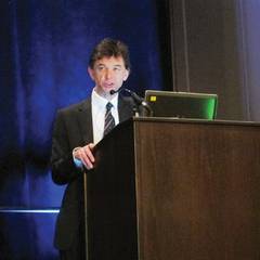
"The feedback we got from patients each time is that the brace made their knee feel more supported, they felt more comfortable, they felt more confident," he said. "One of the things we’re thinking is that perhaps the brace gave patients the ability to use their knee a little bit more comfortably, functionally better than they could previously, and that was enough to start improving muscle strength."
Data are available to show that muscle strength did improve with brace wearing, and a recently completed biomechanical analysis should determine whether biomechanical changes were actually occurring.
Although nonpharmacologic interventions are urgently needed for knee osteoarthritis (OA), little attention has been paid to treatments specifically for the patellofemoral (PF) joint, said Dr. Callaghan, a research physiotherapist at the University of Manchester (England). About 24% of patients with knee OA have symptomatic patellofemoral (PF) OA in the absence of tibiofemoral arthritis, but more importantly, 41% have a combination of tibiofemoral and PF knee OA. Despite this, a recent editorial (Osteoarthritis Cartilage 2011;19:765-7) called the patellofemoral joint "the forgotten joint" in knee OA, he observed.
Nonsurgical treatments include anti-inflammatory medications, physical therapy, and cortisone shots. Arthroscopic debridement, lateral retinacular release, and partial or full knee replacement are reserved for more severe cases.
The one prior study of PF braces showed no effect on pain, but it compared the same brace with and without a patellar strap (Osteoarthritis Cartilage 2011;19:792-800). In clinical practice, the more relevant question is the benefit of PF bracing versus no bracing, Dr. Callaghan observed.
The 126 patients in the trial had a radiographic Kellgren-Lawrence score of 2 or 3 in the PF joint; daily knee pain for the previous 3 months with stair climbing, kneeling, prolonged sitting, or squatting; and tenderness over the lateral or medial patellar facet on palpation or a positive patellar compression test. In the trial, the mean VAS pain score was 64.6 mm, 57% of the participants were women, and the patients’ mean body mass index was 30.8 kg/m2. Their average age was 55.5 years.
Patients were randomly assigned to immediate brace treatment with the Bioskin Patellar Tracking Q Brace (Ossur, Manchester, England) or delayed treatment at 6 weeks. All patients ultimately received 12 weeks of brace, and wore their brace for an average of 7.35 hours per day.
At 6 weeks, Knee Osteoarthritis Outcome Score subscale scores for pain and activities of daily living improved significantly with the brace vs. no brace by an average of –6.70 and –5.64, respectively (8.78 vs. 2.08 and 6.67 vs. 1.03; both P = .02), Dr. Callaghan reported at the meeting, sponsored by the Osteoarthritis Research Society International.
There was a trend toward improved aggregated locomotor function with the PT brace (–2.5 vs. –1.21; P = .065) in the study, which was led by colleague Dr. David Felson, professor of medicine at the University of Manchester and Boston University.
During a discussion of the study, an attendee said that in her practice she avoids using the type of brace used in the study with a cutout over the knee in younger patients with PF pain, because of data suggesting that bracing compresses the patella and increases the contact area between the patella and the trochlear notch, thereby increasing stress rather than changing the patella position or movement. Dr. Callaghan acknowledged that compression could be an issue and said MRI data available on 30 patients are being evaluated for evidence of compression and joint space narrowing.
Arthritis Research UK funded the trial. The authors reported no relevant disclosures.
PHILADELPHIA – A lightweight brace on the patellofemoral joint significantly improves the pain and symptoms of knee osteoarthritis, according to BRACE, the largest patellofemoral bracing trial to date.
After 6 weeks of brace wearing, pain fell an average of 16.87 points on a visual analog scale (VAS) during an activity patients reported as aggravating, compared with no brace (–18.16 vs. –1.29, P less than .001).
A 12-point reduction is considered clinically relevant on the 100-mm VAS pain scale, where 0 is no pain and 100 is worst pain, Michael Callaghan, Ph.D., said at the World Congress on Osteoarthritis.
"The feedback we got from patients each time is that the brace made their knee feel more supported, they felt more comfortable, they felt more confident," he said. "One of the things we’re thinking is that perhaps the brace gave patients the ability to use their knee a little bit more comfortably, functionally better than they could previously, and that was enough to start improving muscle strength."
Data are available to show that muscle strength did improve with brace wearing, and a recently completed biomechanical analysis should determine whether biomechanical changes were actually occurring.
Although nonpharmacologic interventions are urgently needed for knee osteoarthritis (OA), little attention has been paid to treatments specifically for the patellofemoral (PF) joint, said Dr. Callaghan, a research physiotherapist at the University of Manchester (England). About 24% of patients with knee OA have symptomatic patellofemoral (PF) OA in the absence of tibiofemoral arthritis, but more importantly, 41% have a combination of tibiofemoral and PF knee OA. Despite this, a recent editorial (Osteoarthritis Cartilage 2011;19:765-7) called the patellofemoral joint "the forgotten joint" in knee OA, he observed.
Nonsurgical treatments include anti-inflammatory medications, physical therapy, and cortisone shots. Arthroscopic debridement, lateral retinacular release, and partial or full knee replacement are reserved for more severe cases.
The one prior study of PF braces showed no effect on pain, but it compared the same brace with and without a patellar strap (Osteoarthritis Cartilage 2011;19:792-800). In clinical practice, the more relevant question is the benefit of PF bracing versus no bracing, Dr. Callaghan observed.
The 126 patients in the trial had a radiographic Kellgren-Lawrence score of 2 or 3 in the PF joint; daily knee pain for the previous 3 months with stair climbing, kneeling, prolonged sitting, or squatting; and tenderness over the lateral or medial patellar facet on palpation or a positive patellar compression test. In the trial, the mean VAS pain score was 64.6 mm, 57% of the participants were women, and the patients’ mean body mass index was 30.8 kg/m2. Their average age was 55.5 years.
Patients were randomly assigned to immediate brace treatment with the Bioskin Patellar Tracking Q Brace (Ossur, Manchester, England) or delayed treatment at 6 weeks. All patients ultimately received 12 weeks of brace, and wore their brace for an average of 7.35 hours per day.
At 6 weeks, Knee Osteoarthritis Outcome Score subscale scores for pain and activities of daily living improved significantly with the brace vs. no brace by an average of –6.70 and –5.64, respectively (8.78 vs. 2.08 and 6.67 vs. 1.03; both P = .02), Dr. Callaghan reported at the meeting, sponsored by the Osteoarthritis Research Society International.
There was a trend toward improved aggregated locomotor function with the PT brace (–2.5 vs. –1.21; P = .065) in the study, which was led by colleague Dr. David Felson, professor of medicine at the University of Manchester and Boston University.
During a discussion of the study, an attendee said that in her practice she avoids using the type of brace used in the study with a cutout over the knee in younger patients with PF pain, because of data suggesting that bracing compresses the patella and increases the contact area between the patella and the trochlear notch, thereby increasing stress rather than changing the patella position or movement. Dr. Callaghan acknowledged that compression could be an issue and said MRI data available on 30 patients are being evaluated for evidence of compression and joint space narrowing.
Arthritis Research UK funded the trial. The authors reported no relevant disclosures.
AT THE WORLD CONGRESS ON OSTEOARTHRITIS
Major finding: VAS pain scores were reduced by an average of 18.16 points after 6 weeks of patellofemoral bracing vs. 1.29 points without bracing (P less than .001).
Data source: Randomized, open-label study of 126 patients with patellofemoral osteoarthritis.
Disclosures: Arthritis Research UK funded the trial. The authors reported no relevant disclosures.

Studies raise red flag of CVD risk in osteoarthritis
PHILADELPHIA – Concerns are being raised over a possible link between osteoarthritis and an increased risk of cardiovascular disease, with the signal strongest among those with walking disability.
A recent British population-based cohort study reported an excess of premature death from all causes among patients with knee or hip osteoarthritis (standardized mortality ratio, 1.55) as well as disease-specific increases that were particularly striking for cardiovascular disease (SMR, 1.71). Further, OA patients were twice as likely to die if they had a walking disability (BMJ 2011;342:d1165).

This comes on the heels of a systematic review involving seven studies that found moderate evidence of increased mortality in patients with OA (Clin. Exp. Rheumatol. 2008;26:S120-4).
However, a new, population-based study from the Netherlands found no link between OA and future cardiovascular disease (CVD) among adults aged 55 years and older. On the other hand, the presence of disability significantly predicted cardiovascular disease, independent of other CVD risk factors and the presence of OA, Sita Bierma-Zeinstra, Ph.D., reported.
She suggested that OA-related disability and co-morbidity may explain the earlier findings, but many in the audience were not convinced.
Fellow presenter Dr. Peter Jüni, professor of clinical epidemiology at the University of Bern, Switzerland where, the British analysis was performed, said what’s most impressive about their data is that the annual cardiovascular mortality rate of 2% in patients with OA and walking disability mirrors mortality rates observed in patients with an established diagnosis of coronary artery disease.
"We’re talking about a frequency of cardiovascular deaths that is comparable with what you see in cardiological patients who actually deserve invasive treatment," he said at the World Congress on Osteoarthritis, sponsored by Osteoarthritis Research Society International.
Dr. Jüni said randomized trials are needed to better define the association between OA and CVD, but the bottom line is that clinicians need to start looking differently at OA as a prognostic marker and consider including it in their risk assessment and management strategy.
These data need to be brought to the attention of the OA community so clinicians can "monitor the cardiovascular status of these patients because they are at risk, and I don’t think that’s commonly known yet. There is a signal here that we need to pay attention to," commented Dr. Stefan Lohmander of Lund (Sweden) University and coauthor of the recent European League Against Rheumatism (EULAR) recommendations for the nonpharmacological core management of knee and hip OA (Ann. Rheum. Dis. 2013 [doi:10.1136/annrheumdis-2012-202745]).
"You are my hero; I love this work, it’s incredible," attendee Dr. Gillian Hawker, professor of medicine and rheumatology at the University of Toronto, rose to say following Dr. Jüni’s lecture.
She noted that her group has been following a very similar cohort in the Ontario Osteoarthritis study, and that unpublished data confirm the British findings of increased all-cause mortality and CVD mortality, particularly with increasing disability, when assessed using the Western Ontario and McMaster Universities Arthritis Index (WOMAC) disability subscale, but not its pain subscale.
"People think of it [OA] as normal aging, wear and tear, grin and bear it, but if we can make links to mortality or worsening of other conditions like cardiovascular disease and diabetes, which everyone cares about, then maybe we’ll start paying attention to disability from OA," Dr. Hawker said in an interview. "If we can get people to care, then maybe we can get some effective drugs."
One possible explanation for the conflicting results between the two studies is that the British study included patients with a different OA phenotype, including patients with hip or knee OA and Kellgren-Lawrence grades 1-3, whereas the Dutch study also included patients with hand OA and only K-L grades 2-3, Dr. Jüni suggested. The Dutch study also used a different, "considerably softer" CVD composite outcome, he said in an interview.
"The way our study was set up, we couldn’t look at things the same way that they did, but what came out is that risks are particularly pronounced in people with disability and this message doesn’t change," he added.
The Dutch investigators used radiographs and symptoms of knee, hip, and hand OA to classify 4,648 persons in the 1990-1992 cohort of the prospective Rotterdam Study as having any disability versus none. At baseline, patients were free of CVD, 61% were women and their mean age was 67.6 years. Radiographic evidence of knee OA was present in 21%, hip OA in 10%, and hand OA in 30%. Clinical symptoms of OA were seen in 7%, 3%, and 7%, respectively.
After a mean of 14.4 years follow-up, 1,230 cardiovascular events occurred, of which 101 were in the clinical knee OA group.
In a model that adjusted for age and gender, radiographic OA was not related to future CVD, a composite of coronary heart disease or stroke (hazard ratio, 1.00; P = .96); nor was the presence of clinical OA (HR, 1.08; P = .45), said Dr. Bierma-Zeinstra, professor of arthritis and related disorders at Erasmus Medical Center in Rotterdam, the Netherlands.
After researchers further adjusted for the cardiovascular risk factors of diabetes, body mass index, hypertension, total cholesterol/HDL cholesterol ratio, and current smoking, again, no relationship was observed with CVD and radiographic OA (HR, 0.99; P = .92) or clinical OA (HR, 1.09; P = .43). Data were not shown for hand and hip OA, but the results were similar, she said.
"We didn’t see any increased risk for future cardiovascular disease in this particular cohort of people 55 years or older ... but maybe you could see it in younger cohorts or find it in people with new-onset osteoarthritis, but in this particular cohort, we did not see an association," Dr. Bierma-Zeinstra said.
As noted above, the risk of suffering a cardiovascular event was significantly elevated in individuals with a disability in model one and model two (HR, 1.30 and HR, 1.26, respectively; both P less than .001) and in those with a lower-limb disability in model one (HR, 1.22; P =.002) and model two (HR, 1.19; P =.008).
Dr. Jüni and Dr. Bierma-Zeinstra reported no relevant disclosures.
PHILADELPHIA – Concerns are being raised over a possible link between osteoarthritis and an increased risk of cardiovascular disease, with the signal strongest among those with walking disability.
A recent British population-based cohort study reported an excess of premature death from all causes among patients with knee or hip osteoarthritis (standardized mortality ratio, 1.55) as well as disease-specific increases that were particularly striking for cardiovascular disease (SMR, 1.71). Further, OA patients were twice as likely to die if they had a walking disability (BMJ 2011;342:d1165).
This comes on the heels of a systematic review involving seven studies that found moderate evidence of increased mortality in patients with OA (Clin. Exp. Rheumatol. 2008;26:S120-4).
However, a new, population-based study from the Netherlands found no link between OA and future cardiovascular disease (CVD) among adults aged 55 years and older. On the other hand, the presence of disability significantly predicted cardiovascular disease, independent of other CVD risk factors and the presence of OA, Sita Bierma-Zeinstra, Ph.D., reported.
She suggested that OA-related disability and co-morbidity may explain the earlier findings, but many in the audience were not convinced.
Fellow presenter Dr. Peter Jüni, professor of clinical epidemiology at the University of Bern, Switzerland where, the British analysis was performed, said what’s most impressive about their data is that the annual cardiovascular mortality rate of 2% in patients with OA and walking disability mirrors mortality rates observed in patients with an established diagnosis of coronary artery disease.
"We’re talking about a frequency of cardiovascular deaths that is comparable with what you see in cardiological patients who actually deserve invasive treatment," he said at the World Congress on Osteoarthritis, sponsored by Osteoarthritis Research Society International.
Dr. Jüni said randomized trials are needed to better define the association between OA and CVD, but the bottom line is that clinicians need to start looking differently at OA as a prognostic marker and consider including it in their risk assessment and management strategy.
These data need to be brought to the attention of the OA community so clinicians can "monitor the cardiovascular status of these patients because they are at risk, and I don’t think that’s commonly known yet. There is a signal here that we need to pay attention to," commented Dr. Stefan Lohmander of Lund (Sweden) University and coauthor of the recent European League Against Rheumatism (EULAR) recommendations for the nonpharmacological core management of knee and hip OA (Ann. Rheum. Dis. 2013 [doi:10.1136/annrheumdis-2012-202745]).
"You are my hero; I love this work, it’s incredible," attendee Dr. Gillian Hawker, professor of medicine and rheumatology at the University of Toronto, rose to say following Dr. Jüni’s lecture.
She noted that her group has been following a very similar cohort in the Ontario Osteoarthritis study, and that unpublished data confirm the British findings of increased all-cause mortality and CVD mortality, particularly with increasing disability, when assessed using the Western Ontario and McMaster Universities Arthritis Index (WOMAC) disability subscale, but not its pain subscale.
"People think of it [OA] as normal aging, wear and tear, grin and bear it, but if we can make links to mortality or worsening of other conditions like cardiovascular disease and diabetes, which everyone cares about, then maybe we’ll start paying attention to disability from OA," Dr. Hawker said in an interview. "If we can get people to care, then maybe we can get some effective drugs."
One possible explanation for the conflicting results between the two studies is that the British study included patients with a different OA phenotype, including patients with hip or knee OA and Kellgren-Lawrence grades 1-3, whereas the Dutch study also included patients with hand OA and only K-L grades 2-3, Dr. Jüni suggested. The Dutch study also used a different, "considerably softer" CVD composite outcome, he said in an interview.
"The way our study was set up, we couldn’t look at things the same way that they did, but what came out is that risks are particularly pronounced in people with disability and this message doesn’t change," he added.
The Dutch investigators used radiographs and symptoms of knee, hip, and hand OA to classify 4,648 persons in the 1990-1992 cohort of the prospective Rotterdam Study as having any disability versus none. At baseline, patients were free of CVD, 61% were women and their mean age was 67.6 years. Radiographic evidence of knee OA was present in 21%, hip OA in 10%, and hand OA in 30%. Clinical symptoms of OA were seen in 7%, 3%, and 7%, respectively.
After a mean of 14.4 years follow-up, 1,230 cardiovascular events occurred, of which 101 were in the clinical knee OA group.
In a model that adjusted for age and gender, radiographic OA was not related to future CVD, a composite of coronary heart disease or stroke (hazard ratio, 1.00; P = .96); nor was the presence of clinical OA (HR, 1.08; P = .45), said Dr. Bierma-Zeinstra, professor of arthritis and related disorders at Erasmus Medical Center in Rotterdam, the Netherlands.
After researchers further adjusted for the cardiovascular risk factors of diabetes, body mass index, hypertension, total cholesterol/HDL cholesterol ratio, and current smoking, again, no relationship was observed with CVD and radiographic OA (HR, 0.99; P = .92) or clinical OA (HR, 1.09; P = .43). Data were not shown for hand and hip OA, but the results were similar, she said.
"We didn’t see any increased risk for future cardiovascular disease in this particular cohort of people 55 years or older ... but maybe you could see it in younger cohorts or find it in people with new-onset osteoarthritis, but in this particular cohort, we did not see an association," Dr. Bierma-Zeinstra said.
As noted above, the risk of suffering a cardiovascular event was significantly elevated in individuals with a disability in model one and model two (HR, 1.30 and HR, 1.26, respectively; both P less than .001) and in those with a lower-limb disability in model one (HR, 1.22; P =.002) and model two (HR, 1.19; P =.008).
Dr. Jüni and Dr. Bierma-Zeinstra reported no relevant disclosures.
AT THE 2013 OARSI WORLD CONGRESS ON OSTEOARTHRITIS

Meniscal abnormalities linked to poor knee proprioception
PHILADELPHIA – Meniscal abnormality has long been suggested to be a contributor to poor joint proprioception, but evidence to make the case was not available until now.
An analysis of MRI data from 105 patients with knee osteoarthritis (OA) revealed that reduced proprioceptive accuracy was significantly associated with the number of regions with meniscal abnormalities (P = .006) and the extent of abnormality (P = .02).
This was true independent of muscle strength, joint laxity, pain, age, gender, body mass index, or duration of knee complaints. Patients with other neurologic conditions that could compound the results were also excluded from the analysis.
"This is the first study showing that reduced proprioceptive accuracy is associated with medial meniscal abnormalities in knee osteoarthritis," Dr. Martin van der Esch said at the World Congress on Osteoarthritis.
Knee proprioception is important for protection against excessive movement, stabilization during static postures, and coordination of movements. It diminishes with age and in the presence of OA.
The clinical implication of the results could be a predictive one, said Dr. van der Esch, who is with the Amsterdam Rehabilitation Research Center-Reade, the Netherlands.
"Patients with knee OA and meniscal abnormalities are strongly at risk for developing neuromuscular impairments and therefore at risk for developing limitations in daily activities," he said in an interview. "The knowledge has no consequences at the moment for therapeutic interventions."
Meniscal abnormalities can lead to impaired proprioceptive accuracy and knee OA through knee instability and overloading of the medial meniscus. Conversely, knee OA can lead to a meniscal abnormality through weakening of the meniscal structure, thereby reducing proprioceptive accuracy and creating something of a self-perpetuating cycle, he explained at the meeting, sponsored by the Osteoarthritis Research Society International.
To explore these associations, 105 patients with established knee OA in the Amsterdam Osteoarthritis cohort underwent 3-Tesla MRI of the affected knee. The Boston-Leeds OA Knee Scoring (BLOKS) system was used to score the number of regions with a meniscal abnormality and the extent of the abnormality. In the case of bilateral disease, the knee most affecting daily activities was imaged.
Proprioception was measured in a movement detection test using a computer-controlled, motion-detecting device created at the University of Amsterdam. In motion tests, the joint is moved slowly and passively and the patient is asked to detect the start of the movement as quickly as possible.
At baseline, the mean proprioceptive accuracy was 2.9°, mean quadriceps strength 0.89 N-m/kg, and mean joint laxity 6.9°.
Patients had been symptomatic for an average of 11 years, were mostly women (70%), and had an average age of 61.4 years.
MRI-detected medial meniscal abnormalities were found in the anterior horn in 78% of patients, in the meniscal body in 80%, and in the posterior horn in 89.5%. Specifically, tears were present in 5%, 12.5%, and 28.6%, respectively, and maceration in 26.7%, 45%, and 36%.
The mean proprioceptive inaccuracy increased from 1.83°when no region had a meniscal abnormality to 2.09° for one region with an abnormality, 2.57° with two regions, and 3.20° with three regions, indicating that the patient needed more time to detect a movement, Dr. van der Esch said.
Proprioceptive inaccuracy followed a similar pattern with regard to the extent of abnormality: 1.83° with no abnormality, 2.70° with a meniscal signal abnormality, 2.85° with a tear, and 3.19° with maceration.
Members of the audience questioned whether ligamentous laxity may have contributed to poor proprioception, but previous studies have shown no association between laxity and proprioception, Dr. van der Esch said. "So, it’s hard to believe this could be influencing these results," he added.
A follow-up study is underway to find causal relationships between meniscal abnormalities and knee joint proprioception, with additional MRIs of the cohort to be taken at the end of the year, he said.
As for what might be causing the poor knee proprioception, one of the most fitting hypotheses is the existence of low-grade inflammation in the joint, which is responsible for a decrease in mechanoreceptors within the inner layers of the capsule. Another feasible explanation is mechanical destruction of the capsule due to a loss of the meniscus itself, partly by meniscectomy or maceration by degeneration, he said.
Dr. van der Esch reported no relevant disclosures.
AT THE WORLD CONGRESS ON OSTEOARTHRITIS
Major finding: Reduced proprioceptive accuracy was significantly associated with both the number of regions with meniscal abnormalities (P = .006) and the extent of abnormality (P = .02) after adjustment for several confounders.
Data source: Cross-sectional study in 105 patients with established knee osteoarthritis.
Disclosures: Dr. van der Esch reported no relevant disclosures.