User login
Clearer picture emerging of renal impact of SGLT2s
LOS ANGELES – Results from recent trials suggest that
“Despite optimal care around blood pressure control, glycemic control, and control of other risk factors, our patients still have a significant risk of both cardiovascular disease progression and renal disease progression,” David Cherney, MD, said at the World Congress on Insulin Resistance, Diabetes & Cardiovascular Disease. “In fact, when we have a narrow focus on glycemia, there is a lot of additional residual risk, and that A1c lowering by itself does not negate that risk and in fact has very little effect on clinical outcomes. That brings us to the newer hyperglycemic therapies, including the SGLT2 inhibitors. While these agents do indeed block the reabsorption of glucose in the kidney, they also have an effect on other nonglycemic risk factors.”
“Inside the kidney, there are direct effects on reducing intraglomerular hypertension, leading to reductions in proteinuria,” he said. “These agents are interesting because of the way that they influence how the kidney handles sodium. As a consequence, they impact on glomerular hypertension.”
Under normal physiological conditions, humans who become volume depleted or hypotensive experience a reduction in sodium delivery to the kidney by the afferent arteriole, he explained. If less sodium is delivered to the afferent arteriole, less is filtered and delivered to the macula densa, which is the sodium-sensing area of the kidney.
“If less sodium is delivered to the macula densa, less sodium will be reabsorbed, which is an energy-requiring process that leads to the breakdown of ATP [adenosine triphosphate],” Dr. Cherney said. “If less ATP is broken down to adenosine, then less adenosine is produced. Adenosine is a vasoconstrictor in this area. So, under conditions of hypervolemia or hypotension, that’s great, because we want to maintain blood flow to the kidney; that’s a protective autoregulatory response that all of us have called tubular glomerular feedback. It’s through sodium delivery to the macula densa.”
He went on to note that hyperglycemic patients who are not taking an SGLT2 inhibitor experience an increase in sodium absorption proximally, which decreases sodium delivery to the macula densa. As a result, this causes afferent dilation, which leads to a rise in glomerular pressure, glomerular hypertension, hyperfiltration, and an increased risk of renal disease progression.
“This leads to all the effects that we see clinically, including the GFR [glomerular filtration rate] dip and the reduction in proteinuria that these agents cause either when used alone or with an ACE or ARB [angiotensin II receptor blocker],” Dr. Cherney said. “SGLT2s constrict the afferent arterial and reduce glomerular hypertension and proteinuria, whereas ACE inhibitors dilate the efferent arterial, which also reduces glomerular hypertension and proteinuria.”
An analysis of renal data from the multicenter EMPA-REG OUTCOME trial (Empagliflozin, Cardiovascular Outcomes, and Mortality in Type 2 Diabetes) found that the use of empagliflozin was associated with slower progression of kidney disease than was placebo when added to standard care. Empagliflozin was also associated with a significantly lower risk of clinically relevant renal events, including a 40%-50% reduction in microalbuminuria in patients with micro- or macroalbuminuria (N Engl J Med. 2016 Jul 28;375:323-34).
In a recent study of EMPA-REG OUTCOME patients, Dr. Cherney and his associates examined the effects of empagliflozin on the urinary albumin to creatinine ratio in patients with type 2 diabetes and established cardiovascular disease (Lancet Diabetes Endocrinol. 2017 Aug;5[8]:610-21). They found that even in patients with normal albuminuria at baseline, by the end of the trial at about 3 years there was a modest but statistically significant 15% reduction in urinary albumin secretion. “That reduction was greater in patients with microalbuminuria at baseline,” Dr. Cherney said. “There was a more than 40% reduction in microalbuminuric patients, and almost a 50% in patients who had macroalbuminuria at baseline, suggesting that the effect is greater in patients with higher levels of albuminuria.”
Meanwhile, results from the CANVAS program, which integrated data from two trials of more than 10,000 patients with type 2 diabetes and high cardiovascular disease risk, showed that those who received canagliflozin had a 14% reduced risk of 3-point major adverse cardiovascular events (3P-MACE), compared with those who received placebo. (N Engl J Med. 2017 Aug;377:644-57). “There was a curious increased risk of amputation and fracture in the canagliflozin group, which has not been seen in other trials,” Dr. Cherney said. “That certainly merits further thought and investigation, to better understand how significant this risk is.”
Upcoming trials of renal endpoints to look out for, he said, include the CREDENCE study (results expected in 2019), DAPA-CKD, which is in the recruitment stage, and a new outcome study to evaluate the effect of empagliflozin for the treatment of people with chronic kidney disease. “This is an expanding area in the renal and cardiovascular world that we will hear a lot more about in the next 3-5 years,” he said.
Dr. Cherney reported consulting fees and/or honoraria from AstraZeneca, Boehringer Ingelheim, Janssen, Lilly, Merck, Mitsubishi Tanabe, and Sanofi.
EXPERT ANALYSIS FROM WCIRDC 2017
Learn ‘four Ds’ approach to heart failure in diabetes
LOS ANGELES – , such as lung cancer, because diabetes makes the pathophysiology of heart failure worse, according to Mark Kearney, MD.
“Diabetes amplifies the neurohormonal response to heart failure, so it drives progressive heart failure and increases the risk for sudden death,” Dr. Kearney said at the World Congress on Insulin Resistance, Diabetes & Cardiovascular Disease. “As left ventricular function goes down, patients with diabetes have heightened activation of the renin angiotensin system. They have increased left ventricular hypertrophy, and they have increased sympathetic nervous system activation.”
Dr. Kearney, a cardiologist who directs the Leeds Institute of Cardiovascular & Metabolic Medicine at Leeds (England) University, offered a “four Ds” framework that clinicians can use to improve prognosis in these patients.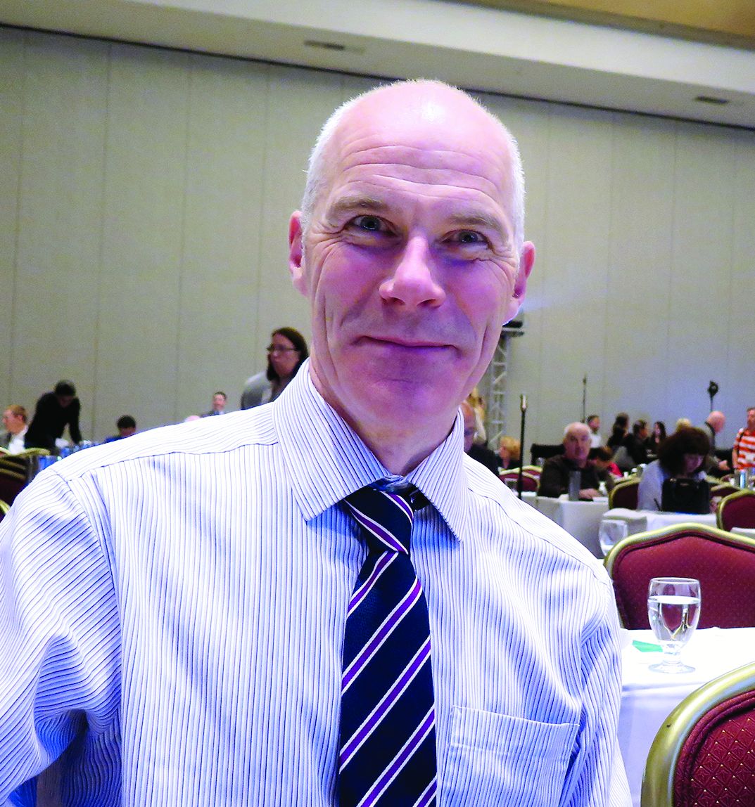
1. Use a loop diuretic to control symptoms. “If the patient has fluid retention, it’s important to get them out of congestive cardiac syndrome as quickly as possible,” he said.
2. Disease modification with beta-blockers and ACE inhibitors, to the maximal dose tolerated. “These are still the mainstays of treatment for patients with heart failure, and they’re even more important in patients with diabetes and heart failure,” he said.
3. Consider device therapy, including defibrillators and resynchronization therapy.4. Optimize diabetes management.
“If you keep these things in your mind as you see a patient with heart failure and diabetes, it will give you a guide to approaching these patients,” said Dr. Kearney, who is also research lead for heart failure services at the University of Leeds. “When I see patients, I’ll check for edema right away. If they have it, I’ll increase the diuretic, and I’ll think about the different steps in the treatment pathway.”
He described heart failure due to systolic dysfunction as a reduction in cardiac output that doesn’t meet the demands of the body, the endpoint of a whole range of insults to the left ventricle.
“The most common cause today is ischemic heart disease; it used to be hypertension,” he said. “People over 40 have a one in five chance of developing heart failure, so it’s really important for all of us to improve outcomes in this terrible syndrome.”
According to a study of nearly 2,000 unselected patients conducted by Dr. Kearney and his associates, those with diabetes and heart failure are more likely to have ischemia, compared with those who have heart failure and no diabetes (75% vs. 58%, respectively), lower hemoglobin (13 g/dL vs. 13.7 g/dL), and worse renal function (eGFR of 51 mL/min per 1.732 m2 vs 57 mL/min per 1.732 m2) (Diab Vasc Dis Res. 2013 Jul;10[4]:330-6).
He added that type 2 diabetes is a sudden death risk equivalent to patients with ischemic heart disease and left ventricular systolic dysfunction (Heart 2016 May 15;102[10]:735-40).
“So, if you have diabetes and the U.K. National Institute for Health and Care Excellence guidelines indication for a defibrillator, your risk of sudden death in 5 years is probably 50%,” Dr. Kearney said. “The best treatment in this case is a prophylactic defibrillator.”
ACE inhibitors are used to protect these patients against cardiac myocyte cell death and vasoconstriction, while beta-blockers are used to protect against the activation of the sympathetic nervous system.
“Often, patients don’t like taking beta-blockers because they say they make them feel tired – when in fact they don’t realize it’s their heart failure that’s making them feel tired,” Dr. Kearney said.
He and his associates examined the effect of different drugs doses on all-cause mortality at 5 years. They found that, among patients with heart failure, reduced ejection fraction, and no diabetes, ramipril conferred a 3% improvement in mortality per milligram. At the same time, the mortality among patients with diabetes and heart failure who did not receive a beta-blocker was about 7%.
However, the absolute gain from beta-blocker use in patients with diabetes and heart failure was three times that of patients without diabetes.
“So, every milligram you increase the dose by, there’s an associated improvement in risk,” Dr. Kearney said. “Over 5 years, comparing the lowest beta-blocker dose to the highest beta-blocker dose, it was 1 year of life gained. So, when I see my patients and they ask about side effects of beta-blockers, I now say to them, ‘The side effects actually make you live longer.’”
He concluded his remarks by noting that while he is not a diabetes expert, it’s clear that diabetes is intimately linked to the pathophysiology of heart failure.
“If you’re insulin resistant, you have hypertension, hyperglycemia, you have inflammation and bone marrow dysfunction – all of which can exacerbate left ventricle dysfunction,” he said. “You have a syndrome in which you have cardiac dysfunction and metabolic dysfunction that conspire to lead to worsening of left ventricular dysfunction.”
Dr. Kearney disclosed that he has been a speaker for Merck.
EXPERT ANALYSIS FROM WCIRDC 2017
How to prioritize CVD reduction in type 2 diabetes
LOS ANGELES – In the opinion of Mikhail N. Kosiborod, MD, the paradigm of treating patients with type 2 diabetes should shift from a narrow focus on hemoglobin A1c control to a broader strategy of reducing cardiovascular risk.
“We already know that the number one killer of patients with diabetes is cardiovascular disease, and we already know that lowering HbA1c as a general strategy does not substantially lower the risk of most important CVD events,” Dr. Kosiborod, a cardiologist at Saint Luke’s Mid-America Heart Institute, Kansas City, Mo., said at the World Congress on Insulin Resistance, Diabetes & Cardiovascular Disease.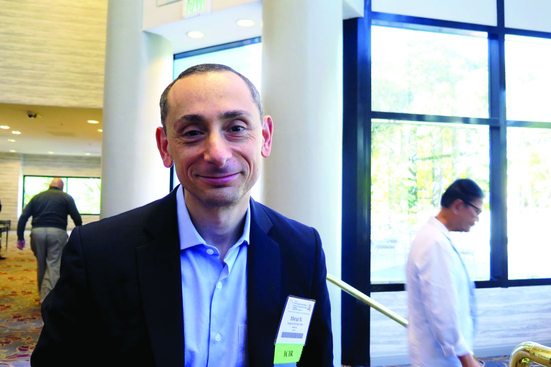
Physicians know that some medications lower the risk of cardiovascular events – including cardiovascular death – substantially, and other drugs don’t. “The bottom line is that we are not talking about ignoring HbA1c, but it’s how you get there that’s important – how you do it and in whom,” Dr. Kosiborod explained.
He pointed to a meta-analysis of four large diabetes trials involving 27,049 participants and 2,370 major vascular events (Diabetologia. 2009 Nov;52[11]:2288-98). It found that the general strategy of targeting more-intensive glucose lowering modestly reduced nonfatal myocardial infarction and increased major hypoglycemia over 4.4 years in people with type 2 diabetes – yet there was no difference in the effect of intensive glucose control on cardiovascular death or hospitalization for heart failure.
“Some point to the benefit of glucose control on the risk of nonfatal myocardial infarction, but that’s a modest benefit,” he said. “It’s observed beyond the randomization phase of clinical trials and takes many years to see it. It’s a large, very long-term investment for a modest reduction in MI risk, with no benefit in death or heart failure. So, when you test intensive glucose control as a general strategy, it has not been successful in reducing cardiovascular complications of type 2 diabetes.”
However, there is now evidence that specific classes of medications, such as sodium-glucose co-transporter-2 (SGLT2) inhibitors and glucagon-like peptide-1 (GLP-1) receptor agonists, initially developed for glucose lowering in type 2 diabetes, can significantly reduce cardiovascular risk within a relatively short time frame.
In EMPA-REG (Empagliflozin Cardiovascular Outcome Event Trial in Type 2 Diabetes Mellitus Patients), the first trial to demonstrate such benefits, all patients had established CVD, compared with 67% of patients in CANVAS (Canagliflozin Cardiovascular Assessment Study), a second RCT program to report cardiovascular outcomes with SGLT2 inhibitors. In the meantime, about 15%-20% of patients in real-world clinical practice have established CVD.
This led Dr. Kosiborod and his associates to launch CVD-REAL (Comparative Effectiveness of Cardiovascular Outcomes in New Users of SGLT2 Inhibitors), a real-world comparative effectiveness study that evaluated hospitalization for heart failure and total mortality among new users of SGLT2 inhibitors, compared with other glucose-lowering drugs.
In all, 154,528 patients in six countries were initiated on an SGLT2 inhibitor, and 154,528 were initiated on other glucose-lowering drugs (Circulation. 2017 May 18. doi: 10/1161/circulationaha.117.029190). The greatest exposure time was observed from canagliflozin (53%) followed by dapagliflozin (42%) and empagliflozin (5%).
The pooled analysis showed that initiation of SGLT2 inhibitors was associated with a significantly lower risk of heart failure events, compared with other glucose-lowering drugs (risk ratio, 0.61; P less than .001). The researchers observed an overall 39% lower risk of heart failure hospitalization, 51% reduction in total death, and 46% reduction in the composite of heart failure hospitalization or death.
“There was no heterogeneity across countries, despite the fact that the health care systems were very different and the prescribing patterns were very different,” he said.
Dr. Kosiborod, who is also professor of medicine at the University of Missouri-Kansas City, noted that 13% of patients from CVD-REAL had established CVD, while 87% did not. When comparing the results within these two key subgroups, “what’s striking is the difference in event rates, stratified by treatment allocation,” he said of the unpublished data.
“If you look at the composite outcome of heart failure or death, you see an almost seven-fold difference in annualized event rates – about 7% per year in patients with established CVD, compared with about 1% per year in the primary prevention cohort,” he explained. “But the relative risk reduction associated with SGLT2 inhibitors versus other glucose-lowering drugs is identical across both patient groups. That’s a good lesson in epidemiology: You can have patients with dramatically different absolute risks, dramatically different absolute risk reductions, and therefore dramatically different numbers needed to treat, but identical relative risk reductions.”
Dr. Kosiborod also pointed out that heart failure is emerging as one of the most important outcomes in trials patents with type 2 diabetes.
“That’s because people with diabetes who develop heart failure have very poor outcomes,” he said. “Among elderly patients with type 2 diabetes who develop new heart failure, there’s less than 25% survival at 5 years. That’s the reason, I think, that if you really want to impact survival and complication rates in people with diabetes, preventing and treating heart failure is one of the surest ways of doing so.
“You shouldn’t just think of the patient in front of you as someone who has an A1c of 7%, 8%, or 9%,” he cautioned. “You should also start thinking of where the patient is on the spectrum of cardiovascular disease, all the way from CVD risk factors only to symptomatic heart failure.”
Some evidence already exists to help clinicians make treatment decisions based on where the patients fall on that spectrum, he continued.
For example, clinical trials have demonstrated that in patients with established atherosclerotic cardiovascular disease, GLP-1 receptor agonists and SGLT2 inhibitors can reduce the risk of cardiovascular events, including, in some cases, cardiovascular death.
“We don’t have a lot of data demonstrating benefit for patients with recent acute coronary syndrome,” he said. “Some compounds have proven to be neutral, but none has been proven to save lives in this patient group.
“Now, we also have data for people with prior stroke that pioglitazone may be beneficial in managing those patients to prevent recurrent stroke and MI, based on the recent IRIS Trial, provided they don’t have heart failure at baseline,” Dr. Kosiborod added. “We don’t have definitive data yet in people with established heart failure, but those studies are ongoing.”
Dr. Kosiborod disclosed that he is a consultant for Amgen, AstraZeneca, Boehringer Ingelheim, Eisai, Glytec, GSK, Intarcia, Merck (Diabetes), Novartis, Novo Nordisk, Sanofi, and ZS Pharma. He has also received research grants from AstraZeneca and Boehringer Ingelheim.
EXPERT ANALYSIS FROM WCIRDC 2017
Three in 10 diabetic patients may have liver fibrosis
LOS ANGELES – For every 10 adult patients with type 2 diabetes, three are likely to have moderate to severe liver fibrosis, according to Kenneth Cusi, MD, FACP, FACE.
“The question is, How are we going to tackle this problem? My academic goal is that we incorporate screening for NASH [nonalcoholic steatohepatitis], or for fibrosis more specifically, in the same way we do for retinopathy or nephropathy [in diabetes], because we do have a way to treat it,” he said at the World Congress on Insulin Resistance, Diabetes & Cardiovascular Disease.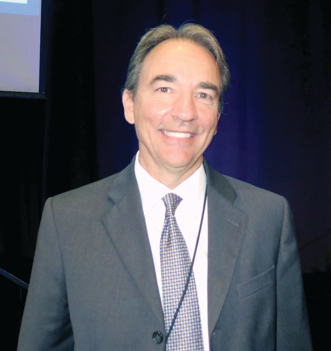
Dr. Cusi, chief of the division of endocrinology, diabetes, and metabolism at the University of Florida, Gainesville, predicted that obesity will become the No. 1 cause of liver transplantation. “It’s a real epidemic; you’re not seeing it because the inflexion of obesity happened just 2 decades ago,” he said. “Patients with diabetes face the greatest risk of fatty liver and of fibrosis. Untreated, it’s the equivalent of having macroalbuminuria. If you do nothing and they don’t die of cardiovascular disease, they’re going to have a good chance of getting fibrosis.”
As part of the large population-based Rotterdam study of individuals aged 45 years and older, researchers found that liver stiffness of 8 kPa or more by transient elastography was present in 5.6% of the study participants and was strongly associated with steatosis and diabetes (Hepatology. 2016;63:138-47). According to Dr. Cusi, individuals who have steatosis without diabetes face a 5%-10% risk of fibrosis, while those with steatosis and diabetes face a 15%-20% risk. “It’s well established in a number of studies that if you have fibrosis, you’re at high risk not only of cirrhosis, but also of hepatocellular carcinoma,” he said. “The key thing is not detecting fat, which is not really the target. The target is if there’s fibrosis or not.” Three ways to assess for fibrosis include MR elastography, transient elastography (which is the most commonly used), and fibrosis marker panels.
Liver fibrosis likely starts with adipose tissue dysfunction, said Dr. Cusi, who authored a review on the pathophysiology of interactions between adipose tissue and target organs in obesity and the resulting clinical implications for the management of nonalcoholic steatohepatitis (Gastroenterology. 2012;142[4]:711-25.e6). “When you have insulin-resistant, sick adipose tissue, that leads to the accumulation of fat in the liver,” he said. “. Even if you get people who are matched for BMIs [body mass indexes] between 30 and 35 kg/m2, there is a spectrum in which some individuals have very insulin-resistant adipose tissue and others less so. I would say that 1 out of 10 are metabolically healthy, and we don’t understand exactly why.”
In a recent cross-sectional analysis of 352 healthy individuals, Dr. Cusi and his associates found that intrahepatic triglyceride (IHTG) accumulation is strongly associated with adipose tissue insulin resistance, supporting the current theory of lipotoxicity as a driver of IHTG accumulation (Hepatology. 2017;65[4]:1132-44). The researchers observed that once IHTG accumulation reaches about 6%, skeletal muscle insulin resistance, hypertriglyceridemia, and low HDL cholesterol become fully established. “The next question is, How does this correlate with NASH?” Dr. Cusi said. “Our take is that there is a threshold effect. Once you have a critical amount of triglycerides in your liver, some individuals are going to activate pathways that are harmful. NASH is not something exclusive to individuals who are obese. Lean people can also develop NASH. The key feature is insulin resistance, not metabolic syndrome. Once you develop a fatty liver, your chances of NASH are comparable to that of an obese individual. The paradox is that lean individuals get a fatty liver, but when they get a fatty liver, they are at risk for NASH and for fibrosis.”
Why lean individuals develop NASH is not fully understood, but Dr. Cusi said he suspects that the problem develops at the mitochondrial level. Results from an unpublished animal model in which mice were fed a high–trans-fat diet for 24 weeks showed that the mice developed steatosis by week 8 and NASH by week 24. The mice had an increase in the tricarboxylic acid (TCA) cycle, which is typical of the NASH period, as well as an increase in ceramides. “Perhaps a unifying hypothesis would be that the development of NASH is linked to inflammation and to insulin signaling,” Dr. Cusi said. “Not surprisingly, it had a number of effects on the mitochondria, and in this animal model it decreases the TCA.” He noted that the biology of fibrosis remains unknown in humans. “What we have been familiar with is the high-triglyceride, low-HDL pattern,” he said. “If you look at how that correlates with the amount of liver fat, it is basically a threshold effect. Once you have steatosis, you don’t see much worse dyslipidemia, which is typical of these patients.”
Recently published guidance from the American Association for the Study of Liver Diseases on the diagnosis and management of nonalcoholic fatty liver disease (NAFLD) suggests that patients require a weight loss of 3%-5% to improve steatosis, but a loss of 7%-10% to improve most histologic features of NASH, including fibrosis (Hepatology. 2018;67[1]:328-57). Exercise alone may prevent or reduce steatosis, but its ability to improve other aspects of liver histology remains unknown. Bariatric surgery can be considered in otherwise eligible obese individuals with NAFLD or NASH. The procedure’s impact on fibrosis is unknown.
The AASLD practice guideline notes that metformin is not recommended for treating NASH in adult patients, but pioglitazone improves liver histology in patients with and without type 2 diabetes with biopsy-proven NASH. “Pioglitazone has had the greatest benefit in terms of treatment effect, compared to placebo,” Dr. Cusi said. “It’s a generic drug; at the VA [Veterans Affairs], it costs 8 cents per tablet. I think that pioglitazone will be to NASH what metformin has been to type 2 diabetes. The most common side effect is weight gain, typically between 4 and 9 lb. Risks and benefits should be discussed with each patient. It should not be used for NAFLD without biopsy-proven NASH.” The guideline goes on to say that it’s currently premature to consider GLP-1 (glucagonlike peptide–1) agonists for treating liver disease in patients with NAFLD or NASH. Meanwhile, vitamin E at 800 IU has been shown to improve liver histology in nondiabetic adults with NASH, but the risks and benefits should be discussed with each patient. Vitamin E is not recommended for NASH in diabetic patients, NAFLD without a liver biopsy, NASH cirrhosis, or cryptogenic cirrhosis.
The AASLD practice guideline also states that the best evidence for using SGLT2 (sodium-glucose cotransporter–2) inhibitors in NAFLD comes from animal studies, which report a reduction in steatosis with and without weight loss. Clinical studies reporting a reduction in steatosis are limited. There are positive observational studies with a reduction in alanine aminotransferase and some studies that have shown a reduction in liver fat. “For me, the best option is to tailor treatment to the pathophysiology of the disease,” Dr. Cusi said. “You reduce fat by weight loss in some way, or you change the biology of fat with a thiazolidinedione.”
Dr. Cusi reported that he has received grant support from the Burroughs Wellcome Fund, the American Diabetes Association, and the National Institutes of Health.
EXPERT ANALYSIS FROM WCIRDC 2017
Three in 10 of your diabetic patients may have liver fibrosis
LOS ANGELES – For every 10 of your adult patients with type 2 diabetes, three are likely to have moderate to severe liver fibrosis, according to Kenneth Cusi, MD, FACP, FACE.
“If in the last 10 patients, you didn’t diagnose anybody with fibrosis, you probably missed it,” he said at the World Congress on Insulin Resistance, Diabetes & Cardiovascular Disease. “The question is, How are we going to tackle this problem? My academic goal is that we incorporate screening for NASH [nonalcoholic steatohepatitis], or for fibrosis more specifically, in the same way we do for retinopathy or nephropathy, because we do have a way to treat it.”
As part of the large population-based Rotterdam study of individuals aged 45 years and older, researchers found that liver stiffness of 8 kPa or more by transient elastography was present in 5.6% of the study participants and was strongly associated with steatosis and diabetes (Hepatology. 2016;63:138-47). According to Dr. Cusi, individuals who have steatosis without diabetes face a 5%-10% risk of fibrosis, while those with steatosis and diabetes face a 15%-20% risk.
“It’s well established in a number of studies that if you have fibrosis, you’re at high risk not only of cirrhosis, but also of hepatocellular carcinoma,” he said. “The key thing is not detecting fat, which is not really the target. The target is if there’s fibrosis or not.” Three ways to assess for fibrosis include MR elastography, transient elastography (which is the most commonly used), and fibrosis marker panels.
Liver fibrosis likely starts with adipose tissue dysfunction, said Dr. Cusi, who authored a review on the pathophysiology of interactions between adipose tissue and target organs in obesity and the resulting clinical implications for the management of nonalcoholic steatohepatitis (Gastroenterology. 2012;142[4]:711-25.e6).
“When you have insulin-resistant, sick adipose tissue, that leads to the accumulation of fat in the liver,” he said. “. Even if you get people who are matched for BMIs [body mass indexes] between 30 and 35 kg/m2, there is a spectrum in which some individuals have very insulin-resistant adipose tissue and others less so. I would say that 1 out of 10 are metabolically healthy, and we don’t understand exactly why.”
In a recent cross-sectional analysis of 352 healthy individuals, Dr. Cusi and his associates found that intrahepatic triglyceride (IHTG) accumulation is strongly associated with adipose tissue insulin resistance, supporting the current theory of lipotoxicity as a driver of IHTG accumulation (Hepatology. 2017;65[4]:1132-44). The researchers observed that once IHTG accumulation reaches about 6%, skeletal muscle insulin resistance, hypertriglyceridemia, and low HDL cholesterol become fully established.
“The next question is, How does this correlate with NASH?” Dr. Cusi said. “Our take is that there is a threshold effect. Once you have a critical amount of triglycerides in your liver, some individuals are going to activate pathways that are harmful. NASH is not something exclusive to individuals who are obese. Lean people can also develop NASH. The key feature is insulin resistance, not metabolic syndrome. Once you develop a fatty liver, your chances of NASH are comparable to that of an obese individual. The paradox is that lean individuals get a fatty liver, but when they get a fatty liver, they are at risk for NASH and for fibrosis.”
Why lean individuals develop NASH is not fully understood, but Dr. Cusi said he suspects that the problem develops at the mitochondrial level.
Results from an unpublished animal model in which mice were fed a high–trans-fat diet for 24 weeks showed that the mice developed steatosis by week 8 and NASH by week 24. The mice had an increase in the tricarboxylic acid (TCA) cycle, which is typical of the NASH period, as well as an increase in ceramides.
“Perhaps a unifying hypothesis would be that the development of NASH is linked to inflammation and to insulin signaling,” Dr. Cusi said. “Not surprisingly, it had a number of effects on the mitochondria, and in this animal model it decreases the TCA.” He noted that the biology of fibrosis remains unknown in humans. “What we have been familiar with is the high-triglyceride, low-HDL pattern,” he said. “If you look at how that correlates with the amount of liver fat, it is basically a threshold effect. Once you have steatosis, you don’t see much worse dyslipidemia, which is typical of these patients.”
Recently published guidance from the American Association for the Study of Liver Diseases on the diagnosis and management of nonalcoholic fatty liver disease (NAFLD) suggests that patients require a weight loss of 3%-5% to improve steatosis, but a loss of 7%-10% to improve most histologic features of NASH, including fibrosis (Hepatology. 2018;67[1]:328-57). Exercise alone may prevent or reduce steatosis, but its ability to improve other aspects of liver histology remains unknown. Bariatric surgery can be considered in otherwise eligible obese individuals with NAFLD or NASH. The procedure’s impact on fibrosis is unknown.
The AASLD practice guideline notes that metformin is not recommended for treating NASH in adult patients, but pioglitazone improves liver histology in patients with and without type 2 diabetes with biopsy-proven NASH.
“Pioglitazone has had the greatest benefit in terms of treatment effect, compared to placebo,” Dr. Cusi said. “It’s a generic drug; at the VA [Veterans Affairs], it costs 8 cents per tablet. I think that pioglitazone will be to NASH what metformin has been to type 2 diabetes. The most common side effect is weight gain, typically between 4 and 9 lb. Risks and benefits should be discussed with each patient. It should not be used for NAFLD without biopsy-proven NASH.”
The guideline goes on to say that it’s currently premature to consider GLP-1 (glucagonlike peptide–1) agonists for treating liver disease in patients with NAFLD or NASH. Meanwhile, vitamin E at 800 IU has been shown to improve liver histology in nondiabetic adults with NASH, but the risks and benefits should be discussed with each patient. Vitamin E is not recommended for NASH in diabetic patients, NAFLD without a liver biopsy, NASH cirrhosis, or cryptogenic cirrhosis.
The AASLD practice guideline also states that the best evidence for using SGLT2 (sodium-glucose cotransporter–2) inhibitors in NAFLD comes from animal studies, which report a reduction in steatosis with and without weight loss. Clinical studies reporting a reduction in steatosis are limited. There are positive observational studies with a reduction in alanine aminotransferase and some studies that have shown a reduction in liver fat. “For me, the best option is to tailor treatment to the pathophysiology of the disease,” Dr. Cusi said. “You reduce fat by weight loss in some way, or you change the biology of fat with a thiazolidinedione.”
Dr. Cusi reported that he has received grant support from the Burroughs Wellcome Fund, the American Diabetes Association, and the National Institutes of Health.
EXPERT ANALYSIS FROM WCIRDC 2017
Shedding light on shift work’s influence on cardiometabolic risk
LOS ANGELES – Current and previous night workers had significantly increased levels of hemoglobin A1c, compared with diurnal workers, preliminary results from an ongoing study showed. The finding sheds further insight into the link between environmental light, circadian rhythms, and metabolic disorders.
“To date, observational studies on bright light have revealed that evening bright light is associated with increased appetite and that bedroom light intensity is correlated with obesity,” Massimo Federici, MD, said at the World Congress on Insulin Resistance, Diabetes & Cardiovascular Disease. “It’s also been reported that artificial light is correlated with type 2 diabetes in the home setting and that daytime light exposure is positively correlated with body mass index. However, no studies have directly investigated the effect of acute light on human glucose metabolism.”
Although few metabolic intervention studies using light have been done, Dr. Federici mentioned three of note. One, in patients with seasonal affective disorder and type 2 diabetes, showed reduced insulin requirements after light therapy (Lancet. 1992;339[8800]:1065-6). Another, a short-term study of 25 obese subjects treated with 5,000 lux bright light therapy in addition to exercise, showed reduced body fat after 6 weeks (Obesity 2007; 15[7]:1749-57). A third, in 34 obese subjects who were exposed to 1,300 lux bright light every morning for 3 weeks, showed a small but significant reduction in fat mass (Obes Facts 2013;6:28-38).
As part of an ongoing project known as EuRhythDia, researchers including Dr. Federici set out to identify metabolic and molecular variables associated with shift work, and to test the effect of a lifestyle intervention that comprised light exposure, exercise, and melatonin. He presented unpublished results from one aspect of the trial: a cross-sectional analysis of 273 nurses divided into one of three groups: 64 diurnal workers (DW), 111 active night shift workers (aNW), and 98 prior night shift workers (pNW). Those with diabetes or taking oral antidiabetic drugs were excluded from the study.
The analysis showed that nurses in the pNW group were significantly older, at a mean of 39.7 years, than those in the DW group, whose mean age was 37 years, and the aNW group, who averaged 36.1 years. Those in the pNW group also had a significantly greater body mass index, compared with their counterparts in the aNW and DW groups (a mean of 25.7 kg/m2, vs. 24.8 and 23.7, respectively) as well has a higher mean waist circumference (a mean of 87.2 cm, vs. 84.6 cm and 82 cm).
The mean HbA1c was higher in the nurses with prior and active night shift work, at 5.3% each, than in the diurnal workers (5.1%, P less than .001).
When Pittsburgh Sleep Quality Index scores were used to evaluate sleep quality independent of work status, more than half of the study subjects (163) were classified as being “good sleepers,” while 110 were considered to be “bad sleepers.” Bad sleepers had a significantly higher mean HbA1c level compared with good sleepers (5.3% vs. 5.2%). Bad sleepers also had higher levels of HDL cholesterol (a mean of 60.8 mg/dL vs. 56.3 mg/dL).
Dr. Federici highlighted preliminary findings from a study of 32 aNW subjects who were assigned to treatment with warm light therapy at 1,000 lux for 30 minutes at 30 cm every morning for 3 months. They observed a mild improvement in the area under the curve of the oral glucose tolerance test at 24 weeks (12 weeks’ washout after 12 weeks of light therapy). “However, the effect was obtained not at the end of the intervention but at the end of the washout period,” he said.
He called for more studies going forward that take into account the effect of seasons as well as the effects of diet and exercise.
Dr. Federici disclosed that he receives editorial fees from Springer Nature group.
EXPERT ANALYSIS FROM WCIRDC 2017
Elevated CRP and mortality risk differs by gender, race
LOS ANGELES – The relationship between elevated C-reactive protein concentrations and increased all-cause mortality risk varies by gender and by race/ethnicity, an analysis of national data showed.
“Opportunities exist to discuss the importance of this marker and the relationship with mortality risk,” study author M. Ryan Richardson said in an interview following the World Congress on Insulin Resistance, Diabetes & Cardiovascular Disease. “Although we do not fully understand all of the mechanisms that underlie this harmful relationship, our results add to the limited evidence available to those working within the clinical setting.”
A total of 4,383 adults between the ages of 30 and 79 years were included in the analysis. After the researchers adjusted for age, race, education, smoking, alcohol consumption, cardiovascular disease, waist circumference, and aerobic physical activity, they observed a significantly higher risk for all-cause mortality in non-Hispanic black males (hazard ratio, 2.04) and Mexican-American females (HR, 2.24). “We were surprised that this relationship was also independent of measured waist circumference and any volume of reported aerobic physical activity, which are both acknowledged as strong mediator variables in this relationship,” Mr. Richardson said. The HR in non-Hispanic white males approached but did not reach statistical significance (HR, 1.32).
He acknowledged certain limitations of the study, including its cross-sectional design. “We cannot make causal inferences based on this data,” he said.
The researchers reported having no financial disclosures.
SOURCE: M. Ryan Richardson et al.
REPORTING FROM WCIRDC 2017
Key clinical point:
Major finding: The risk for all-cause mortality was significantly higher in non-Hispanic black men and in Mexican-American females (HR of 2.04 and 2.24, respectively).
Study details: An analysis of 4,383 adults who participated in NHANES 1999-2006.
Disclosures: The researchers reported having no financial disclosures.
Source: M. Ryan Richardson et al.
Expert discusses the role of salt and fructose in diabetes
LOS ANGELES – Sugar consumption has been implicated as a risk factor for the development of diabetes since at least the 1920s, but high salt intake may also increase the risk for obesity and prediabetes by stimulating fructose production in the liver.
“We think about high-salt diets as being associated with hypertension, but if you put people on a high-salt diet, you can induce insulin resistance within 5 or 10 days,” Richard J. Johnson, MD, said at the World Congress on Insulin Resistance, Diabetes & Cardiovascular Disease. “There is a fair amount of published data suggesting that a high salt intake, defined as greater than 150 mmol per day, is associated with hypertension, insulin resistance, obesity, and metabolic syndrome. In fact, it’s been reported that obese people are slightly hyperosmolar and tend to have elevated vasopressin levels. Interestingly, sugar stimulates vasopressin production.”
Furthermore, the mice on high-salt diets became insulin resistant. “Their fasting glucose went up, their fasting insulin went up, and they developed marked fatty liver, obesity, abdominal fat, and hypertension,” Dr. Johnson added. The discovery supports the notion that osmolality is the mechanism by which salt drives blood pressure, not volume expansion. “And I think that’s going to turn out to be important in obesity and metabolic syndrome as well,” he said.
Dr. Johnson, a professor of medicine in the department of renal diseases and hypertension at the University of Colorado at Denver, Aurora, discussed the role of role of sugar and fructose in the development of diabetes as well. He noted that annual sugar consumption in the United States rose from about 4 pounds per person in 1700 to about 150 pounds per person today. About one-third of current sugar intake comes from soft drinks.
“The reason why we think that fructose is a good candidate for playing a role in the diabetes epidemic is because, when you give an animal water drinking combined with fructose, they will rapidly start increasing their energy intake,” he said. “They become lethargic and less active, and they gain weight.”
He and his colleagues have demonstrated that when rats are fed fructose over time, they become leptin resistant (Am J Physiol. 2008 Nov;295:R1370-5). “Not only that, it’s been shown in animals and humans that, if you feed people fructose over time, fructose will decrease resting energy expenditure,” he said. “Our work and that of others has shown that fructose stimulates weight gain by stimulating energy intake. It does so by inducing leptin resistance. It also works in the brain to stimulate dopamine and to drive food intake that way as well.”
Fructose also impairs fatty acid oxidation and reduces energy expenditure, he continued. In one human trial, in which men consumed 200g of fructose for 2 weeks, Dr. Johnson and his associates found that 25% of them developed features of metabolic syndrome (Int J Obes. 2010;34[3]:454-61). “Just think about how long it takes to get obese or metabolic syndrome,” he said. “We think in terms of years, but this was a 2-week study! This means there’s something special about fructose that seems to drive metabolic syndrome.”
A key player in the process appears to be an enzyme in the liver known as fructokinase, which metabolizes fructose so rapidly that it causes ATP depletion. “Normally, when glucose is metabolized, ATP levels stay normal in the cell because if you start to consume too much ATP in the initial phosphorylation, there’s a feedback mechanism,” Dr. Johnson said. “But not so for fructose; it’s a runaway train, and it activates a nucleotide degradation pathway, which we call the energy depletion pathway. This seems to be what is critical for fructose effects.”
Further evaluation of that pathway led him and his associates to discover that lowering uric acid reduced fatty liver formation in fructose-fed rats (PLOS One 2012 Oct. 24. doi:10.1371/journal.pone.0047948). “We started looking at how this worked, and we found that, when we put uric acid on liver cells, that they actually stimulated fat accumulation,” he said. “We showed that in an in vitro system, and we found that both fructose and uric acid stimulate oxidative stress in the mitochondria. It’s very specific. You can actually block the production of oxidative stress with allopurinol, a drug that lowers uric acid. We’ve been building a case that this pathway is involved in a lot of mechanisms that lead to obesity, insulin resistance, and hypertension.”
Dr. Johnson concluded by noting that not all calories are created equal. “Some additives, like salt, might be playing a role in metabolic syndrome, obesity, and diabetes,” he said. “We think that sugar and high fructose corn sugar are the major causes driving metabolic syndrome. High-glycemic carbs are working primarily through fructose to induce [insulin resistance]. We think that salt may accelerate this pathway as well.”
He disclosed that he holds patents and patent applications related to this work and that he has launched a start-up company trying to develop inhibitors of fructose metabolism.
Dr. Johnson reported having no conflicts of interest related to this article.
EXPERT ANALYSIS AT WCIRDC 2017
Intranasal insulin, ketogenic diet may benefit Alzheimer’s patients
LOS ANGELES – Growing evidence from basic science and preclinical studies demonstrates that
In addition, brain insulin resistance in Alzheimer’s disease (AD) is associated with increased cerebral hyperglycemia, reduced cerebral glucose utilization, reduced blood flow, and reduced accumulation of amyloid and tau.
When that process goes awry, several pathologic processes linking insulin resistance (IR) and Alzheimer’s disease occur, including impaired proteostasis (oligomeric beta amyloid, microtubule-associated tau, and oligomeric insulin); hyperglycemia-induced toxicity and reduced glucose utilization; mitochondrial dysfunction; and vascular dysfunction, Dr. Craft continued.
In the past 5 years, researchers have developed ways to measure expression levels of insulin resistance markers like insulin receptor substrate 1 (IRS-1) pSer–positive neurons, Dr. Craft said. Others have found that increased IRS-1 pSer is associated with paired helical filaments (PHFs) tau in mild cognitive impairment (MCI) and AD, and that increased IRS-1 pSer in neutrally derived plasma exomes increased in AD years before onset and in type 2 diabetes. “We can look at the neurons themselves and what we see is that this IR marker colocalizes with tau,” Dr. Craft said. “It’s not very common in normal folks, but as you progress through the stages of MCI to AD, it becomes more common. So there’s a progressive increase in IR markers that associates with neurons in tau.” Other imaging studies have shown that homeostatic model assessment IR predicts gray matter atrophy, reduced blood flow, and amyloid deposition in middle-aged adults.
One way to overcome IR in AD is to increase insulin availability in the brain. Intranasal administration of insulin is a novel method being tested by Dr. Craft and her associates. “This is not inhaled insulin; it does not target the lungs,” she explained. “It’s insulin administered with a very specialized device that targets the olfactory cleft in the upper nasal passages. Virtually none of the insulin is deposited in the lungs or nasopharyngeally.” The approach is modeled on the notion that there are pathways from the olfactory perivascular spaces to the brain by which peptides can travel readily by bulk flow. “They reach the brain within minutes,” she said. “It’s a way of delivering peptides to the brain that bypasses the blood-brain barrier.”
In a published study, she and her associates randomized 104 adults with MCI or AD to receive 20 IU insulin, 40 IU insulin, or placebo twice daily for 4 months (Arch Neurol. 2012;61[1]:29-38). Tests performed at baseline and at 4 months included cognitive evaluation based on story recall and the ADAS-Cog (Alzheimer’s Disease Assessment Scale–Cognitive subscale); function based on the Dementia Rating Severity Scale, FDG-PET (positron emission tomography with 18fluorodeoxyglucose), and cerebrospinal fluid biomarkers. “We showed that the 20-IU dose of intranasal insulin improved memory quite substantially (P less than .05),” Dr. Craft said. “It also improved glucose utilization as assessed by FDG-PET. We also saw changes in spinal fluid biomarkers of amyloid in a favorable direction. Most recently, we looked at the exosomal indicator of IR (IRS-1 pSer), and what we saw quite remarkably was a reduction in the same condition that the memory improved. This gives us hope that we have a marker of whether or not we’re having a positive impact.”
She and her research team recently finished a phase 2 pilot study of regular insulin vs. long-acting insulin detemir, to determine if a longer-acting agent with longer exposure would have greater efficacy (J Alzheimers Dis. 2017;57[4]:1325–34). In all, 36 participants were randomized to receive placebo, 40 IU of insulin detemir, or 40 IU of regular insulin daily for 4 months, administered with a nasal delivery device. The investigators found that only the group treated with 40 IU regular insulin had better memory after 2 and 4 months, compared with placebo (P less than .03). Regular insulin treatment was also associated with preserved volume on MRI. “The normal pattern is for AD patients to lose brain volume rather rapidly,” Dr. Craft commented. “We see that abolished by the insulin treatment, which suggests to us that we’re able to stave off this disease-related mechanism.” She and her associates are currently conducting a phase 3 clinical trial with regular insulin and a phase 3 trial with rapid-acting insulin that are expected to be completed in the summer of 2018.
Dr. Craft spent the last few minutes of her presentation discussing the ketogenic diet as a nonpharmacologic approach to preventing or treating brain insulin resistance and AD. “I think the power of diet is underestimated, both in terms of causing disease and potentially modulating it,” she said. Her research team just completed a study of 87 middle-aged adults who were randomized to a Western diet or a healthy diet. The Western diet was high in saturated fat, sugar, and salt. The healthy diet was low in saturated fat, sugar, and salt, but the macronutrient composition of both diets was the same. “It was a eucaloric diet with normal calorie intake; no weight change, so trying to understand the integrated effect of the Western diet,” she said. “All food was prepared by us and delivered to the patients two times per week.” Patients with type 2 diabetes, patients with hypertension, and those who were on statins were excluded from the study.
The researchers observed pronounced diet-induced changes in cerebral blood flow, all which favored the healthy diet group. “The Western diet reduced blood flow, and the healthy diet increased blood blow in the hippocampus, which is critical for memory, as in some other regions that are known to be affected in AD,” Dr. Craft said. “We saw an effect on memory as well, with the healthy diet improving memory and the Western diet reducing it. Both of these effects were significant, so 4 weeks on a diet such as this is sufficient to modulate key aspects of brain function.”
More recently, Dr. Craft and her colleagues have been evaluating the effects of what they term the modified Mediterranean ketogenic diet (MMKD). “It does allow for higher carbohydrate consumption, compared with a traditional ketogenic diet, but they still have to stay under 10% a day,” she said. “We have an emphasis on healthy fats. We send everybody home with extra virgin olive oil. We think it gives us extra compliance and the potential for long-term nutrition.” She explained that the diet increases plasma and CNS ketone bodies, beta-hydroxybutyrate, acetoacetate, and acetone, which serves as preferred alternative fuel for the brain. “If the brain has a choice between glucose and ketones, it will choose ketones,” Dr. Craft said. “It can use them more easily.”
Ketone bodies are derived from hepatic fatty acid oxidation and readily diffuse across the blood-brain barrier into the brain. They are also synthesized in the brain by astrocytes, and they appear to have direct neuroprotective effects. “Ketone bodies may be beneficial because they may correct the hyperglycemic state and reduce glucose utilization in the brain in AD years prior to symptom onset,” Dr. Craft said. “They may correct neuronal hyperexcitability and preclinical seizures in presymptomatic and early stages of AD; they restore the balance between inhibitory and excitatory neurotransmitters like GABA [gamma-aminobutyric acid] and glutamate.”
In an unpublished, 16-week study, Dr. Craft and her associates randomized 16 patients to a Mediterranean ketogenic diet or to an American Heart Association low-fat diet. Lumbar punctures and brain imaging were performed before and after diet intervention. By the end of 6 weeks, they observed significant increases in ketones and in HDL cholesterol level in the MMKD group, compared with the AHA diet group, as well as significant decreases in trigylcerides and HbA1c level. “I would say that we improved the peripheral metabolic profile with the ketogenic diet,” Dr. Craft said. They also observed significant improvements from baseline in memory, spinal fluid AD biomarkers, and mitochondrial respiration.
“One of the things we’re appreciating is the role of insulin in a host of activities in the brain,” she concluded. “Disrupting those activities can have dire consequences on brain function that may lead to a neurological milieu that lends itself to pathological aging conditions like Alzheimer’s. Several large ongoing trials are poised to validate results of smaller studies, elucidate underlying mechanisms, and provide new therapeutic targets. It’s an exciting time.”
Dr. Craft’s research is supported by the National Institute on Aging and the Alzheimer’s Association Zenith Program. Intranasal delivery devices were provided by Kurve Technology.
AT WCIRDC 2017