User login
Cardiovascular Research Foundation: Transcatheter Cardiovascular Therapeutics (TCT)
TCT: Lower MI, thrombosis, higher bleeding with extended DAPT in patients with everolimus-eluting stents
SAN FRANCISCO – Continued thienopyridine therapy beyond 1 year in patients treated with an everolimus-eluting stent was associated with significantly lower rates of myocardial infarction and thrombosis, but an increased risk of bleeding, study results have shown.
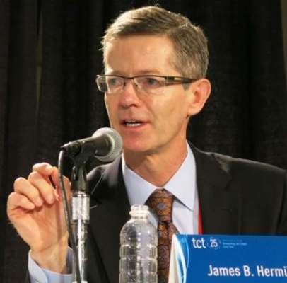
Dr. James B. Hermiller of St. Vincent´s Medical Group, Indianapolis, presented the data at the Transcatheter Cardiovascular Therapeutics annual meeting. The results were also published online (JACC: Cardiovasc Interv 2015. doi: 10.1016/j.jcin.2015.10.001).
In a post hoc analysis of a subset of patients from the DAPT (Dual Antiplatelet Therapy) study, Dr. James B. Hermiller Jr. and his colleagues evaluated major adverse cardiac and cerebrovascular event (MACCE) and bleeding events in 4,703 patients who’d been treated with an everolimus-eluting stent and 1 year of therapy with thienopyridine and aspirin before being randomly assigned to 18 additional months of the same DAPT, or an additional 18 months of aspirin plus placebo.
The original DAPT Study, a multisite, international, double-blinded, trial of 25,682 coronary stent patients was done to evaluate the efficacy and safety of various combinations of types of drug-eluting stents plus DAPT.
Results from the subanalysis of the everolimus-eluting stent group were that at 12-30 months post percutaneous intervention, those given additional DAPT had significantly reduced thrombosis (P =.04) and MI (P =.01), compared with aspirin-only controls, but the DAPT test cohort had more moderate to severe bleeding complications (P = .01). Composite death, MI, or stroke rates were also better in the placebo group (P = .42), Dr. Hermiller said at the meeting sponsored by the Cardiovascular Research Foundation.
The continuous-DAPT group had a stent thrombosis rate of 0.3%, compared with 0.7% of the 12-month–only group (hazard ratio, 0.38). The rate of MI in the test group was 2.11% vs. 3.2% in controls (HR, 0.63). The DAPT test group had a 2.5% rate of increased bleeding vs. 1.3% in the placebo group (HR, 1.79).
Patients in the subanalysis were typically older than across all original study groups – typically, men in their early 60s who were more likely to have been treated with clopidogrel (84.4% vs. 48.2%), and to have a slightly lower body mass index, although they were still obese (30.3 vs. 30.8 kg/m2).
The subanalysis group also had a slightly higher rate of history of cancer (P less than .001), a data point that Dr. Hermiller and his coauthors noted in the study accounted for the DAPT continuation group’s significantly increased all-cause mortality, compared with the placebo group (2.2% vs. 1.1%, P =.02).
In an interview, Dr. Spencer B. King, president of the Heart and Vascular Institute of St. Joseph’s Health System in Georgia, and comoderator of the session where the data were presented, also emphasized this finding’s importance, since “cancer increases bleeding risk and is one of the conditions in which prolonged DAPT should usually be avoided.”
The most frequent cause of noncardiovascular death in the DAPT-continuation group was cancer: 18 of the test group vs. 4 in the placebo group (P =.002); 3 of the study group’s cancer deaths involved bleeding. Overall, the second most common cause of death was bleeding, which occurred more in the DAPT test group than in placebo (P = .04).
Dr. Hermiller and his coauthors wrote that the therapeutic window for continued thienopyridine therapy “may be narrow” since the number needed to treat to benefit for stent thrombosis was 235 over 18 months. For MI it was 98. The number needed to treat to harm for moderate or severe bleeding was 84.
“Therefore, meticulous assessment of bleeding risk should always impact decisions regarding thienopyridine therapy duration.”
However, they also noted that current data suggest a net benefit to continuing therapy with absolute reductions in stent thrombosis or MI of about 0.2%, an effect that was exceeded in the everolimus-eluting stent patient subset.
Dr. Hermiller is a consultant for Abbott Vascular, Boston Scientific, Medtronic, and St. Jude Medical. The study also was sponsored by the Harvard Clinical Research Institute Harvard Clinical Research Institute and funded by Abbott, Boston Scientific Corporation, Cordis Corporation, Medtronic, Bristol-Myers Squibb Company/Sanofi Pharmaceuticals Partnership, Eli Lilly and Company, and Daiichi Sankyo Company Limited and the U.S. Department of Health & Human Services.
On Twitter @whitneymcknight
SAN FRANCISCO – Continued thienopyridine therapy beyond 1 year in patients treated with an everolimus-eluting stent was associated with significantly lower rates of myocardial infarction and thrombosis, but an increased risk of bleeding, study results have shown.
Dr. James B. Hermiller of St. Vincent´s Medical Group, Indianapolis, presented the data at the Transcatheter Cardiovascular Therapeutics annual meeting. The results were also published online (JACC: Cardiovasc Interv 2015. doi: 10.1016/j.jcin.2015.10.001).
In a post hoc analysis of a subset of patients from the DAPT (Dual Antiplatelet Therapy) study, Dr. James B. Hermiller Jr. and his colleagues evaluated major adverse cardiac and cerebrovascular event (MACCE) and bleeding events in 4,703 patients who’d been treated with an everolimus-eluting stent and 1 year of therapy with thienopyridine and aspirin before being randomly assigned to 18 additional months of the same DAPT, or an additional 18 months of aspirin plus placebo.
The original DAPT Study, a multisite, international, double-blinded, trial of 25,682 coronary stent patients was done to evaluate the efficacy and safety of various combinations of types of drug-eluting stents plus DAPT.
Results from the subanalysis of the everolimus-eluting stent group were that at 12-30 months post percutaneous intervention, those given additional DAPT had significantly reduced thrombosis (P =.04) and MI (P =.01), compared with aspirin-only controls, but the DAPT test cohort had more moderate to severe bleeding complications (P = .01). Composite death, MI, or stroke rates were also better in the placebo group (P = .42), Dr. Hermiller said at the meeting sponsored by the Cardiovascular Research Foundation.
The continuous-DAPT group had a stent thrombosis rate of 0.3%, compared with 0.7% of the 12-month–only group (hazard ratio, 0.38). The rate of MI in the test group was 2.11% vs. 3.2% in controls (HR, 0.63). The DAPT test group had a 2.5% rate of increased bleeding vs. 1.3% in the placebo group (HR, 1.79).
Patients in the subanalysis were typically older than across all original study groups – typically, men in their early 60s who were more likely to have been treated with clopidogrel (84.4% vs. 48.2%), and to have a slightly lower body mass index, although they were still obese (30.3 vs. 30.8 kg/m2).
The subanalysis group also had a slightly higher rate of history of cancer (P less than .001), a data point that Dr. Hermiller and his coauthors noted in the study accounted for the DAPT continuation group’s significantly increased all-cause mortality, compared with the placebo group (2.2% vs. 1.1%, P =.02).
In an interview, Dr. Spencer B. King, president of the Heart and Vascular Institute of St. Joseph’s Health System in Georgia, and comoderator of the session where the data were presented, also emphasized this finding’s importance, since “cancer increases bleeding risk and is one of the conditions in which prolonged DAPT should usually be avoided.”
The most frequent cause of noncardiovascular death in the DAPT-continuation group was cancer: 18 of the test group vs. 4 in the placebo group (P =.002); 3 of the study group’s cancer deaths involved bleeding. Overall, the second most common cause of death was bleeding, which occurred more in the DAPT test group than in placebo (P = .04).
Dr. Hermiller and his coauthors wrote that the therapeutic window for continued thienopyridine therapy “may be narrow” since the number needed to treat to benefit for stent thrombosis was 235 over 18 months. For MI it was 98. The number needed to treat to harm for moderate or severe bleeding was 84.
“Therefore, meticulous assessment of bleeding risk should always impact decisions regarding thienopyridine therapy duration.”
However, they also noted that current data suggest a net benefit to continuing therapy with absolute reductions in stent thrombosis or MI of about 0.2%, an effect that was exceeded in the everolimus-eluting stent patient subset.
Dr. Hermiller is a consultant for Abbott Vascular, Boston Scientific, Medtronic, and St. Jude Medical. The study also was sponsored by the Harvard Clinical Research Institute Harvard Clinical Research Institute and funded by Abbott, Boston Scientific Corporation, Cordis Corporation, Medtronic, Bristol-Myers Squibb Company/Sanofi Pharmaceuticals Partnership, Eli Lilly and Company, and Daiichi Sankyo Company Limited and the U.S. Department of Health & Human Services.
On Twitter @whitneymcknight
AT TCT 2015
Key clinical point: The window of extended DAPT therapy may be narrow in patients stented with an everolimus-eluting stent.
Major finding: Patients randomly assigned to 18 months of thienopyridine plus aspirin after 1 year of DAPT had significantly reduced thrombosis (P = .04) and MI (P = .01) vs. placebo plus aspirin but more bleeding (P = .01).
Data source: Subanalysis of 4,703 patients from a double-blind, international, multisite, randomized, controlled trial of 25,682 patients.
Disclosures: Dr. Hermiller is a consultant for Abbott Vascular, Boston Scientific, Medtronic, and St. Jude Medical. The study also was sponsored by the Harvard Clinical Research Institute Harvard Clinical Research Institute and funded by Abbott, Boston Scientific Corporation, Cordis Corporation, Medtronic, Bristol-Myers Squibb Company/Sanofi Pharmaceuticals Partnership, Eli Lilly and Company, and Daiichi Sankyo Company Limited and the U.S. Department of Health & Human Services.

VIDEO: Bioresorbable vascular scaffolds serve niche patients
SAN FRANCISCO – Results from the ABSORB III pivotal trial of the Absorb bioresorbable vascular scaffold will likely soon bring the device to the U.S. market, where it initially will be appropriate for a minority of patients with coronary artery disease, Dr. Daniel I. Simon said in an interview during the Transcatheter Cardiovascular Therapeutics annual meeting.
“I suspect its penetration will be no different than in Europe, where it’s used on about 10%-15% of patients, for certain niche patients. There will be target populations,” said Dr. Simon, professor and chief of cardiovascular medicine at University Hospitals Case Medical Center in Cleveland.
One attractive coronary candidate for treatment with bioresorbable vascular scaffolds would be a middle-aged man with diffuse and distal stenoses in his left anterior descending coronary artery and disease that’s not especially suitable for coronary bypass surgery that if stented would likely result in 70 mm or more in stent length, posing a high long-term risk for restenosis. In such patients devices that disappear after 3 years are attractive to avoid what might otherwise be a long stretch of metal, Dr. Simon said at the meeting sponsored by the Cardiovascular Research Foundation.
Dr. Simon has been an advisor to Medtronic and Heart Flow and he was an investigator on the ABSORB III trial, sponsored by Abbott Vascular.
The video associated with this article is no longer available on this site. Please view all of our videos on the MDedge YouTube channel
On Twitter @mitchelzoler
EXPERT ANALYSIS FROM TCT 2015
TCT: Bivalirudin no better than heparin for preventing post-TAVR bleeding
SAN FRANCISCO – The reversible direct thrombin inhibitor bivalirudin, compared with unfractionated heparin, did not reduce the rate of major bleeding at 48 hours after transcatheter aortic valve replacement, in the randomized, open label, phase IIIb BRAVO 3 trial.
Bivalirudin, which has a half-life of 25 minutes, has been shown to reduce major bleeding in the setting of percutaneous coronary intervention when compared with other regimens, but its safety and efficacy as compared with that of unfractionated heparin was unknown. In BRAVO 3 (The effect of BivaliRudin on Aortic Valve Intervention Outcomes trial), the rate of major bleeding at 48 hours, defined as Bleeding Academic Research Consortium (BARC) 3b or greater, was 6.9% in 404 patients treated with bivalirudin vs. 9.0% in 398 patients treated with heparin (relative risk, 0.77), Dr. Thierry Lefevre reported at the Transcatheter Cardiovascular Therapeutics annual meeting.
Further, the rate of net adverse clinical outcomes (NACE), including all-cause mortality, myocardial infarction, stroke, and major bleeding at up to 30 days was 14.4% in the bivalirudin group vs. 16.1% in the heparin group (relative risk, 0.89), Dr. Lefevre of Institut Hospitalier Jacques Cartier, Massy, France, said at the meeting, which was sponsored by the Cardiovascular Research Foundation.
The differences on both measures failed to meet statistical significance. With respect to bleeding at 48 hours, bivalirudin did not meet superiority, and with respect to cardiovascular events at 30 days, the prespecified noninferiority hypothesis was met.
No difference was seen between the groups for secondary endpoints, including bleeding defined according to various other bleeding scales and other BARC types.
The findings were published simultaneously in the Journal of the American College of Cardiology (J Am Coll Cardiol. 2015;Oct 15. doi: 10.1016/j.jacc.2015.10.003).
Heparin should remain the standard of care, especially given its lower cost compared to the cost of bivalirudin, Dr. Lefevre said.
However, Dr. Lefevre noted, major bleeding remains an important concern in transcatheter aortic valve replacement (TAVR), and “bivalirudin may be used as an alternative to heparin during TAVR in patients who cannot be treated with UFH.”
BRAVO III participants were adults with aortic stenosis who were at high surgical risk and who were scheduled for TAVR via transfemoral access. They were enrolled at 31 centers in seven countries throughout Europe and North America.
Those randomized to the bivalirudin group received an initial bolus of 0.75 mg/kg followed by a continuous infusion at a rate of 1.75 mg/kg per hour in those with an estimated glomerular filtration rate (eGFR) of 60 mL/min or greater, 1.4 mg/kg per hour in those with eGFR of 30-59 mL/min, and 1.0 mg/kg per hour in those with eGFR lower than 30 mL/min.
Heparin dosing and administration included a recommended target activated clotting time of greater than 25 seconds. Protamine was used for reversal at the end of the procedure based on standard local institution practice.
Session moderator Dr. Ajay J. Kirtane said that “one of the reasons this trial is exciting is that there has been a hypothesis that bivalirudin could reduce bleeding compared to heparin alone in PCI patients. While this is a much bigger access site, you don’t necessarily see these trends. On the other hand, when you have a bigger access site there is potential for increased signal, but this study is still somewhat underpowered.”
The Medicines Company provided funding for the BRAVO 3 trial to the Icahn School of Medicine at Mount Sinai, New York. Dr. Lefevre disclosed ties with Boston Scientific, Directflow, Edwards, Symetis and Medtronic.
AT TCT 2015
Key clinical point: The reversible direct thrombin inhibitor bivalirudin did not reduce the rate of major bleeding at 48 hours after transcatheter aortic valve replacement, compared with unfractionated heparin, in the BRAVO 3 trial.
Major finding: The rate of major bleeding at 48 hours was 6.9% vs. 9.0% in patients treated with bivalirudin vs. heparin (relative risk, 0.77).
Data source: A randomized, open-label, phase IIIb study of 802 patients.
Disclosures: The Medicines Company provided funding for the BRAVO 3 trial to the Icahn School of Medicine at Mount Sinai, New York. Dr. Lefevre disclosed ties with Boston Scientific, Directflow, Edwards, Symetis and Medtronic.
TCT: Early intervention cut mortality in severe, asymptomatic AS
SAN FRANCISCO - Early valve replacement may be in the best interest of asymptomatic patients with severe aortic stenosis, possibly halving their 5-year risk of death, based on data from the CURRENT AS registry study.
Compared to watchful waiting, early surgical intervention also reduced by 81% the risk of hospitalization for heart failure, Dr. Tomohiko Taniguchi said at the Transcatheter Cardiovascular Therapeutics annual meeting. The study was simultaneously published in the Journal of the American College of Cardiology (Am Coll Cardiol. 2015. doi: 10.1016/j.jacc.2015.10.001).
Observation has been the byword for asymptomatic patients with severe aortic stenosis (AS). The American College of Cardiology recommends a conservative approach to the asymptomatic AS patient, but acknowledges the disorder inevitably progresses in nearly all patients. In the ACC’s 2014 treatment guidelines, survival during the asymptomatic phase is similar to that of age-matched controls when patients are carefully followed.
But the CURRENT AS registry results suggest that “the long-term outcome of asymptomatic patients with severe aortic stenosis was dismal when they were managed conservatively in real clinical practice,” Dr. Taniguchi said during a press briefing at the meeting, which was sponsored by the Cardiovascular Research Foundation.
“If you’re watching and waiting, and you wait for sudden death, then that is a problem,” said Dr. Ajay J. Kirtane, who moderated the briefing. Early intervention “potentially changes the game because we do have a less-invasive procedure we can offer – transcatheter aortic valve replacement (AVR),” said Dr. Kirtane of New York-Presbyterian Hospital.
In the CURRENT AS study, severe AS was considered a peak aortic jet velocity over 4.0 m/s, or a mean aortic pressure gradient greater than 40 mm Hg, or an aortic valve area less than 1.0 cm2. The registry includes 3,815 patients; Dr. Taniguchi of Kyoto University reported outcomes for a propensity-score matched cohort of 582 patients, 291 in the initial AVR group and 291 in the conservatively managed group. There was no treatment randomization; treatment decisions were made at the clinical level.
Patients in the matched cohort were in their early 70s; in 80%, the AS etiology was degenerative. The mean aortic pressure gradient was 54 mm Hg in the early intervention group and 45 mm Hg in the watchful waiting group. In 79% of the early intervention group and in 54% of the watchful waiting group, the mean aortic pressure gradient was below 40 mm Hg.
Among the patients who underwent AVR despite being asymptomatic, most (63%) had at least one surgical indication, including severe AS (41%), left ventricular dysfunction (7%), rapid hemodynamic progression (11%), or active infective endocarditis (0.3%). Other cardiac surgery indications were present in 8%. Many of the conservatively managed patients (41%) did eventually require AVR, Dr. Taniguchi noted.
By the end of the 5-year follow up period, 26% of the conservative therapy group and 15% of the early AVR group had died – a significant difference (hazard ratio, 0.64; P = .02).
The coprimary endpoint of heart failure hospitalization was also significantly more common among the conservatively treated group (19.9% vs. 3.8%; HR, 0.19; P less than .001).
The early intervention group also did significantly better on several secondary clinical outcomes, including cardiovascular death (10% vs. 18.6%), aortic valve-related death (5.3% vs. 13.5%), sudden death (3.6% vs. 5.8%), and emerging symptoms (3.2% vs. 46.3%).
Dr. Taniguchi had no financial disclosures. The study was sponsored by Kyoto University.
The findings make a good case for carefully considering which patients might benefit more from early intervention than from close observation. They also suggest a place for less invasive valve replacement rather than watchful waiting in some asymptomatic patients.
 |
Dr. Jeffrey Popma |
I think this finding is fabulous, and raises the question of whether the ventricle cares even if the patient doesn’t care. Should we be intervening earlier with transcatheter aortic valve replacement? A less-invasive therapy early on may have benefits.
Dr. Jeffrey J. Popma is professor of medicine at Harvard Medical School and director of interventional cardiology at the Beth Israel Deaconess Medical Center, both in Boston.
The findings make a good case for carefully considering which patients might benefit more from early intervention than from close observation. They also suggest a place for less invasive valve replacement rather than watchful waiting in some asymptomatic patients.
|
Dr. Jeffrey Popma |
I think this finding is fabulous, and raises the question of whether the ventricle cares even if the patient doesn’t care. Should we be intervening earlier with transcatheter aortic valve replacement? A less-invasive therapy early on may have benefits.
Dr. Jeffrey J. Popma is professor of medicine at Harvard Medical School and director of interventional cardiology at the Beth Israel Deaconess Medical Center, both in Boston.
SAN FRANCISCO - Early valve replacement may be in the best interest of asymptomatic patients with severe aortic stenosis, possibly halving their 5-year risk of death, based on data from the CURRENT AS registry study.
Compared to watchful waiting, early surgical intervention also reduced by 81% the risk of hospitalization for heart failure, Dr. Tomohiko Taniguchi said at the Transcatheter Cardiovascular Therapeutics annual meeting. The study was simultaneously published in the Journal of the American College of Cardiology (Am Coll Cardiol. 2015. doi: 10.1016/j.jacc.2015.10.001).
Observation has been the byword for asymptomatic patients with severe aortic stenosis (AS). The American College of Cardiology recommends a conservative approach to the asymptomatic AS patient, but acknowledges the disorder inevitably progresses in nearly all patients. In the ACC’s 2014 treatment guidelines, survival during the asymptomatic phase is similar to that of age-matched controls when patients are carefully followed.
But the CURRENT AS registry results suggest that “the long-term outcome of asymptomatic patients with severe aortic stenosis was dismal when they were managed conservatively in real clinical practice,” Dr. Taniguchi said during a press briefing at the meeting, which was sponsored by the Cardiovascular Research Foundation.
“If you’re watching and waiting, and you wait for sudden death, then that is a problem,” said Dr. Ajay J. Kirtane, who moderated the briefing. Early intervention “potentially changes the game because we do have a less-invasive procedure we can offer – transcatheter aortic valve replacement (AVR),” said Dr. Kirtane of New York-Presbyterian Hospital.
In the CURRENT AS study, severe AS was considered a peak aortic jet velocity over 4.0 m/s, or a mean aortic pressure gradient greater than 40 mm Hg, or an aortic valve area less than 1.0 cm2. The registry includes 3,815 patients; Dr. Taniguchi of Kyoto University reported outcomes for a propensity-score matched cohort of 582 patients, 291 in the initial AVR group and 291 in the conservatively managed group. There was no treatment randomization; treatment decisions were made at the clinical level.
Patients in the matched cohort were in their early 70s; in 80%, the AS etiology was degenerative. The mean aortic pressure gradient was 54 mm Hg in the early intervention group and 45 mm Hg in the watchful waiting group. In 79% of the early intervention group and in 54% of the watchful waiting group, the mean aortic pressure gradient was below 40 mm Hg.
Among the patients who underwent AVR despite being asymptomatic, most (63%) had at least one surgical indication, including severe AS (41%), left ventricular dysfunction (7%), rapid hemodynamic progression (11%), or active infective endocarditis (0.3%). Other cardiac surgery indications were present in 8%. Many of the conservatively managed patients (41%) did eventually require AVR, Dr. Taniguchi noted.
By the end of the 5-year follow up period, 26% of the conservative therapy group and 15% of the early AVR group had died – a significant difference (hazard ratio, 0.64; P = .02).
The coprimary endpoint of heart failure hospitalization was also significantly more common among the conservatively treated group (19.9% vs. 3.8%; HR, 0.19; P less than .001).
The early intervention group also did significantly better on several secondary clinical outcomes, including cardiovascular death (10% vs. 18.6%), aortic valve-related death (5.3% vs. 13.5%), sudden death (3.6% vs. 5.8%), and emerging symptoms (3.2% vs. 46.3%).
Dr. Taniguchi had no financial disclosures. The study was sponsored by Kyoto University.
Resorbable coronary scaffolds require ‘leap of faith’
Results reported earlier this month at the Transcatheter Cardiovascular Therapeutics annual meeting for the Absorb bioresorbable vascular scaffold made by Abbott Vascular showed the device was statistically noninferior for treating selected coronary stenoses, compared with Xience, a standard of care drug-eluting metallic stent, in the ABSORB III trial, designed as the study to get Absorb onto the U.S. market.
Given the way such trials develop now, after consultation with the Food and Drug Administration, it seems very likely that proving noninferiority against the best metallic-stent competitor will mean that, sometime in 2016, the Absorb bioresorbable scaffold will become the first U.S. routinely available coronary device built to dissolve away after a period of time, about 3 years in this case.
That means that sometime in the next year, U.S. interventional cardiologists, patients, other collaborating physicians, and payers will have to start deciding whether the potential advantages of using a stentlike device that eventually disappears is worth a probable uptick in price as well as certain other limitations. One of the key variables is that for the moment the advantages remain mostly hypothetical.
Dr. Gregg W. Stone, chairman of the ABSORB III trial and one of the leading advocates for U.S. development of bioresorbable vascular scaffolds (BVS) told me about some of the hoped-for benefits that BVS could provide by leaving the coronaries. Imaging data have already suggested that it improves vasomotion, and other possible benefits include restored vascular adaptability, stimulated plaque regression, and reduced late polymer reactions and neoatherosclerosis. “This is all hypothetical, and we won’t know whether the promise is a reality until we have the 5-year results from ABSORB IV, which won’t be until 6-7 years from now,” Dr. Stone said.
Of course, he added, other, more modest benefits are already real: It’s been proven that the BVS really goes away after about 3 years, which makes noninvasive imaging of coronaries where a BVS was placed easier; and it also frees side-branch arteries, as well as the treated segment itself, from the metal jacket of a stent. And some patients like the idea of a disappearing stent for “personal, religious, or cultural reasons,” he said.
Balancing this are the device’s very real limitations. Because a BVS is stiffer and less maneuverable than is a metallic stent, it isn’t appropriate for heavily calcified lesions, tortuous arteries, chronic total occlusions, true bifurcations, or bypass grafts. Also, the width of the struts on the Absorb BVS make it unsuitable for use in coronary arteries less than 2.5 mm in diameter, a limitation borne out by the ABSORB III results, which showed a much higher rate of target-lesion failure and stent thrombosis, compared with the Xience stent in the 19% of lesions that sneaked into the study in coronaries narrower than 2.5 mm. Also, for the time being, BVS is targeted only for patients with stable coronary disease or patients who have stabilized following an acute coronary syndrome event and not for patients with an acute MI or unstable acute coronary syndrome.
Dr. Stone estimated that, collectively, these clinical and anatomic limitations exclude roughly half the patients who undergo percutaneous interventions today. “The sweet spot may be young patients with relatively noncomplex lesions.”
There are more restrictions, based on what using a BVS means for the operator. Not only is the device harder to deliver to a specific coronary site, but it requires “more lesion preparation, more accurate sizing, and more frequent postdilitation.” Operators “need to realize that if they use a BVS, they can’t get away with as much as they can with state-of-the-art metallic stents,” Dr. Stone said. In fact, he felt that the ABSORB III results showed “we could do better with more training and more attention to detail.” In the future, operators “will need to work harder and pay attention to procedural factors.”
The increased technical demands posed by a BVS and the possibility that not all of the devices were placed optimally in the trial may have led to another source of doubt about Absorb: its performance relative to Xience.
At the meeting, Dr. Stone reported results from a meta-analysis of the four randomized controlled trials that have now reported 1-year outcomes data for the Absorb BVS compared with Xience: ABSORB III (with 2,008 patients), ABSORB II (502 patients), ABSORB Japan (400 patients), and ABSORB China (480 patients).
The meta-analysis confirmed two concerning trends also seen in the results from ABSORB III by itself: a strong trend toward an increased risk of 1-year stent thrombosis with Absorb, at 1.3%, compared with a 0.6% risk with Xience (P = .08); and a statistically significant excess of target-vessel MI with Absorb, at 5.1%, compared with 3.3% with Xience (P = .04). Results from both the meta-analysis and from ABSORB III by itself also showed numerically higher rates of target-lesion failure – the primary efficacy endpoint – with Absorb, although not enough of a difference to undermine fulfilling the prespecified definition of noninferiority in ABSORB III.
“I think we can do better if people pay better attention to their technique.” Dr. Stone said. But it remains to be seen whether the inferior performance of Absorb relative to a metallic stent can be overcome with better training and technique, and if so, whether operators will be willing to take the extra steps required to overcome the challenges of the device. Some physicians “will focus on the fact that target-lesion failure was higher with Absorb, and they will want to wait to see whether Absorb actually improves hard outcomes,” he acknowledged. “Some will focus on the promise; other will say ‘show me the data.’ ”
One of his collaborators on ABSORB III, Dr. Dean J. Kereiakes, said that opting for Absorb will require a “leap of faith.”
“Absorb has limitations,” Dr. Stone concluded. “Physician and patient opinions will vary, and the device is not for every patient and every lesion.”
Physicians will soon need to start deciding exactly which patients in their practice, if any, are good candidates for a BVS.
On Twitter @mitchelzoler

Polymer-free drug-coated stent matches drug-eluting stent at 5 years
A polymer-free, drug-coated stent was as safe and effective as a conventional drug-eluting stent at 5 years in the treatment of de novo coronary lesions, a first-in-man study has shown.
A stent coated with a lower dose of the drug Biolimus A9 did not reach the same efficacy rate as the higher dose.
The results may change practice when treating complex patients at high risk for bleeding, according to lead investigator, Dr. Ricardo A. Costa of the Instituto Dante Pazzanese de Cardiologia, São Paulo. The results were presented the results at this year’s Transcatheter Cardiovascular Therapeutics annual meeting, and simultaneously published online (JACC Cardiovasc Interv. 2015. doi: 10.1016/j.jcin.2015.09.008).
The lipophilic Biolimus A9 is intended for use with a proprietary polymer-free, stainless steel stent (BioFreedom, Biosensors International) that is structured to hold the drug on its abluminal surface. The drug was developed to avoid the chronic inflammation and local toxicity associated with the durable polymers used in first-generation drug-eluting stents. It was also intended to reduce the duration of dual-antiplatelet therapy, which often can surpass 6 months and is contraindicated in patients at high risk for bleeding.
In this first-in-man study conducted at four sites across Germany, 182 patients with de novo lesions were randomly assigned to treatment with a standard-dose drug-coated stent, a low-dose drug-coated stent, or to receive a first-generation paclitaxel-eluting stent.
About two-thirds of all study participants were men in their mid- to late-60s and 70s with stable angina. Prior percutaneous intervention had been performed in a third of the standard-dose drug-coated stent group, 44% of the low-dose group, and 46% of the control group.
Angiographic follow-up at 4-months showed that in-stent late lumen loss, a surrogate for neointimal hyperplasia, in both drug-coated cohorts was significantly lower than in the drug-eluting stent group (0.08 mm and 0.12 mm vs. 0.37 mm, respectively; P less than .0001 for regular dose vs. drug-eluting stent; P = .002 for low-dose vs. drug-eluting stent).
Angiographic follow-up at 1 year showed in-stent late lumen loss was 0.17 mm in the drug-coated stent group vs. 0.35 mm in the drug-eluting stent group (P = .001 for noninferiority; P = .11 for superiority); although at 0.22 mm, the low-dose group did not achieve noninferiority (P = .21).
At 5-year follow-up in 175 of the original 182 patients, there were no significant differences in major adverse cardiac events between the standard-dose (23.8%) and low-dose (26.4%) drug-coated stents and the drug-eluting stent (20.3%), or in clinically-indicated target lesion revascularization, at 10.8%, 13.4%, and 10.2%, respectively. In addition, there were no reported incidences of stent thrombosis across the cohorts.
The results of this trial support those from the LEADERS FREE trial, also presented at this year’s TCT annual meeting, which indicated that the drug-coated stent with only 1 month of dual antiplatelet therapy was safe and effective in complex patients at high risk for post-PCI bleeding.
Dr. Costa and his coinvestigators wrote that although their data were based on patients with simple, discrete lesions, they hope their findings lead to larger studies, and they concluded that the drug-coated stent “may offer less dependence on prolonged dual antiplatelet therapy than polymer-coated drug-eluting stents, while maintaining efficacy; thus, it may be suitable for those at high risk for bleeding.”
This trial was underwritten by Biosensors International. Dr. Costa has received speakers fees from Biosensors, Medtronic, and Daiichi-Sankyo. Three coinvestigators disclosed ties to Bristol-Myers Squibb/Sanofi, the Medicines Company, Lilly/Daiichi-Sankyo, Abbott Vascular, AstraZeneca, Regado Biosciences, and Johnson & Johnson.
On Twitter @whitneymcknight
AT TCT 2015
Key clinical point: A polymer-free, drug-coated stent demonstrated safety and efficacy rates comparable with a conventional drug-eluting stent at 5 years in the treatment of de novo coronary lesions.
Major finding: Late lumen loss at 1 year was 0.17 mm in the drug-coated stent vs. 0.35 mm in the drug-eluting stent (P = .001 for noninferiority). Major adverse event rates at 5 years were also similar.
Data source: A multisite, prospective, randomly assigned, 182 person, first-in-man study in Germany.
Disclosures: This trial was underwritten by Biosensors International.
TCT: Paclitaxel-coated balloon delivers durable SFA patency
SAN FRANCISCO – Treatment of femoropopliteal arterial disease with a paclitaxel-coated balloon produced durable, 2-year benefits compared with conventional balloon angioplasty during extended follow-up of the pivotal trial that led to U.S. approval of this drug-coated balloon.
The durability of the benefit first seen after 1 year when follow-up continued out to 2 years was an important finding that distinguishes the IN.PACT Admiral paclitaxel-covered balloon used in the current study from the first and only other drug-covered balloon (DCB) approved for U.S. practice, the Lutonix 035 DCB.
“Not all drug-coated balloons are the same,” Dr. John R. Laird said while reporting the IN.PACT Admiral DCB results at the Transcatheter Cardiovascular Therapeutics annual meeting.

Although both the IN.PACT Admiral and Lutonix 035 DCB have paclitaxel coatings, the two devices differ by paclitaxel dose density on the balloon’s surface (3.5 mcg/mm2 and 2.0 mcg/mm2, respectively), type of excipient (carrier) used, and the balloon coating, noted Dr. Laird, professor and medical director of the Vascular Center at the University of California, Davis in Sacramento.
After the first year, primary patency ran 82% among the 220 patients randomized to the DCB and 52% in patients treated with percutaneous transluminal angioplasty, a statistically significant 30 percentage point difference in favor of the DCB. After 2 years, the rates were 79% in the DCB arm and 50% with a conventional balloon. “We saw no late catch-up that reduced the patency rate,” said Dr. Laird.
The INPACT SFA I(Randomized Trial of IN.PACT Admiral Drug Coated Balloon vs. Standard PTA for the Treatment of SFA and Proximal Popliteal Arterial Disease) trial enrolled 331 patients at 57 centers in the United States and Europe. Researchers reported the study’s primary efficacy and safety endpoints with 1-year follow-up earlier this year (Circulation. 2015 Feb 3;131:495-502). Concurrent with Dr. Laird’s report at the meeting, the 2-year results appeared online (J Amer Coll Card. 2015.doi:10.1016/j.jacc.2015.09.063).
Dr. Laird acknowledged that some types of stents also have shown good efficacy for treating femoropopliteal disease, but he had reservations about placing a stent when the DCB option exists.
“A lot of people have the sense that if we can avoid placing a stent in a femoral artery it helps preserve future treatment options for the patient. The problem with a stent is that once in-stent restenosis occurs in a leg artery, then the chances of getting a good result with an intravascular approach are poor,” Dr. Laird said at the meeting, sponsored by the Cardiovascular Research Foundation.
One potentially concerning finding from the 2-year follow-up was a statistically significant excess of all-cause mortality in the patients who received the DCB, with 16 deaths in the DCB arm and 1 death in the control, angioplasty arm. Dr. Laird dismissed the clinical importance of the finding, noting that all the deaths in the DCB arm had been independently adjudicated with none judged related to the device or procedure. In addition, the deaths occurred an average of 560 days following the procedure.
On Twitter @mitchelzoler
The IN.PACT Admiral paclitaxel-covered balloon provides a powerful new tool for treating superficial femoral artery and popliteal artery disease that works better than does a conventional balloon and avoids using a stent.
Not all drug-coated balloons (DCBs) are alike, even if they use the same antiproliferative drug, paclitaxel. The evidence suggests that the IN.PACT Admiral drug-coated balloon is superior to the performance of the Lutonix 035 DCB, although this has only been assessed in separate studies and not as a head-to-head comparison.
 |
Dr. Gary Gershony |
Another option for treating superficial femoropopliteal disease is with any of a variety of stents. I think the general feeling among peripheral-artery specialists is that it’s better for patients to avoid having a stent permanently in their leg when other, equally-good options are available to try first. Sometimes placing a stent is unavoidable to produce a substantially better revascularization outcome, for example when a dissection occurs or for treating a significant residual stenosis.
The IN.PACT Admiral DCB has not yet been tested on complex or calcified lesions so its performance in those settings is not yet know. The basic message from this 2-year follow-up is that this paclitaxel-coated balloon had better results out to 2-years than a conventional balloon for lesions that were not especially complex and with an average length of 9 cm. For many patients with lesions like these a DCB is a good option because it may produce a durable result while maintaining the option to use a stent later if necessary.
Vascular specialists have been concerned about longer-term follow-up of the results from the IN.PACT SFA trial to see if a signal appeared of catchup restenosis between years 1 and 2. The results showed no evidence of this. It is reassuring to see this DCB technology can produce an effect that’s durable for 2 years without leaving behind a permanent implant. It strengthens the case for this particular DCB but should not be extrapolated to all drug-coated balloons or to all types of femoropopliteal lesions.
Dr. Gary Gershony is an interventional cardiologist and medical director of cardiovascular research, education and technology at John Muir Cardiovascular Institute of John Muir Health in Concord, Calif. He had no relevant disclosures. He made these comments as a discussant for the report and in an interview.
The IN.PACT Admiral paclitaxel-covered balloon provides a powerful new tool for treating superficial femoral artery and popliteal artery disease that works better than does a conventional balloon and avoids using a stent.
Not all drug-coated balloons (DCBs) are alike, even if they use the same antiproliferative drug, paclitaxel. The evidence suggests that the IN.PACT Admiral drug-coated balloon is superior to the performance of the Lutonix 035 DCB, although this has only been assessed in separate studies and not as a head-to-head comparison.
|
Dr. Gary Gershony |
Another option for treating superficial femoropopliteal disease is with any of a variety of stents. I think the general feeling among peripheral-artery specialists is that it’s better for patients to avoid having a stent permanently in their leg when other, equally-good options are available to try first. Sometimes placing a stent is unavoidable to produce a substantially better revascularization outcome, for example when a dissection occurs or for treating a significant residual stenosis.
The IN.PACT Admiral DCB has not yet been tested on complex or calcified lesions so its performance in those settings is not yet know. The basic message from this 2-year follow-up is that this paclitaxel-coated balloon had better results out to 2-years than a conventional balloon for lesions that were not especially complex and with an average length of 9 cm. For many patients with lesions like these a DCB is a good option because it may produce a durable result while maintaining the option to use a stent later if necessary.
Vascular specialists have been concerned about longer-term follow-up of the results from the IN.PACT SFA trial to see if a signal appeared of catchup restenosis between years 1 and 2. The results showed no evidence of this. It is reassuring to see this DCB technology can produce an effect that’s durable for 2 years without leaving behind a permanent implant. It strengthens the case for this particular DCB but should not be extrapolated to all drug-coated balloons or to all types of femoropopliteal lesions.
Dr. Gary Gershony is an interventional cardiologist and medical director of cardiovascular research, education and technology at John Muir Cardiovascular Institute of John Muir Health in Concord, Calif. He had no relevant disclosures. He made these comments as a discussant for the report and in an interview.
SAN FRANCISCO – Treatment of femoropopliteal arterial disease with a paclitaxel-coated balloon produced durable, 2-year benefits compared with conventional balloon angioplasty during extended follow-up of the pivotal trial that led to U.S. approval of this drug-coated balloon.
The durability of the benefit first seen after 1 year when follow-up continued out to 2 years was an important finding that distinguishes the IN.PACT Admiral paclitaxel-covered balloon used in the current study from the first and only other drug-covered balloon (DCB) approved for U.S. practice, the Lutonix 035 DCB.
“Not all drug-coated balloons are the same,” Dr. John R. Laird said while reporting the IN.PACT Admiral DCB results at the Transcatheter Cardiovascular Therapeutics annual meeting.
Although both the IN.PACT Admiral and Lutonix 035 DCB have paclitaxel coatings, the two devices differ by paclitaxel dose density on the balloon’s surface (3.5 mcg/mm2 and 2.0 mcg/mm2, respectively), type of excipient (carrier) used, and the balloon coating, noted Dr. Laird, professor and medical director of the Vascular Center at the University of California, Davis in Sacramento.
After the first year, primary patency ran 82% among the 220 patients randomized to the DCB and 52% in patients treated with percutaneous transluminal angioplasty, a statistically significant 30 percentage point difference in favor of the DCB. After 2 years, the rates were 79% in the DCB arm and 50% with a conventional balloon. “We saw no late catch-up that reduced the patency rate,” said Dr. Laird.
The INPACT SFA I(Randomized Trial of IN.PACT Admiral Drug Coated Balloon vs. Standard PTA for the Treatment of SFA and Proximal Popliteal Arterial Disease) trial enrolled 331 patients at 57 centers in the United States and Europe. Researchers reported the study’s primary efficacy and safety endpoints with 1-year follow-up earlier this year (Circulation. 2015 Feb 3;131:495-502). Concurrent with Dr. Laird’s report at the meeting, the 2-year results appeared online (J Amer Coll Card. 2015.doi:10.1016/j.jacc.2015.09.063).
Dr. Laird acknowledged that some types of stents also have shown good efficacy for treating femoropopliteal disease, but he had reservations about placing a stent when the DCB option exists.
“A lot of people have the sense that if we can avoid placing a stent in a femoral artery it helps preserve future treatment options for the patient. The problem with a stent is that once in-stent restenosis occurs in a leg artery, then the chances of getting a good result with an intravascular approach are poor,” Dr. Laird said at the meeting, sponsored by the Cardiovascular Research Foundation.
One potentially concerning finding from the 2-year follow-up was a statistically significant excess of all-cause mortality in the patients who received the DCB, with 16 deaths in the DCB arm and 1 death in the control, angioplasty arm. Dr. Laird dismissed the clinical importance of the finding, noting that all the deaths in the DCB arm had been independently adjudicated with none judged related to the device or procedure. In addition, the deaths occurred an average of 560 days following the procedure.
On Twitter @mitchelzoler
AT TCT 2015
Key clinical point: Two-year follow-up of paclitaxel-coated balloon treatment of femoropopliteal lesions showed durable and substantially better patency, compared with conventional balloon treatment.
Major finding: Two-year primary patency rate was 79% after treatment with the IN.PACT Admiral balloon and 50% with a conventional balloon.
Data source: INPACT SFA 1, a multicenter, randomized trial with 331 enrolled patients.
Disclosures: INPACT SFA I was sponsored by Medtronic, the company that markets the IN.PACT Admiral drug-coated balloon. Dr. Laird has been a consultant to Medtronic as well as to Bard, Abbott Vascular, Boston Scientific and Cordis. He also owns stock in several device companies.
TCT: Extended follow-up supports PFO closure in cryptogenic stroke
SAN FRANCISCO – Closure of the patent foramen ovale (PFO) by way of a small investigational medical device inserted via the leg vein is more effective than is medical management for reducing recurrent cryptogenic ischemic stroke, according to long-term outcomes in the prospective, randomized, multicenter RESPECT trial.
At a mean follow-up of more than 5 years, significantly fewer recurrent cryptogenic strokes occurred among 499 patients randomized to undergo PFO closure by way of the AMPLATZER PFO Occluder device (St. Jude Medical) than among 481 patients randomized to receive guideline-directed medical treatment (10 vs. 19; hazard ratio, 0.460), reported Dr. John D. Carroll, professor of medicine at the University of Colorado at Denver, Aurora, and director of interventional cardiology at the University of Colorado Hospital, Denver. He presented the findings at the Transcatheter Cardiovascular Therapeutics annual meeting.

When only those under age 60 years were looked at, the PFO closure group also experienced significantly fewer recurrent strokes of any mechanism; the number of strokes among 475 patients younger than age 60 years in that group was 12, compared with 22 in 463 patients under age 60 years in the medical management group (HR, 0476), Dr. Carroll reported at the meeting, which was sponsored by the Cardiovascular Research Foundation.
Among patients with substantial shunt or atrial septal aneurysm, the benefits were even more pronounced, with a 75% relative risk reduction for recurrent cryptogenic strokes; 4 strokes occurred in 319 such patients in the PFO closure group, compared with 13 strokes in 301 patients in the medical management group (HR, 0.245).
There were no cases of intraprocedure stroke, device embolization, device thrombosis, or device erosion, Dr. Carroll said.
“The treatment effect is fully manifest in the types of strokes for which PFO closure is intended,” he said, adding that the superiority of the AMPLATZER device over guideline-directed medical therapy was “substantial and sustained.”
“The procedure and the device now have long-term data showing their safety,” he said, noting that the number of patients with recurrent strokes that were not PFO mediated underscores another important take-home lesson: “There has to be attention to other modification of stroke risk factors.”
The event-driven RESPECT trial began enrolling patients in 2003, and primary results of the trial were analyzed when 25 primary endpoint events had been observed and adjudicated. A total of 980 patients aged 18-60 years (mean of 45.9 years) were enrolled at 69 sites within 270 days of a prior cryptogenic stroke. No significant benefit was seen with PFO closure in the intention-to-treat population, but closure was superior to medical therapy alone in the prespecified per-protocol and as-treated analysis.
The current analysis with extended follow-up of up to 10 years with more than 5,000 patient-years of follow-up, making RESPECT the largest trial of its type with the longest follow-up, confirms the benefit of PFO closure for reducing the risk of recurrent PFO-related strokes, Dr. Carroll said.
In response to concerns that the primary endpoint in the original trial was not met, Dr. Carroll noted that with long-term follow-up, especially in a trial with so many younger patients, recurrent strokes from other mechanisms that can’t be prevented by PFO closure are inevitable; one in five study subjects is now over the age of 60 years, and the risk of such strokes is thus increased.
“The confounding impact of other etiologies of stroke was seen,” he said, explaining that an ASCOT (A Severity Characterization of Trauma) analysis was used to stratify patients based on risk etiologies, and since that analysis was not prespecified, an additional analysis was performed to look at recurrent strokes in those under age 60 years – the target age group in the study.
“And here, once again, we see by this separate sensitivity analysis, the superiority of PFO closure,” he said, adding that this is “an important lesson in terms of the age group.”
Doing an ASCOT analysis and an age censoring was key to revealing the significant and clinically important risk reduction for the kinds of strokes PFO closure prevents, he explained.
That 600 patients remained in the trial at 5 years is remarkable, he added. “This adds knowledge.”
Session moderator Dr. Ajay J. Kirtane, TCT codirector and director of interventional cardiology fellowship training at NewYork-Presbyterian Hospital/Columbia University Medical Center, summarized the findings: “If you have a stroke and you have a PFO, does this randomized trial show that you will reduce recurrent stroke risk by closing it? The answer is ‘no.’ But, if you happen to be a young patient where it’s likely to be cryptogenic, or, if you happen to have features that would predispose you to that, there are clear data, at least in my mind, that it would make sense, based on these results, to close the PFO.”
The RESPECT trial was funded by St. Jude Medical. Dr. Carroll reported serving on the steering committee for the RESPECT trial, with compensation paid to the University of Colorado.
SAN FRANCISCO – Closure of the patent foramen ovale (PFO) by way of a small investigational medical device inserted via the leg vein is more effective than is medical management for reducing recurrent cryptogenic ischemic stroke, according to long-term outcomes in the prospective, randomized, multicenter RESPECT trial.
At a mean follow-up of more than 5 years, significantly fewer recurrent cryptogenic strokes occurred among 499 patients randomized to undergo PFO closure by way of the AMPLATZER PFO Occluder device (St. Jude Medical) than among 481 patients randomized to receive guideline-directed medical treatment (10 vs. 19; hazard ratio, 0.460), reported Dr. John D. Carroll, professor of medicine at the University of Colorado at Denver, Aurora, and director of interventional cardiology at the University of Colorado Hospital, Denver. He presented the findings at the Transcatheter Cardiovascular Therapeutics annual meeting.
When only those under age 60 years were looked at, the PFO closure group also experienced significantly fewer recurrent strokes of any mechanism; the number of strokes among 475 patients younger than age 60 years in that group was 12, compared with 22 in 463 patients under age 60 years in the medical management group (HR, 0476), Dr. Carroll reported at the meeting, which was sponsored by the Cardiovascular Research Foundation.
Among patients with substantial shunt or atrial septal aneurysm, the benefits were even more pronounced, with a 75% relative risk reduction for recurrent cryptogenic strokes; 4 strokes occurred in 319 such patients in the PFO closure group, compared with 13 strokes in 301 patients in the medical management group (HR, 0.245).
There were no cases of intraprocedure stroke, device embolization, device thrombosis, or device erosion, Dr. Carroll said.
“The treatment effect is fully manifest in the types of strokes for which PFO closure is intended,” he said, adding that the superiority of the AMPLATZER device over guideline-directed medical therapy was “substantial and sustained.”
“The procedure and the device now have long-term data showing their safety,” he said, noting that the number of patients with recurrent strokes that were not PFO mediated underscores another important take-home lesson: “There has to be attention to other modification of stroke risk factors.”
The event-driven RESPECT trial began enrolling patients in 2003, and primary results of the trial were analyzed when 25 primary endpoint events had been observed and adjudicated. A total of 980 patients aged 18-60 years (mean of 45.9 years) were enrolled at 69 sites within 270 days of a prior cryptogenic stroke. No significant benefit was seen with PFO closure in the intention-to-treat population, but closure was superior to medical therapy alone in the prespecified per-protocol and as-treated analysis.
The current analysis with extended follow-up of up to 10 years with more than 5,000 patient-years of follow-up, making RESPECT the largest trial of its type with the longest follow-up, confirms the benefit of PFO closure for reducing the risk of recurrent PFO-related strokes, Dr. Carroll said.
In response to concerns that the primary endpoint in the original trial was not met, Dr. Carroll noted that with long-term follow-up, especially in a trial with so many younger patients, recurrent strokes from other mechanisms that can’t be prevented by PFO closure are inevitable; one in five study subjects is now over the age of 60 years, and the risk of such strokes is thus increased.
“The confounding impact of other etiologies of stroke was seen,” he said, explaining that an ASCOT (A Severity Characterization of Trauma) analysis was used to stratify patients based on risk etiologies, and since that analysis was not prespecified, an additional analysis was performed to look at recurrent strokes in those under age 60 years – the target age group in the study.
“And here, once again, we see by this separate sensitivity analysis, the superiority of PFO closure,” he said, adding that this is “an important lesson in terms of the age group.”
Doing an ASCOT analysis and an age censoring was key to revealing the significant and clinically important risk reduction for the kinds of strokes PFO closure prevents, he explained.
That 600 patients remained in the trial at 5 years is remarkable, he added. “This adds knowledge.”
Session moderator Dr. Ajay J. Kirtane, TCT codirector and director of interventional cardiology fellowship training at NewYork-Presbyterian Hospital/Columbia University Medical Center, summarized the findings: “If you have a stroke and you have a PFO, does this randomized trial show that you will reduce recurrent stroke risk by closing it? The answer is ‘no.’ But, if you happen to be a young patient where it’s likely to be cryptogenic, or, if you happen to have features that would predispose you to that, there are clear data, at least in my mind, that it would make sense, based on these results, to close the PFO.”
The RESPECT trial was funded by St. Jude Medical. Dr. Carroll reported serving on the steering committee for the RESPECT trial, with compensation paid to the University of Colorado.
AT TCT 2015
Key clinical point: Closure of the patent foramen ovale by way of a small investigational medical device inserted via the leg vein is more effective than is medical management for reducing recurrent cryptogenic ischemic stroke.
Major finding: Significantly fewer recurrent cryptogenic strokes occurred in the PFO closure group vs. the medical therapy group (10 vs. 19; HR, 0.460).
Data source: The prospective, randomized, multicenter RESPECT trial involving 980 patients.
Disclosures: St. Jude Medical funded the trial. Dr. Carroll reported serving on the steering committee for the RESPECT trial, with compensation paid to the University of Colorado.

TCT: Immobilized leaflets on bioprosthetic aortic valves trigger concern
SAN FRANCISCO – The newly discovered issue of reduced leaflet motion and possible thrombus on bioprosthetic aortic heart valves, called by one expert “an imaging observation of uncertain clinical significance,” nonetheless drew lots of attention at the Transcatheter Cardiovascular Therapeutics annual meeting. Reduced leaflet motion was the focus of the meeting’s opening session as well as a specially scheduled press conference.
Much of the attention dealt with clarifying the situation and calling for calm after patient concerns were aroused by a report on Oct. 5 that examination of detailed CT scans from small series of patients who had recently undergone aortic valve replacement showed reduced-motion or immobilized valve leaflets on some of the bioprosthetic valves. The pattern of the finding, made using four-dimensional CT imaging, indicated that reduced-motion leaflets did not occur, and possibly even resolved, when patients were on anticoagulant therapy, suggesting that leaflet immobilization involved thrombus. Also, reduced-motion leaflets appeared following both transcatheter aortic valve replacement (TAVR) and surgical aortic valve replacement (SAVR), said Dr. Raj R. Makkar.

Dr. Makkar summarized his CT findings in several talks during the meeting and also in a report published a few days before the meeting (N Engl J Med. 2015 Oct 5. doi: 10.1056/NEJMoa1509233).
“We started with what we thought was an imaging artifact and established that is it real. We also established with reasonable certainty that it is related to thrombus,” said Dr. Makkar, professor at the University of California, Los Angeles, and director of the Cardiovascular Interventional Center at Cedars-Sinai Medical Center in Los Angeles. The evidence also indicates that this is a class effect that occurs with all types of TAVR systems as well as surgically placed valves.
What the evidence so far does not indicate is that patients with reduced-motion leaflets face any clinical consequence nor need for routine CT imaging of a newly-placed TAVR or SAVR valve. Also no need for routine anticoagulant therapy instead of standard treatment with dual antiplatelet therapy for several months following placement of a bioprosthetic aortic valve. “We should not make the leap that following TAVR, everyone should be on an anticoagulant” because anticoagulant treatment carries it own risks, said Dr. Makkar, who noted that roughly a quarter of TAVR patients receive anticoagulant treatment because of another indication, such as atrial fibrillation.
“The study did not show a temporal or causal relationship between the imaging findings and stroke. That needs emphasis,” commented Dr. Susheel Kodali, codirector of the Heart Valve Center at the Center for Interventional Vascular Therapy at Columbia University in New York. The possible link between leaflet immobility and strokes or other neurologic events “warrants further study,” as the data that Dr. Makkar reported involved a total of only six strokes or transient ischemic attacks. Data from all the TAVR trials and registries reported so far showed “no late signal of stroke,” said Dr. Kodali, who added that SAVR had a 30-year record of net benefit for appropriate patients.

“Is valve-leaflet thickening an important controversy or much ado about nothing?” wondered Dr. Martin B. Leon, director of the Center for Interventional Vascular Therapy of Columbia University.
“Patients should not feel at risk, and there is no need to do anything differently” for the time being in routine practice, commented Dr. Jeffrey J. Popma, professor at Harvard Medical School and director of interventional cardiology at Beth Israel Deaconess Medical Center, both in Boston.

Dr. Makkar said that in the days following the publication of his report, he had “a lot of phone calls and time spent allaying anxiety in patients and reassuring them.”
One reason why these leaflet-motion abnormalities may have shown up on CT examinations recently is that “the cameras have gotten better,” said Dr. Jonathon A. Leipsic, codirector of advanced cardiac imaging at the Providence Health Care Heart Center at St. Paul’s Hospital in Vancouver. Dr. Leipsic also highlighted that with state-of-the-art CT images, immobilized leaflets are easy to identify.

Despite that, Dr. Popma stressed that standardized imaging protocols are needed going forward to produce reliable incidence data.
The data that Dr. Makkar reported came from a review of four-dimensional CT imaging done on 187 replacement aortic valves, usually within 3 months of placement. Images for 55 aortic valves came from the device-approval trial for a new TAVR system, taken 30 days after patients underwent TAVR with any of three types of systems. The images showed reduced leaflet motion in 22 valves (40%).
CT images for another 132 valves came from a Cedar’s-Sinai registry and a second, independent registry maintained in Denmark. CT images showed that 17 (13%) of the replaced aortic valves showed a leaflet-motion abnormality, including two valves placed using SAVR. Half the registry patients had undergone CT imaging within 88 days of valve replacement. The only signal of a clinical outcome linked with reduced-motion leaflets was a small increase in the incidence of transient ischemic attacks, but Dr. Makkar cautioned that transient ischemic attacks “are hard to adjudicate.”
Dr. Makkar’s report was “a small but important study, one of the first reports of this phenomenon. You don’t want to lose sight of all the evidence of patient benefit” from aortic valve replacement, stressed Dr. Kodali at the meeting, sponsored by the Cardiovascular Research Foundation. “This needs to be investigated further, probably by a Food and Drug Administration–mandated trial with CT imaging.”
“Aortic valves are lifesaving devices. The last thing that should happen is patients not getting their aortic valves replaced” when their condition demands it, Dr. Makkar said.
The PORTICO IDE study and RESOLVE registry were funded by St. Jude. Dr. Makkar has received honoraria and research support from St. Jude, lecture fees from Edwards Lifesciences, research grants from Edwards and Medtronic, and has an equity interest in Entourage. Dr. Kodali has been a consultant to Edwards Lifesciences and Claret Medical and has an equity interest in Thubrikar Aortic Valve. Dr. Leon has been a consultant to Edwards. Dr. Popma has been a consultant to Abbott Laboratories, Boston Scientific, and St. Jude, and he has been a speaker for and received grants from Medtronic, Dr. Leipsic has been a consultant to Edwards and Heartflow and received grants from Edwards, Neovasc, and Tendyne.
On Twitter @mitchelzoler
SAN FRANCISCO – The newly discovered issue of reduced leaflet motion and possible thrombus on bioprosthetic aortic heart valves, called by one expert “an imaging observation of uncertain clinical significance,” nonetheless drew lots of attention at the Transcatheter Cardiovascular Therapeutics annual meeting. Reduced leaflet motion was the focus of the meeting’s opening session as well as a specially scheduled press conference.
Much of the attention dealt with clarifying the situation and calling for calm after patient concerns were aroused by a report on Oct. 5 that examination of detailed CT scans from small series of patients who had recently undergone aortic valve replacement showed reduced-motion or immobilized valve leaflets on some of the bioprosthetic valves. The pattern of the finding, made using four-dimensional CT imaging, indicated that reduced-motion leaflets did not occur, and possibly even resolved, when patients were on anticoagulant therapy, suggesting that leaflet immobilization involved thrombus. Also, reduced-motion leaflets appeared following both transcatheter aortic valve replacement (TAVR) and surgical aortic valve replacement (SAVR), said Dr. Raj R. Makkar.
Dr. Makkar summarized his CT findings in several talks during the meeting and also in a report published a few days before the meeting (N Engl J Med. 2015 Oct 5. doi: 10.1056/NEJMoa1509233).
“We started with what we thought was an imaging artifact and established that is it real. We also established with reasonable certainty that it is related to thrombus,” said Dr. Makkar, professor at the University of California, Los Angeles, and director of the Cardiovascular Interventional Center at Cedars-Sinai Medical Center in Los Angeles. The evidence also indicates that this is a class effect that occurs with all types of TAVR systems as well as surgically placed valves.
What the evidence so far does not indicate is that patients with reduced-motion leaflets face any clinical consequence nor need for routine CT imaging of a newly-placed TAVR or SAVR valve. Also no need for routine anticoagulant therapy instead of standard treatment with dual antiplatelet therapy for several months following placement of a bioprosthetic aortic valve. “We should not make the leap that following TAVR, everyone should be on an anticoagulant” because anticoagulant treatment carries it own risks, said Dr. Makkar, who noted that roughly a quarter of TAVR patients receive anticoagulant treatment because of another indication, such as atrial fibrillation.
“The study did not show a temporal or causal relationship between the imaging findings and stroke. That needs emphasis,” commented Dr. Susheel Kodali, codirector of the Heart Valve Center at the Center for Interventional Vascular Therapy at Columbia University in New York. The possible link between leaflet immobility and strokes or other neurologic events “warrants further study,” as the data that Dr. Makkar reported involved a total of only six strokes or transient ischemic attacks. Data from all the TAVR trials and registries reported so far showed “no late signal of stroke,” said Dr. Kodali, who added that SAVR had a 30-year record of net benefit for appropriate patients.
“Is valve-leaflet thickening an important controversy or much ado about nothing?” wondered Dr. Martin B. Leon, director of the Center for Interventional Vascular Therapy of Columbia University.
“Patients should not feel at risk, and there is no need to do anything differently” for the time being in routine practice, commented Dr. Jeffrey J. Popma, professor at Harvard Medical School and director of interventional cardiology at Beth Israel Deaconess Medical Center, both in Boston.
Dr. Makkar said that in the days following the publication of his report, he had “a lot of phone calls and time spent allaying anxiety in patients and reassuring them.”
One reason why these leaflet-motion abnormalities may have shown up on CT examinations recently is that “the cameras have gotten better,” said Dr. Jonathon A. Leipsic, codirector of advanced cardiac imaging at the Providence Health Care Heart Center at St. Paul’s Hospital in Vancouver. Dr. Leipsic also highlighted that with state-of-the-art CT images, immobilized leaflets are easy to identify.
Despite that, Dr. Popma stressed that standardized imaging protocols are needed going forward to produce reliable incidence data.
The data that Dr. Makkar reported came from a review of four-dimensional CT imaging done on 187 replacement aortic valves, usually within 3 months of placement. Images for 55 aortic valves came from the device-approval trial for a new TAVR system, taken 30 days after patients underwent TAVR with any of three types of systems. The images showed reduced leaflet motion in 22 valves (40%).
CT images for another 132 valves came from a Cedar’s-Sinai registry and a second, independent registry maintained in Denmark. CT images showed that 17 (13%) of the replaced aortic valves showed a leaflet-motion abnormality, including two valves placed using SAVR. Half the registry patients had undergone CT imaging within 88 days of valve replacement. The only signal of a clinical outcome linked with reduced-motion leaflets was a small increase in the incidence of transient ischemic attacks, but Dr. Makkar cautioned that transient ischemic attacks “are hard to adjudicate.”
Dr. Makkar’s report was “a small but important study, one of the first reports of this phenomenon. You don’t want to lose sight of all the evidence of patient benefit” from aortic valve replacement, stressed Dr. Kodali at the meeting, sponsored by the Cardiovascular Research Foundation. “This needs to be investigated further, probably by a Food and Drug Administration–mandated trial with CT imaging.”
“Aortic valves are lifesaving devices. The last thing that should happen is patients not getting their aortic valves replaced” when their condition demands it, Dr. Makkar said.
The PORTICO IDE study and RESOLVE registry were funded by St. Jude. Dr. Makkar has received honoraria and research support from St. Jude, lecture fees from Edwards Lifesciences, research grants from Edwards and Medtronic, and has an equity interest in Entourage. Dr. Kodali has been a consultant to Edwards Lifesciences and Claret Medical and has an equity interest in Thubrikar Aortic Valve. Dr. Leon has been a consultant to Edwards. Dr. Popma has been a consultant to Abbott Laboratories, Boston Scientific, and St. Jude, and he has been a speaker for and received grants from Medtronic, Dr. Leipsic has been a consultant to Edwards and Heartflow and received grants from Edwards, Neovasc, and Tendyne.
On Twitter @mitchelzoler
EXPERT ANALYSIS FROM TCT 2015
Key clinical point: CT imaging of recently placed bioprosthetic aortic valves showed several cases of leaflets with reduced motion, suggesting possible clinical consequences.
Major finding: CT imaging showed reduced leaflet motion in 22 of 55 (40%) trial patients and 17 of 132 (13%) registry patients.
Data source: An observational study of CT images collected on 187 patients who had undergone aortic valve replacement from the PORTICO IDE study (55 patients), and the RESOLVE and SAVORY registries (132 total patients).
Disclosures: The PORTICO IDE study and RESOLVE registry were funded by St. Jude. Dr. Makkar has received honoraria and research support from St. Jude, lecture fees from Edwards Lifesciences, research grants from Edwards and Medtronic, and has an equity interest in Entourage.

TCT: FORMA system tested in severe tricuspid regurgitation
The investigational FORMA system seems safe and may be effective in patients with NYHA Class III/IV heart failure and severe tricuspid valve regurgitation, based on 13 first-in-human cases.
A Canadian surgical team employed the FORMA system (Edwards Lifesciences) as compassionate use therapy for a set of patients with inoperable tricuspid regurgitation. The device was successfully deployed in 12 of the 13 patients, according to data presented at the Transcatheter Cardiovascular Therapeutics annual meeting. There were no deaths or major clinical complications in any of the patients.
A report on seven of these patients was simultaneously published in the Journal of the American College of Cardiology. All of the patients had severe tricuspid regurgitation and heart failure; before surgery, six had a New York Heart Association (NYHA) Functional Classification of III/IV. By 30 days after the procedure, all had improved to NYHA II, wrote Dr. Francisco Campelo-Parada of the Quebec Heart and Lung Institute, the paper’s primary author. Peripheral edema declined and all patients experienced functional improvement, as well.
According to Edwards Lifesciences, the FORMA device uses a foam-filled polymer balloon spacer to reduce tricuspid regurgitation by occupying the regurgitant orifice area and providing a surface for the coaptation of the valve’s native leaflets. Implantation is performed via the left axillary vein.
Patients in the series were a mean of 76 years old. All had severe tricuspid regurgitation. The mean maximal vena contracta was 15.5 mm.
Six had coronary artery disease and five had previously undergone open heart surgery. Additionally, two had previously undergone mitral valve surgery and two had undergone aortic valve surgery. Pulmonary hypertension was present in five. Five patients also had persistent atrial fibrillation. Six had renal insufficiency, with one patient on dialysis. The baseline furosemide dose was 80 mg/day.
All procedures were performed under general sedation and fluoroscopic guidance, with postprocedural positioning checked by cardiac-CT and/or a chest x-ray. The mean postop stay was 4 days.
Tricuspid regurgitation was reduced by at least 1 degree in all patients during the operation; four patients had an immediate 2-degree reduction, reclassifying their regurgitation as mild. Two experienced new-onset atrial fibrillation, and one had several episodes of nonsustained ventricular tachycardia that was managed with beta-blockers.
At the first clinical follow-up 30 days after surgery, all but one patient had an improvement to Class II NYHA status.
Two patients were able to reduce their diuretic dosage; there were no other medication changes. Peripheral edema declined in the entire cohort. Tricuspid regurgitation was graded as moderate in all patients.
There were also associated improvements in quality of life, based on scores on the Kansas City Cardiomyopathy Questionnaire, which increased from 59 before surgery to 86 after surgery. Exercise capacity as measured by the 6-Minute Walk Test improved from 297 meters to 326 meters.
The authors suggested that the 15-mm spacer used in the FORMA device was not well-matched with the mean 15.5-mm vena contracta size in the cohort. Better outcomes might be possible if a larger spacer were available.
“Despite good device positioning, complete coaptation was not achieved, resulting in significant residual degree of postprocedural tricuspid regurgitation,” they said. “Also, the very advanced stage of the disease in most patients may have played a role in the mild reduction at 30 days.”
Despite the rather mild, 1-degree improvement, patients did make considerable improvements in heart failure and functional status. Therefore, the team recommended further study for FORMA, with an eye toward optimizing patient selection.
“Specific criteria for quantifying right ventricular dysfunction and pulmonary hypertension, along with novel quantitative echocardiographic imaging criteria may be required,” they said. “It is conceivable that larger than the currently available spacer sizes may be required to improve echocardiographic results in patients with large noncoaptation defects and vena contracta.”
Dr. Campelo-Parada had no financial disclosures with regard to the device.
FROM TCT 2015
Key clinical point: The investigational FORMA system seems safe and may be effective in patients with NYHA Class III/IV heart failure and severe tricuspid valve regurgitation.
Major finding: The improved heart failure from NYHA Class III/IV to Class II in six of seven patients with severe tricuspid valve regurgitation.
Data source: The device has been used in 13 patients thus far, under compassionate use allowance.
Disclosures: Edwards Lifesciences manufactures and is investigating the device. Dr. Campelo-Parada had no disclosures.