User login
Woman Worried Skin Change May Produce Scars
Answer
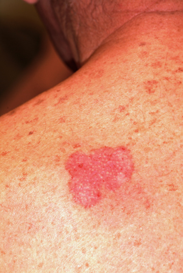
The correct answer is discoid lupus (choice “d”), which is generally confined to sun-exposed areas, tends to be chronic, and eventually causes scarring. Psoriasis (choice “a”) was unlikely, given the lack of corroborative findings over an extended period and the negative personal or family history—and because scarring would be quite unusual with that disease.
Basal cell carcinoma (BCC; choice “b”) is an unlikely choice because it is usually accompanied by significant sun damage and it does not typically wax and wane. Actinic keratoses (choice “c”) can come and go, but as with BCC, it would almost certainly be seen in the context of more widespread sun damage on the face.
Discussion
A 3-mm punch biopsy was performed to confirm the diagnosis and rule out the other items in the differential. Discoid lupus erythematosus (DLE) usually demonstrates a classic set of histologic findings, confirming this common—and usually benign—form of lupus that favors the nose, ears, and neck but can be seen on any chronically sun-exposed area. Cancer/precancerous skin changes were definitely worth considering, given the chronicity of the condition.
Although DLE is quite common, outside dermatology it is seldom diagnosed initially. Instead, the natural inclination is treat the condition as “some sort of infection.” DLE often presents with annular, scaly lesions. This is potentially misleading, unless you look for two particular features (both of which, alas, were missing in this case!): The lesions often demonstrate atrophic, whitish centers, and when the scale is gently peeled off the lesions, you can see that the scale was plugging the follicular orifices, in effect dilating them.
Since systemic lupus erythematosus (SLE) can present with lesions of DLE, the initial labs should include an antinuclear antibody test. Approximately 5% of purely cutaneous DLE will evolve into SLE. The more extensive and serious the discoid eruption, the more likely is the presence of SLE.
DLE can also involve the scalp, where it can eventuate in focal or widespread scarring alopecia. Less often, it is seen on the lips, tongue, or other areas of oral mucosa.
Treatment
With early, mild cases of DLE, better sun protection and topical steroid creams are all the treatment needed. But in more advanced cases involving scarring, oral therapy with hydroxychloroquine (6.5 mg/kg) is also necessary—assuming results of baseline lab testing (eg, comprehensive metabolic panel, complete blood count) and ophthalmologic examination are normal.
After three months of therapy with hydroxychloroquine (200 mg bid), this patient’s condition improved dramatically. The dosage was then reduced to once daily for another three months, at which point she’ll stop. She will repeat her eye exam and blood work at that time and continue to use sun protection, since the potential for recurrence is high.
Answer
The correct answer is discoid lupus (choice “d”), which is generally confined to sun-exposed areas, tends to be chronic, and eventually causes scarring. Psoriasis (choice “a”) was unlikely, given the lack of corroborative findings over an extended period and the negative personal or family history—and because scarring would be quite unusual with that disease.
Basal cell carcinoma (BCC; choice “b”) is an unlikely choice because it is usually accompanied by significant sun damage and it does not typically wax and wane. Actinic keratoses (choice “c”) can come and go, but as with BCC, it would almost certainly be seen in the context of more widespread sun damage on the face.
Discussion
A 3-mm punch biopsy was performed to confirm the diagnosis and rule out the other items in the differential. Discoid lupus erythematosus (DLE) usually demonstrates a classic set of histologic findings, confirming this common—and usually benign—form of lupus that favors the nose, ears, and neck but can be seen on any chronically sun-exposed area. Cancer/precancerous skin changes were definitely worth considering, given the chronicity of the condition.
Although DLE is quite common, outside dermatology it is seldom diagnosed initially. Instead, the natural inclination is treat the condition as “some sort of infection.” DLE often presents with annular, scaly lesions. This is potentially misleading, unless you look for two particular features (both of which, alas, were missing in this case!): The lesions often demonstrate atrophic, whitish centers, and when the scale is gently peeled off the lesions, you can see that the scale was plugging the follicular orifices, in effect dilating them.
Since systemic lupus erythematosus (SLE) can present with lesions of DLE, the initial labs should include an antinuclear antibody test. Approximately 5% of purely cutaneous DLE will evolve into SLE. The more extensive and serious the discoid eruption, the more likely is the presence of SLE.
DLE can also involve the scalp, where it can eventuate in focal or widespread scarring alopecia. Less often, it is seen on the lips, tongue, or other areas of oral mucosa.
Treatment
With early, mild cases of DLE, better sun protection and topical steroid creams are all the treatment needed. But in more advanced cases involving scarring, oral therapy with hydroxychloroquine (6.5 mg/kg) is also necessary—assuming results of baseline lab testing (eg, comprehensive metabolic panel, complete blood count) and ophthalmologic examination are normal.
After three months of therapy with hydroxychloroquine (200 mg bid), this patient’s condition improved dramatically. The dosage was then reduced to once daily for another three months, at which point she’ll stop. She will repeat her eye exam and blood work at that time and continue to use sun protection, since the potential for recurrence is high.
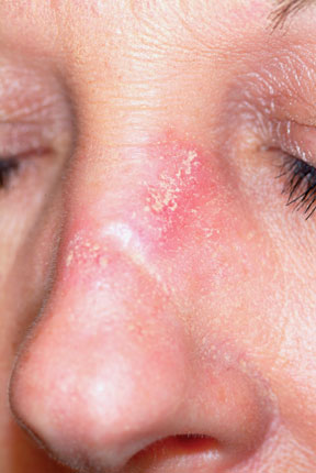
A 45-year-old nurse self-refers for evaluation of changes on her nose and upper forehead. These began about five years ago but in the past few months have become alarming in their progression. The condition is largely asymptomatic and wanes a bit each winter, but overall it has grown considerably over the years—now threatening to produce scarring. The patient denies having any other significant medical issues (ie, joint pain, fever, or malaise). She has never had skin cancer but does acknowledge getting “too much sun” every summer while gardening. She denies any history of foreign travel. As a nurse, she has worked in cardiology exclusively. There is no known family history of skin disease. On exam, the lesions are fairly impressive—especially on the nasal bridge, where there is focal, significant erosion surrounded by peeling skin, all on an erythematous base that is roughly 3 cm in its maximum dimension and polygonal in shape. Despite these changes, surface adnexae (pores, follicles, skin lines) on this lesion are largely intact, except on the superior margin, where definite scarring is seen. Similar changes are seen at the forehead/scalp interface, but with some small areas of scarring alopecia noted focally. Overall, the patient’s skin is fair, but with little overt evidence of sun damage. Her elbows, knees, scalp, and fingernails are free of pathologic changes.

40 Year Old "Wart" Suddenly Changes
Answer
Digital mucous cysts (choice “c”), also known as myxoid cysts, are quite common and are found often on fingers and occasionally on toes. But they are almost always located on the distal dorsal portion of the digit, between the cuticle and distal interphalangeal joint—close enough, in many cases, to compress the nail matrix, which leads to a longitudinal trough in the nail plate. Surface erosion of these lesions is unusual but is occasionally seen.
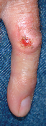
However, given the history, appearance, and especially the location of this patient’s lesion, this is the one diagnosis our patient almost certainly did not have. The others, discussed below, were all real possibilities, and since “common things occur commonly,” squamous cell carcinoma was the most likely.
Discussion
With this clinical picture, cancer is assumed until proven otherwise. In that regard, the presence of a longstanding wart in this location is especially significant, since human papillomavirus (HPV) is known to be potentially oncogenic. The patient’s heavily sun-damaged skin adds another layer of risk for malignant transformation.
The only way to sort through this differential diagnosis was to perform a shave biopsy, which confirmed the diagnosis of squamous cell carcinoma (SCC)—one that showed evidence of arising from a long-standing wart. The patient was referred for Mohs micrographic surgery, for two reasons: (1) this location does not lend itself to simple excision and closure, both because of the paucity of adjacent skin and because of the potential for damage to the underlying tendons, nerves, and blood supply, and (2) SCCs of nonsolar causation (besides HPV, these include ionizing radiation, arsenic, and chronic ulcers) have more potential for metastasis than do the far more common sun-caused SCCs. This is all the more reason to obtain adequate margins. Often in such cases, irradiation of the site is also done, postoperatively.
Had the biopsy not shown clear evidence of cancer, the other items in the differential diagnosis would have come into play. This would have necessitated an additional biopsy, this time to obtain tissue for acid-fast bacilli and bacterial and fungal cultures.
As of this writing, the patient is awaiting Mohs surgery. He will have to be followed closely for at least a year to watch for any signs of metastasis. With an intact immune system, his prognosis is excellent.
Answer
Digital mucous cysts (choice “c”), also known as myxoid cysts, are quite common and are found often on fingers and occasionally on toes. But they are almost always located on the distal dorsal portion of the digit, between the cuticle and distal interphalangeal joint—close enough, in many cases, to compress the nail matrix, which leads to a longitudinal trough in the nail plate. Surface erosion of these lesions is unusual but is occasionally seen.
However, given the history, appearance, and especially the location of this patient’s lesion, this is the one diagnosis our patient almost certainly did not have. The others, discussed below, were all real possibilities, and since “common things occur commonly,” squamous cell carcinoma was the most likely.
Discussion
With this clinical picture, cancer is assumed until proven otherwise. In that regard, the presence of a longstanding wart in this location is especially significant, since human papillomavirus (HPV) is known to be potentially oncogenic. The patient’s heavily sun-damaged skin adds another layer of risk for malignant transformation.
The only way to sort through this differential diagnosis was to perform a shave biopsy, which confirmed the diagnosis of squamous cell carcinoma (SCC)—one that showed evidence of arising from a long-standing wart. The patient was referred for Mohs micrographic surgery, for two reasons: (1) this location does not lend itself to simple excision and closure, both because of the paucity of adjacent skin and because of the potential for damage to the underlying tendons, nerves, and blood supply, and (2) SCCs of nonsolar causation (besides HPV, these include ionizing radiation, arsenic, and chronic ulcers) have more potential for metastasis than do the far more common sun-caused SCCs. This is all the more reason to obtain adequate margins. Often in such cases, irradiation of the site is also done, postoperatively.
Had the biopsy not shown clear evidence of cancer, the other items in the differential diagnosis would have come into play. This would have necessitated an additional biopsy, this time to obtain tissue for acid-fast bacilli and bacterial and fungal cultures.
As of this writing, the patient is awaiting Mohs surgery. He will have to be followed closely for at least a year to watch for any signs of metastasis. With an intact immune system, his prognosis is excellent.

An 87-year-old man presents for evaluation of a lesion on his left fifth finger that has grown and become ulcerated in the past five months. There is almost no pain in the lesion, which the patient insists was a “wart” for the 40 years prior to these recent changes. Over the years, he has treated his wart with acids, curettement, and liquid nitrogen, to no effect. A recent seven-day course of cephalexin (500 mg tid) also failed to help. Additional history taking reveals that the patient is a farmer who spent his entire life working outdoors every day, year-round. There is no history of immunosuppression. His medications include metoprolol and hydrochlorothiazide. Closer inspection of the lesion reveals a 9-mm, centrally eroded nodule overlying the dorsal proximal interphalangeal joint. The lesion is quite firm on palpation, with a surface that looks and feels smooth and is nontender, with minimal redness. There are no palpable nodes in the epitrochlear or axillary locations. Elsewhere, the patient’s skin is remarkably sun-damaged, with numerous actinic keratoses, solar lentigines, and solar atrophy evident, especially on his hands.
Rash Emerges After 18 Holes of Golf
ANSWER
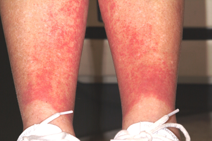
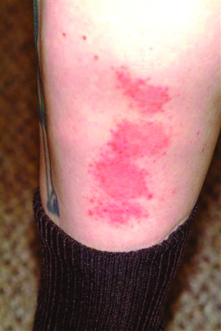
The correct answer is golfer’s vasculitis (choice “d”), which favors older patients who spend extended periods on their feet in hot weather.
Schamberg’s disease (choice “a”), a type of capillaritis, presents with nonblanchable, true purpura that are classically described and seen as having a peculiar brown color (which has been called cinnamon or cayenne pepper).
Those who experience a true contact dermatitis (choice “b”) almost invariably itch and would most likely present with vesiculation of the skin surface.
Leukocytoclastic vasculitis (LCV; choice “c”) presents as a nonblanchable purpuric condition that, on biopsy, demonstrates classic findings of red blood cell (RBC) extravasation from venules damaged by neutrophils.
DISCUSSION
“Golfer’s vasculitis” has been described in nongolfers who are older and who have spent considerable time on their feet in hot weather. Fair-goers, amusement park patrons, and hikers are just as likely to develop it, but it was first studied in golfers—and at first, it was thought to represent a sensitivity to chemical grass treatment.
However, the lack of symptoms and vesiculation (blistering) suggested otherwise, and biopsies of the affected skin confirmed gravity-related hyperemia with mild extravasation of RBCs. They also failed to show any signs of contact dermatitis. The sharply defined linear inferior border of the rash is clearly caused by the compressive effects of socks, which prevent the leakage of RBCs.
The other items in the differential were rightly considered—particularly LCV, which can be associated with conditions such as hypersensitivity reactions to medications or can be a presenting sign of lupus and rheumatoid arthritis, among several other possibilities. But the key differentiating finding was the highly blanchable nature of this patient’s condition, in marked contrast to the nonblanchable, purpuric nature of classic LCV.
Often enough, blanchability is partial, or at least questionable, and a punch biopsy is necessary to clarify the picture. When histologic signs of LCV are found, blood work is necessary to rule out similar damage to “end organs,” such as kidneys and liver, as well as to attempt to establish the causative trigger.
ANSWER
The correct answer is golfer’s vasculitis (choice “d”), which favors older patients who spend extended periods on their feet in hot weather.
Schamberg’s disease (choice “a”), a type of capillaritis, presents with nonblanchable, true purpura that are classically described and seen as having a peculiar brown color (which has been called cinnamon or cayenne pepper).
Those who experience a true contact dermatitis (choice “b”) almost invariably itch and would most likely present with vesiculation of the skin surface.
Leukocytoclastic vasculitis (LCV; choice “c”) presents as a nonblanchable purpuric condition that, on biopsy, demonstrates classic findings of red blood cell (RBC) extravasation from venules damaged by neutrophils.
DISCUSSION
“Golfer’s vasculitis” has been described in nongolfers who are older and who have spent considerable time on their feet in hot weather. Fair-goers, amusement park patrons, and hikers are just as likely to develop it, but it was first studied in golfers—and at first, it was thought to represent a sensitivity to chemical grass treatment.
However, the lack of symptoms and vesiculation (blistering) suggested otherwise, and biopsies of the affected skin confirmed gravity-related hyperemia with mild extravasation of RBCs. They also failed to show any signs of contact dermatitis. The sharply defined linear inferior border of the rash is clearly caused by the compressive effects of socks, which prevent the leakage of RBCs.
The other items in the differential were rightly considered—particularly LCV, which can be associated with conditions such as hypersensitivity reactions to medications or can be a presenting sign of lupus and rheumatoid arthritis, among several other possibilities. But the key differentiating finding was the highly blanchable nature of this patient’s condition, in marked contrast to the nonblanchable, purpuric nature of classic LCV.
Often enough, blanchability is partial, or at least questionable, and a punch biopsy is necessary to clarify the picture. When histologic signs of LCV are found, blood work is necessary to rule out similar damage to “end organs,” such as kidneys and liver, as well as to attempt to establish the causative trigger.

When an asymptomatic rash appeared rather suddenly on both of her legs, a 54-year-old woman sought medical evaluation by her primary care provider. He diagnosed contact dermatitis and prescribed triamcinolone cream 0.1%. Forty-eight hours later, with no signs of improvement evident, the patient seeks and is granted a same-day appointment with dermatology. The patient denies any previous occurrences of such a rash and further denies having joint pain, fever, or malaise. She had not taken any new medications prior to the rash’s onset; furthermore, she only occasionally uses OTC medicines. A thorough history reveals that two days before the rash appeared, on one of the first hot days of the summer (with a temperature above 95°F), the woman played 18 holes of golf. The rash itself is strikingly red and affects both lower legs symmetrically, from mid-calf to just above the ankles. There, it ends abruptly with a linear, transverse border. There is no tenderness or increased warmth appreciated on palpation, nor is there any nodularity, vesiculation, or other disruption of the skin’s surface. Distinct, complete blanching is noted on firm digital palpation.
Spreading Lesions on Sun-damaged Skin
ANSWER
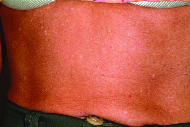
The correct answer is idiopathic guttate hypomelanosis (choice “d”), a common condition primarily seen on sun-damaged patients, but often mistaken for tinea versicolor (choice “b”).
Vitiligo (choice “a”) can present with “confetti” lesions, but would more likely lead to complete loss of pigment in most of the lesions, not the partial loss seen in this patient. Biopsy is indicated in questionable cases.
Tinea versicolor, equally common, is caused by a commensal yeast called Malassezia furfur that feeds on sebum, which is why it favors the oily parts of the body (back and chest, primarily). It is almost never seen on the legs, which have the fewest oil glands on the body. In addition, tinea versicolor is, by its nature, an epidermal process, leading to the formation of a fine KOH-positive scale.
Since lupus (choice “c”) is a form of vasculitis, the associated inflammation can lead to pigment loss, especially in darker-skinned patients. Without a more likely explanation for these lesions, a biopsy might well have been indicated.
DISCUSSION
Biopsy might have suggested any number of other diseases that can also present with hypomelanosis, such as cutaneous T-cell lymphoma or sarcoid. But idiopathic guttate hypomelanosis is far more common, and the patient’s age, gender, and history of chronic UV damage all lend themselves perfectly to this diagnosis.
Idiopathic guttate hypomelanosis, for unknown reasons, tends to appear in women at earlier ages than in men (usually about a decade younger). For either gender, treatment is problematic once the lesions have fully developed. Early in the process, the obvious remedy is better sun protection. Lasers, retinoids, liquid nitrogen, and anti-inflammatory creams have all been tried with little success.
ANSWER
The correct answer is idiopathic guttate hypomelanosis (choice “d”), a common condition primarily seen on sun-damaged patients, but often mistaken for tinea versicolor (choice “b”).
Vitiligo (choice “a”) can present with “confetti” lesions, but would more likely lead to complete loss of pigment in most of the lesions, not the partial loss seen in this patient. Biopsy is indicated in questionable cases.
Tinea versicolor, equally common, is caused by a commensal yeast called Malassezia furfur that feeds on sebum, which is why it favors the oily parts of the body (back and chest, primarily). It is almost never seen on the legs, which have the fewest oil glands on the body. In addition, tinea versicolor is, by its nature, an epidermal process, leading to the formation of a fine KOH-positive scale.
Since lupus (choice “c”) is a form of vasculitis, the associated inflammation can lead to pigment loss, especially in darker-skinned patients. Without a more likely explanation for these lesions, a biopsy might well have been indicated.
DISCUSSION
Biopsy might have suggested any number of other diseases that can also present with hypomelanosis, such as cutaneous T-cell lymphoma or sarcoid. But idiopathic guttate hypomelanosis is far more common, and the patient’s age, gender, and history of chronic UV damage all lend themselves perfectly to this diagnosis.
Idiopathic guttate hypomelanosis, for unknown reasons, tends to appear in women at earlier ages than in men (usually about a decade younger). For either gender, treatment is problematic once the lesions have fully developed. Early in the process, the obvious remedy is better sun protection. Lasers, retinoids, liquid nitrogen, and anti-inflammatory creams have all been tried with little success.

A few years ago, a woman first noticed lesions developing on her legs. The patient sought care from her primary care provider; however, despite a number of treatment regimens, including topical clotrimazole and terbinafine creams, the lesions have not only failed to resolve but have also grown in number and spread to other areas of her body. The woman, now 48, is referred to dermatology for evaluation of her persistent but asymptomatic condition. The patient claims that her health is otherwise excellent, although she has seasonal allergies, was a long-time smoker until two months ago, and has just begun estrogen replacement therapy. She is especially concerned that the lesions have started to appear on the skin of her abdomen. She specifically denies shortness of breath, joint pain, fever, unexplained weight loss, or cough. An examination of her skin reveals a multitude of 2- to 6-mm partially depigmented, roughly round macules uniformly distributed on her legs, arms, and trunk. The lesions, which average about 3 mm, have no palpable component and no observable scale or underlying induration. There is a concentration of them on the anterior tibial areas, as well as on the dorsal forearms; however, none are seen on her face, and the volar surfaces of her forearms are almost completely spared. Significantly, the patient’s exposed skin is tremendously sun-damaged, evidenced by a deep brown color and a weathered, wrinkled look, with many telangiectasias and brown to tan–orange macules on her face.
It "Must" Be a Fungal Infection—Right?
ANSWER
The correct answer is ED&C (choice “a”); see below for discussion. Imiquimod cream (choice “b”) is indicated in such cases, although it is not without shortcomings as a treatment choice (more information below).
Given the size of the lesion, one could argue that two-stage excision (choice “c”), in which two-thirds of the lesion is removed initially, the wound allowed to heal, then the rest of the lesion removed later, would be a good option. Again, however, it is not perfect.
Mohs surgery (choice “d”) is the only option offered that takes care of the entire lesion in one step, with microscopically controlled margins to ensure complete removal and primary closure all in one session.
DISCUSSION
There are at least five types of BCC; of these, the superficial BCC is the one that looks the least like a skin cancer. The back is one of the more common areas for a BCC to appear, where it is often mistaken for fungal infection. The latter condition is actually quite uncommon on the back; furthermore, this patient had no particular reason to be exposed to such an organism or to be susceptible to it. Lack of response to treatment for fungal infection was further evidence against such a diagnosis.
Knowing that BCCs can look like this is a priceless piece of information. Corroboration for the diagnosis of this sun-caused skin cancer comes readily from the presence of sun damage elsewhere on the skin. Superficial BCCs are so common that biopsy is often unnecessary, although insurance providers frequently require it before excision can be done.
ED&C, a perfectly acceptable mode of treatment for many minor BCCs, is not a good choice in this area and with a lesion of this size, as it is likely to produce a large, nonhealing, raw wound covered by inappropriate granulation (so-called pyogenic granuloma).
Treatment with imiquimod cream, two to four times a week for at least a month, has the potential to be curative, but involves the creation of a large oozing site while it’s working. Even after several months’ treatment on a lesion of this size, biopsies may still be necessary to check for residual cancer. In selected patients and lesions, cases in which surgery is problematic, imiquimod can be an excellent treatment choice.
Two-stage excision does ensure the complete removal of the lesion but has the obvious disadvantage of taking several weeks and requiring two surgery sessions.
Even if the patient is sent for Mohs surgery, closure will be problematic with a lesion this size and in this keloid-prone area.
But job #1 in such cases is to make a correct diagnosis, then select from among the various treatment choices. Without a biopsy, that diagnosis is uncertain. But without the suspicion of BCC, biopsy may not appear to be indicated.
A 50-year-old man has had a nonhealing but otherwise asymptomatic lesion on his back for more than three years. The lesion has slowly grown, despite the use of a number of treatments, including OTC and prescription antifungal creams and OTC steroid creams. The consensus from his primary care providers is that this must represent some form of fungal infection, although they are at a loss to explain why it has not responded to treatment for that condition. The patient presents to dermatology because his wife insisted on referral to a specialist when the lesion finally became focally eroded around its margins. Further history taking reveals that there are no pets or children at home, there is no personal history of atopy, and the patient has never worked with livestock. For 30 years, he worked outdoors for the power company, but he is now assigned to a desk job. He is not immunosuppressed, and his only medication is for hypertension. During the physical examination, you observe an arciform, faintly and focally eroded border surrounding a 5-cm pink patch on the patient’s left upper back. Closer inspection reveals prominent pilosebaceous units confined by well-defined borders, with a background of uniformly atrophic, glassy-looking telangiectatic skin. No scale is seen on or around the lesion. Elsewhere, you see a moderate number of solar lentigines across the patient’s shoulders and note that his posterior neck has a deeply lined appearance. The rest of his trunk and face have similar lesions, although none is especially worrisome. To confirm your clinical suspicions, you perform a shave biopsy on the central portion of the lesion, which shows superficial basal cell carcinoma (BCC). Treatment options are then discussed at length with the patient.

Family Thinks Man has Worms
ANSWER
The correct answer is nummular eczema (choice “a”), an extremely common condition, especially in older men. Had this been psoriasis (choice “b”), the lesions would not have been so pruritic and would have displayed the characteristic thick, whitish scaling on a salmon pink base—and that diagnosis would, in all likelihood, have been corroborated by signs of psoriasis elsewhere on the patient’s body.
A fungal infection (choice “c”) can be difficult to treat but probably would have responded at least in part to one of the many medications the man tried. Other factors that spoke against this diagnosis were the negative KOH preparations and the apparent lack of specific sources (eg, pets or other animals) for such an infection.
Bowen’s disease (choice “d”) represents superficial epidermal squamous cell carcinoma—the round, scaly lesions of which are often mistaken for the other choices in this differential. But Bowen’s lesions will be fixed (permanent) instead of waxing and waning as this patient’s lesions did, will not itch to any appreciable extent, and will be found almost exclusively on chronically sun-damaged skin (which was not evident in this patient).
DISCUSSION
Nummular eczema (NE) is an extremely common condition with unclear origins. We often see it in the context of older patients with dry skin made drier by bathing habits, swimming, or use of hot tubs. But many cases appear without these conditions, causing much conjecture about potential triggers, such as contact versus irritant dermatitis. Indeed, the multiplicity of topical medications applied by this patient was probably making things worse, as is often the case.
NE typically manifests acutely, but can persist for months, worrying some patients a great deal. If need be, a 4-mm punch biopsy will rule out the other suspects in the differential diagnosis. Then the lesions can be treated, as in this case, with a class 1 or 2 topical steroid ointment or emollient cream, as well as advice about the need to limit the length of time spent in, and the temperature of, showers or hot tubs.
As with so many dermatologic conditions, patients with NE are greatly reassured just knowing about all the conditions they don’t have. And as is often the case for providers, just knowing a given condition such as NE exists and is common is a priceless asset.

A 70-year-old man self-refers to dermatology for evaluation of very itchy lesions that have been present for at least three months. The patient reports that the lesions have persisted despite the use of “everything but the kitchen sink”—meaning several OTC topical preparations, including those containing benzocaine, neomycin/polymyxin/bacitracin, and diphenhydramine, as well as “every antifungal cream I could buy.” The man has also tried pouring alcohol and peroxide over the lesions, again to no good effect. The lesions appeared in the midst of a long, cold winter with very low humidity; they itch terribly, causing the patient to scratch them vigorously. The lesions, which come and go, concern him not only because of the pruritus, but because his family is convinced he has some sort of “worms.” Further history taking reveals that the patient is not taking any new medications in addition to his simvastatin and metoprolol. He is not atopic and has no new pets, but he does admit to being “addicted” to sitting in his new hot tub. There is no history of gastric ulcers or dyspepsia, no joint pain, and no family history of psoriasis. Examination of the patient’s legs, particularly his calves, shows discrete and confluent round, scaly plaques of a striking reddish orange hue. They range from 1 to 5 cm in diameter. KOH prep of samples taken from two different lesions is negative for fungal elements. Elsewhere, the knees, elbows, nails, and scalp are free of any significant skin changes. There are no signs of chronic sun damage.
Is Patient With Fingernail Changes a "Contagious Threat"?
ANSWER
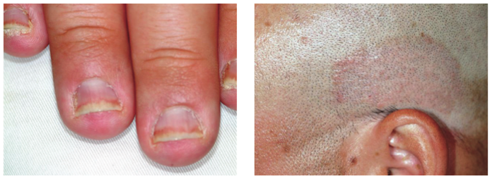
The correct answer is psoriasis (choice “d”), which often involves nail and skin changes such as those seen in this case. There was no particular reason to think this patient had an infection of any kind, but, as is often the case, the original provider considered only one diagnostic possibility.
DISCUSSION
Nail disease can be problematic for both patient and provider. Arguably the biggest issue with it, initially, is “the one-item differential”—the idea that all nail disease can be classified as “fungal infection” simply because neither the patient nor the provider considers any other explanation for the changes seen. This case illustrates that dilemma nicely.
Psoriasis affects more than 2% of the US population, making it a common skin disorder. As this case illustrates, it may affect several areas with diverse but predictable morphologic presentations, all appearing at the same time.
The key diagnostic points from this case include:
• Stress is a known trigger for psoriasis vulgaris.
• The patient’s nails failed to respond to terbinafine treatment.
• There was no obvious source of fungal infection (eg, contact with animals) or reason for him to have a yeast infection (eg, diabetes) or develop mold “infection” in the nails (eg, trauma from digging in soil).
• Fungal infections are about 18 times less likely to occur in the fingernails than in the toenails.
• A unifying explanation for simultaneous nail and skin changes was needed.
• The nail and skin changes seen here are quite typical for psoriasis.
Most important, though, is the need to overcome the urge to hop on the “fungal bandwagon” by first considering other diagnostic possibilities.
Continue for the treatment of this condition >>
TREATMENT
Unfortunately, there is no good treatment for this patient’s nails. However, there is an excellent chance that his scalp psoriasis will respond to short-term use of fluocinolone 0.05% cream.
This patient was also counseled about the role of stress in his condition and the benefits of modest increases in UV exposure. During follow-up, it will be important to ask about joint pain, since up to 30% of psoriasis patients will eventually develop psoriatic arthritis.
If the patient’s current treatment does not produce sufficient results, other options include calcipotriene ointment, oral methotrexate, or even one of the biologics (etanercept or adalimumab).
ANSWER
The correct answer is psoriasis (choice “d”), which often involves nail and skin changes such as those seen in this case. There was no particular reason to think this patient had an infection of any kind, but, as is often the case, the original provider considered only one diagnostic possibility.
DISCUSSION
Nail disease can be problematic for both patient and provider. Arguably the biggest issue with it, initially, is “the one-item differential”—the idea that all nail disease can be classified as “fungal infection” simply because neither the patient nor the provider considers any other explanation for the changes seen. This case illustrates that dilemma nicely.
Psoriasis affects more than 2% of the US population, making it a common skin disorder. As this case illustrates, it may affect several areas with diverse but predictable morphologic presentations, all appearing at the same time.
The key diagnostic points from this case include:
• Stress is a known trigger for psoriasis vulgaris.
• The patient’s nails failed to respond to terbinafine treatment.
• There was no obvious source of fungal infection (eg, contact with animals) or reason for him to have a yeast infection (eg, diabetes) or develop mold “infection” in the nails (eg, trauma from digging in soil).
• Fungal infections are about 18 times less likely to occur in the fingernails than in the toenails.
• A unifying explanation for simultaneous nail and skin changes was needed.
• The nail and skin changes seen here are quite typical for psoriasis.
Most important, though, is the need to overcome the urge to hop on the “fungal bandwagon” by first considering other diagnostic possibilities.
Continue for the treatment of this condition >>
TREATMENT
Unfortunately, there is no good treatment for this patient’s nails. However, there is an excellent chance that his scalp psoriasis will respond to short-term use of fluocinolone 0.05% cream.
This patient was also counseled about the role of stress in his condition and the benefits of modest increases in UV exposure. During follow-up, it will be important to ask about joint pain, since up to 30% of psoriasis patients will eventually develop psoriatic arthritis.
If the patient’s current treatment does not produce sufficient results, other options include calcipotriene ointment, oral methotrexate, or even one of the biologics (etanercept or adalimumab).

After finishing a month-long course of terbinafine (250 mg), a 27-year-old man is dismayed to see that the fingernail changes he first noted a year ago have not improved. He had been told in no uncertain terms by his primary care provider that his fingernail condition—and accompanying scalp rash—represented fungal infection. When topical antifungal creams failed to help his scalp condition, the oral medication was prescribed. Now it, too, appears to have been unhelpful. At this point, the patient requests referral to dermatology. His primary concern is that he represents a “contagious threat” to his wife and children, although none of them has shown any signs of this condition. The patient denies any skin problems prior to the nail changes and the scalp rash that manifested shortly afterward, which occurred more than a year ago. Shortly before the onset of these problems, he lost his job and had to replace it with two lower-paying part-time jobs. He denies any family history of skin disease or joint pain. There are no new pets in the house, and the patient does not work with animals. Examination reveals that the distal portions of all 10 fingernails are uniformly dystrophic and mildly onycholytic and have numerous longitudinal dark streaks. Several fingernails also have tiny scattered pits in them. The toenails are unaffected. Elsewhere, the man is observed to have salmon-pink scaly patches on the scalp and over both ears, and smaller, round plaques on the forehead, with prominent white tenacious scale. KOH prep of these latter lesions is negative for fungal elements.
A "Spider Bite" That Wasn't
ANSWER
The one unreasonable choice is a 10-day course of rifampin (choice “a”). One potential explanation for the lack of response to two different antibiotics would be the probability that there was no infection to begin with. See below for further explanation.
DISCUSSION
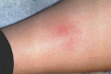
Erythema nodosum (EN; choice “c”) is the presumptive diagnosis in cases such as this one. The “erythemas” (multiforme, annulare centrifigum, etc) are all secondary reactions to antigenic triggers—such as herpes simplex virus, strep, or other organisms—and not primary conditions.
EN is a type of panniculitis that can be triggered by a number of conditions (eg, sarcoidosis), medicines (eg, penicillin), or organisms (eg, group A β streptococcus). It can also, as in this case, accompany inflammatory bowel disease, paralleling the flares and remissions of Crohn’s or ulcerative colitis and responding to treatment for those diseases. This is why part of the workup for EN includes evaluation for bowel disease.
The absence of breaks in the skin in such cases also speaks loudly for EN, since it involves only the adipose tissues and spares the skin. In this case, given the dearth of other suspicious items in the differential, a presumptive diagnosis of EN was quite reasonable.
Deep punch biopsy (choice “b”), which must include significant adipose tissue (5 mm or surgical wedge), can be crucial in questionable cases. It can show septal panniculitis (as opposed to lobular, which is more consistent with erythema induratum), as well as characteristic Miescher’s granulomas.
A chest radiograph (choice “d”), while not especially indicated in this case, is likewise an intelligent choice. Both tuberculosis and sarcoidosis can lead to radiographic changes and trigger EN.
If local infection were truly suspected as the direct cause of this patient’s lesions, an additional punch biopsy or two might serve to provide sufficient material for aerobic bacterial culture, acid-fast culture, and deep fungal culture—all of which are possible, though unlikely, explanations. Literally, the last thing we’d do is throw yet another antibiotic at this patient, who was in need of one simple thing: a correct diagnosis that would dictate proper treatment.
TREATMENT
A letter was dictated and sent to the gastroenterologist the patient was scheduled to see, to alert him as to our findings. If his workup
(H&P, stool cultures for salmonella, yersinia, and campylobacter, colonoscopy with biopsy) fails to pinpoint a particular trigger for her EN, additional labs will be needed. These would include antistreptolysin-O titer and tuberculosis and coccidioidomycosis skin testing, as well as actual biopsy of the lesions.
As of this writing, the patient’s GI appointment was pending, but the likely outcome will be the diagnosis of Crohn’s disease or ulcerative colitis. Treatment for either of these conditions will quiet down her EN. Often, however, EN is idiopathic and must be treated as a stand-alone diagnosis, with NSAIDs, antimalarials, or potassium iodide.
ANSWER
The one unreasonable choice is a 10-day course of rifampin (choice “a”). One potential explanation for the lack of response to two different antibiotics would be the probability that there was no infection to begin with. See below for further explanation.
DISCUSSION
Erythema nodosum (EN; choice “c”) is the presumptive diagnosis in cases such as this one. The “erythemas” (multiforme, annulare centrifigum, etc) are all secondary reactions to antigenic triggers—such as herpes simplex virus, strep, or other organisms—and not primary conditions.
EN is a type of panniculitis that can be triggered by a number of conditions (eg, sarcoidosis), medicines (eg, penicillin), or organisms (eg, group A β streptococcus). It can also, as in this case, accompany inflammatory bowel disease, paralleling the flares and remissions of Crohn’s or ulcerative colitis and responding to treatment for those diseases. This is why part of the workup for EN includes evaluation for bowel disease.
The absence of breaks in the skin in such cases also speaks loudly for EN, since it involves only the adipose tissues and spares the skin. In this case, given the dearth of other suspicious items in the differential, a presumptive diagnosis of EN was quite reasonable.
Deep punch biopsy (choice “b”), which must include significant adipose tissue (5 mm or surgical wedge), can be crucial in questionable cases. It can show septal panniculitis (as opposed to lobular, which is more consistent with erythema induratum), as well as characteristic Miescher’s granulomas.
A chest radiograph (choice “d”), while not especially indicated in this case, is likewise an intelligent choice. Both tuberculosis and sarcoidosis can lead to radiographic changes and trigger EN.
If local infection were truly suspected as the direct cause of this patient’s lesions, an additional punch biopsy or two might serve to provide sufficient material for aerobic bacterial culture, acid-fast culture, and deep fungal culture—all of which are possible, though unlikely, explanations. Literally, the last thing we’d do is throw yet another antibiotic at this patient, who was in need of one simple thing: a correct diagnosis that would dictate proper treatment.
TREATMENT
A letter was dictated and sent to the gastroenterologist the patient was scheduled to see, to alert him as to our findings. If his workup
(H&P, stool cultures for salmonella, yersinia, and campylobacter, colonoscopy with biopsy) fails to pinpoint a particular trigger for her EN, additional labs will be needed. These would include antistreptolysin-O titer and tuberculosis and coccidioidomycosis skin testing, as well as actual biopsy of the lesions.
As of this writing, the patient’s GI appointment was pending, but the likely outcome will be the diagnosis of Crohn’s disease or ulcerative colitis. Treatment for either of these conditions will quiet down her EN. Often, however, EN is idiopathic and must be treated as a stand-alone diagnosis, with NSAIDs, antimalarials, or potassium iodide.

Sore “red bumps” began to appear on this 31-year-old woman’s posterior calves six months ago. They persisted despite a course of cephalexin (500 mg qid for 10 days), prescribed by her primary care provider for “spider bites,” and a few weeks later, a course of double-strength sulfamethoxazole/trimethoprim (bid for 10 days), for “staph infection.” When the patient is seen by dermatology, her health history is good, although she mentions a pending appointment with a gastroenterologist to investigate lower abdominal cramping and diarrhea, both of which started several months ago. There has been no weight loss, nausea, or vomiting. The patient is unaware of any bites that could explain the red patches on her legs and denies any breaks in the affected skin. No lesions are evident elsewhere on her body. There is no personal or family history of skin disease, and the patient used no OTC or prescription medication prior to the appearance of the lesions. Examination reveals several deep subcutaneous nodules palpable in the calf, ranging in size from 1.5 to 2.5 cm. There is no broken skin, nor are there other skin changes overlying the lesions, except erythema and induration. There is modest tenderness on palpation, as well as a modest increase in warmth. No nodes are palpable in the groin on that side. Examination of the patient’s skin elsewhere shows no notable changes.
Six-month Facial Rash Continues to Burn
ANSWER
The correct answer is perioral dermatitis (POD; choice “c”), an extremely common dermatosis that often begins in the perinasal area, spreading downward with time.
Impetigo (choice “a”) does not bear much resemblance to POD and would have responded quite well to the course of cephalexin.
Lupus (choice “b”) belongs in the differential, given the patient’s gender and the condition’s chronicity; however, this condition typically prefers the lateral face, usually causes focal atrophy, and does not manifest with pustules and papules.
Had the patient not responded to medication for POD, a punch biopsy would have been a reasonable step to rule out lupus and psoriasis (choice “d”), which can present in a similar manner, though seldom as the sole manifestation of that disease.
DISCUSSION
POD is an eruption of unknown etiology that demonstrates the clinical features of several other conditions (including eczema, seborrhea, and acne). It is therefore often difficult to diagnose—especially when it has been present for so long and has been treated with several medications.
More than 90% of POD cases occur in women ages 24 to 50, which suggests a possible role for hormones, makeup, or facial products. Yet none of these factors has been specifically implicated in the condition’s genesis.
Indeed, virtually every woman with POD presents with a history of having changed her makeup and other facial products, often many times, to no good effect. Stress has also been suggested as a contributing factor, and indeed appears, to many dermatology providers, to be the culprit; however, this has yet to be proven.
What is known is that in a large percentage of cases, the chronic application of topical steroids either brings on POD or worsens it, thereby necessitating questions about steroid use. Many a patient with seborrhea has successfully treated that disease with topical steroids only to create POD—which she naturally treats with more and more steroid.
The histologic picture of POD is similar to rosacea. But unlike POD, the latter spares the concave areas of the face. Like rosacea, POD responds so readily to oral tetracycline (alternatives including minocycline, amoxicillin, and erythromycin) that treatment success essentially verifies the diagnosis. Simply knowing that POD is utterly common is extremely helpful in the development of any differential of facial eruptions.
This particular patient responded quite well to a course of tetracycline (500 mg bid for a month, then 500 qd for another month), at the end of which her skin was totally clear. While her lack of response to topical antibiotics is typical of POD, many women with this condition actually report significant irritation with these products.

A 39-year-old woman is distraught about the rash that has been present on her face for more than six months. The rash continues to burn and feel raw, despite the use of topical preparations of metronidazole and clindamycin, as well as a seven-day course of cephalexin (500 mg bid). These had been given for diagnoses of acne, then rosacea, and finally, seborrhea. In desperation, the patient has stopped using all makeup, changed brands of facial tissue, and—at the suggestion of her sister—gone on a strict diet of only fresh, raw food. None of these measures has helped, although the condition has waxed and waned a bit. The patient, who is employed as a physical therapist, denies any previous or current use of topical steroids, but states that early on she used a number of OTC products on her face, including triple-antibiotic ointment and numerous moisturizers. Aside from mild dyslipidemia, the patient claims to be in good health, with no history of atopy, eczema, or psoriasis. Initial questioning about her stress level reveals little, but on deeper probing, the patient admits to a great deal of stress and trouble sleeping, which she attributes to job insecurity for both herself and her husband. Moreover, she has noted that her facial condition worsens with spikes in her level of anxiety. Entering the exam room, you are struck by the florid nature of the patient’s condition, especially the redness. But there are also scattered tiny pustules in the eruption, which is largely confined to the perinasal, nasolabial, and perioral areas. Scaling is scattered about the rash. Additional questioning leads to the revelation that the rash originally began in the perinasal area, only later spreading to its current distributive pattern. No rash is observed in the patient’s brows or in or behind the ears, and there is no appreciable dandruff on her scalp. Her elbows, knees, and fingernails show no signs of disease.
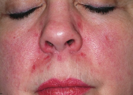
Diagnosis Confirmed: What's Next?
ANSWER
The one inappropriate choice—and therefore, the correct answer—is ED&C (choice “a”). Of all the choices, this option has the greatest potential for an unacceptable scar and for a recurrence of the cancer. All the other choices are entirely appropriate.
DISCUSSION
Had this been a far older patient with little concern about appearance, ED&C would have been a perfectly acceptable choice. But this patient made her expectations clear enough, effectively ruling out such an approach. A plastic surgeon (choice “b”) could be expected to not only obtain adequate margins around this cancer, but to be able to close the defect with an acceptable cosmetic outcome. Mohs surgeons (choice “d”) have the unique ability to examine the excised tissue margins at the surgical bedside, closing only when those margins are determined microscopically to be clear. They also have the ability to close larger defects in difficult areas such as this, obtaining an acceptable final result.
Had the patient or situation contraindicated surgery, at least two options would remain. The first is imiquimod (choice “c”) cream, which has a proven ability to destroy basal cell carcinomas (when applied several times a week over a period of six weeks to three months). Uncertain results can, and often do, necessitate a biopsy to confirm total destruction, and the patient has to endure weeks of redness and irritation at the site during treatment.
The other option is radiation therapy (choice “e”), an extremely effective nonsurgical alternative. However, it requires multiple treatment sessions over several weeks and can damage surrounding skin.
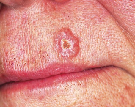
About one year ago, this 48-year-old woman noted the appearance of a tiny papule on her left maxilla. It has grown in the interim, becoming of considerable concern to the patient and her husband. They live in the same high desert country where they both were born and raised. As a result, they have been extensively exposed to a great deal of sunlight, and that exposure has been poorly tolerated in both of them. Several family members have been diagnosed with skin cancer, a fact that finally prompts the patient to have this lesion evaluated. She presents to her primary care provider, who in turn refers her to dermatology. The patient is otherwise fairly healthy. However, she is overweight and has hypertension, which is controlled with medication. On examination, the patient’s red hair, blue eyes, and fair skin are noted, along with signs of past sun damage, including solar lentigines and telangiectasias. The lesion in question is a fairly impressive planar 7.5-mm glassy, telangiectatic nodule with a central area of erosion. It is located on the lower left maxilla, about 3 mm above the vermillion border. The lesion is not tender to palpation but is quite firm. Systematic evaluation of the rest of the patient’s face, ears, and neck reveals no other suspicious findings. Shave biopsy is performed, with the results verifying the suspected diagnosis of basal cell carcinoma. The patient is then presented with treatment options—at which point she expresses great concern over the cosmetic outcome of the lesion’s removal and her desire to be rid of all the cancer. Given these concerns, as well as the diagnosis in question, treatment options might reasonably include all except which of the following?
