User login
Have pedometer, will travel
Advise your patients to use a pedometer, set a step goal, and keep a step diary. This simple intervention takes only a few moments and is effective in increasing patients’ physical activity and decreasing both body-mass index (BMI) and systolic blood pressure.1
Strength of recommendation
A: Based on a meta-analysis of randomized controlled trials (RCTs) and observational studies
Bravata DM, Smith-Spangler C, Sundaram V et al. Using pedometers to increase physical activity and improve health: a systematic review. JAMA 2007; 298:2296–2304.
Illustrative case
Your first 4 patients this morning were a 50-year-old woman with metabolic syndrome, a 62-year-old obese man with high blood pressure, a 44-year-old woman with depression, and a 75-year-old man with a recent admission for myocardial infarction. In addition to managing their medications and reviewing lab results, you have already spent a lot of time discussing the benefits of exercise with each of these patients.
As you prepare to talk with your next patient—a 28-year-old woman with a BMI of 29 whose chief complaint is “wants to lose weight”—you wonder if there are any simple, brief, effective interventions to help your patients increase their physical activity.
BACKGROUND: A long way to go
Although there is no evidence that simply advising patients to walk has any effect, primary care physicians frequently recommend walking as a form of exercise—it is free, requires no special equipment, and is readily accessible to most motivated patients.
The Centers for Disease Control and Prevention recommends that adults engage in moderate physical activity for at least 30 minutes a day, at least 5 days per week.2 Yet 40% of adults do not engage in any leisure-time physical activity. This percentage is higher in women (43%), African-Americans (52%), and Hispanics (54%).3
The health benefits of exercise are clear. Regular physical activity has been shown to decrease overweight and obesity.4 It has also been shown to improve control of type 2 diabetes5 and hypertension.6 Frequent exercise is associated with a decreased mortality rate.7 Walking has been shown to decrease the risk of cardiovascular events in women, regardless of BMI.8
Walking has similarly been shown to decrease overall mortality among men.9 Cardiovascular fitness has also been shown to decrease mortality in adults over 60, even in the absence of weight loss.10
CLINICAL CONTEXT: USPSTF: Advice alone won’t kick-start exercise
We realize, of course, that most of our adult patients could benefit from regular exercise. Exercise is included in the treatment guidelines for overweight/obesity, hypertension, type 2 diabetes, metabolic syndrome, cardiovascular disease, chronic pain, peripheral vascular disease, and depression.11

PURLs EDITOR
Bernard Ewigman. MD, MSPH
Department of Family Medicine
The University of Chicago
be.editor@gmail.com
At last, the humble pedometer gives us a brief intervention for physical exercise that works. yes, we need more research for lots of reasons (always), but this Purl gives us a practical tool that can be recommended in a few minutes, consistent with the realities of daily practice.
The outcomes from this intervention are not dramatic. No lives were saved, no catastrophic diseases averted. yet regular exercise is so fundamentally important to just feeling good and having energy for daily life, not to mention lowering blood pressure and weight.
My guess is that this could become a handy recommendation used daily in family medicine and other primary care practices.
I am interested to know whether you already recommend pedometers to your patients. If not, does this seem like a worthwhile change in your practice?
On a personal note, I made a New year’s resolution to increase my physical activity. as soon as I finish this commentary, I am ordering a pedometer.
However, few office-based interventions have been shown to lead to increased physical activity. Patients sometimes resist making lifestyle changes, and providers are uncertain how to effectively promote physical activity. Furthermore, counseling patients to exercise without a specific intervention has not been shown to lead to long-term increases in physical activity. The US Preventive Services Task Force (USPSTF) finds there is insufficient evidence to recommend behavioral counseling alone for exercise, citing the lack of evidence for long-term efficacy.12,13
STUDY SUMMARY: Pedometer users walked 2491 additional steps
This meta-analysis included 26 RCTs and observational studies of pedometer use in adult outpatients that reported a change in the number of steps walked per day. The 2767 participants in these studies were 85% women, with a mean age of 49. In the 7 studies that reported race, 93% of patients were white. At baseline, most participants were overweight, with normal blood pressure (mean 129/79 mm Hg) and relatively well-controlled lipid levels (mean total cholesterol 198 mg/dL, HDL 52 mg/dL, LDL 113 mg/dL). The mean baseline activity level was 7473 steps per day (range 2140–12,371). Duration of interventions ranged from 3 to 104 weeks, with a mean of 18 weeks. Sixteen of the studies used the Yamax pedometer, which has been validated for accuracy and reliability.
Participants in the RCTs who used pedometers increased their physical activity by 2491 steps per day more than controls. After excluding 1 study with a much higher increase in physical activity than the others, the increase was 2004 steps per day (95% confidence interval [CI], 878–3129; P<.001). In the observational studies, participants walked 2183 steps per day more than they had at baseline (95% CI, 1571–2796; P<.001). Overall, pedometer users increased their number of steps by 27% over baseline.
Step goal and step diary
Only studies that included a step goal and required participants to keep a step diary showed a significant increase in physical activity with pedometer use. There were no differences in outcomes based on duration of the intervention, inclusion of physical activity counseling, or the brand of pedometer used.
BMI and BP improved; lipids, glucose did not
Intervention participants had a statistically significant decrease in BMI of 0.38, which was associated with older age (P=.001), having a step goal (P=.04), and longer duration of the intervention (P=.07, trend). Intervention participants also had a significant decrease in systolic blood pressure of 3.8 mm Hg and diastolic blood pressure of 0.3 mm Hg (TABLE 1), which was associated with greater systolic blood pressure at baseline (P=.009).
There were no significant differences in serum lipids or fasting serum glucose in the studies that reported these variables.1
TABLE 1
Pre- and post-intervention body mass index and blood pressure
| body-mass index | 18 (562) | 30 (3.4) | –0.38 (–0.05 to –0.72) | .03 |
| systolic blood pressure | 12 (468) | 129 (7.5) | –3.8 (–1.7 to –5.9) | <.001 |
| diastolic blood pressure | 12 (468) | 79 (4.5) | –0.3 (0.02 to–0.46) | .001 |
WHAT’S NEW?: Weight loss without dieting
This study is the first large meta-analysis to show that pedometer use is an effective intervention for promoting physical activity. Another recent meta-analysis shows that pedometer use is also effective for short-term weight loss, even in the absence of dietary changes.14
Pedometers and goal-setting are simple, relatively inexpensive ways to help patients become physically active. According to systematic reviews,15,16 telephone-based programs, encouraging stair use, and creating exercise space are other effective interventions to promote physical activity. Some of these interventions are at least as effective as pedometers; however, only encouraging stair walking and pedometer use are practical office-based interventions.
CAVEATS: Price and quality
A 2004 Consumer Reports article ranked pedometers by accuracy, ease of use, and features.17 Accurate step counts allow patients and physicians to assess whether step goals are being met. Pedometers are more accurate when recording fast walking (2.5–3.0 mph), compared with slow walking. Pedometers may therefore be less accurate in the elderly, very obese, or those who walk slowly.18
TABLE 2
Consumer Reports top-rated pedometers17
| Omron healthcare HJ-112 | $28.45* |
| Freestyle Tracer | $15.99* |
| New lifestyles NL-2000 | $59.95† |
| * Price from www.pedometersusa.com, accessed December 12, 2007. | |
| † Price from newlifestyles.com, accessed December 12, 2007. | |
| Omron, Freestyle, Yamax, Walk4Life, and New Lifestyles have been shown to be reliable brands.19,20 | |
Negotiate the goal, patient keeps diary
Remember that patients must be counseled to set a step goal and keep a step diary. Most patients will have an initial step goal between 6000 and 10,000 steps per day. The step goal should be individualized to each patient’s current level of activity and gradually increased as activity level increases.
Schedule monthly or semi-monthly follow-up visits to evaluate progress towards activity or weight loss goals and to re-evaluate the step goal. Before beginning an exercise regimen, including walking, patients must be healthy enough for physical activity. In some cases, patients will need stress testing or other evaluation before using a pedometer to increase activity.
CHALLENGES TO IMPLEMENTATION: Time-wise
Counseling patients on the use of pedometers, and coaching them to set an appropriate step goal and keep a step diary, will take up time during the office visit, but it should be a brief intervention and therefore feasible.21
Omron Healthcare HJ-112

FreeStyle Tracer

New Lifestyles NL-2000
Organizing your office staff to assist you, and using a patient handout containing the basic information on pedometers, could reduce the demands on your time. Including information from the 2004 Consumer Reports article and Web sites with pedometers prices (such as www.pedometersusa.com and newlifestyles.com) should provide a good start for those patients who want more information.
PURLs methodology
This study was selected and evaluated using FPIN’s Priority Updates from the Research Literature (PURL) Surveillance System methodology. The criteria and findings leading to the selection of this study as a PURL can be accessed at www.jfponline.com/purls.
1. Bravata DM, Smith-Spangler C, Sundaram V, et al. Using pedometers to increase physical activity and improve health: a systematic review. JAMA 2007;298:2296-2304.
2. Centers for Disease Control and Prevention/National Center for Health Statistics website. FASTATS: Exercise/physical activity. Available at www.cdc.gov/nchs/fastats/exercise.htm. Accessed January 22, 2008.
3. US Department of Health and Human Services. Office of Disease Prevention and Health Promotion. Healthy People 2010, Available at www.health.gov/healthypeople. Accessed January 22, 2008.
4. Miller WC, Koceja DM, Hamilton EJ. A meta-analysis of the past 25 years of weight loss research using diet, exercise or diet plus exercise intervention. Int J Obes Rel Metabolic Disord 1997;21:941-947.
5. Sigal RJ, Kenny GP, Boulé NG, et al. Effects of aerobic training, resistance training, or both on glycemic control in type 2 diabetes: a randomized trial. Ann Intern Med 2007;147:357-369.
6. Stewart KJ, Bacher AC, Turner KL, et al. Effect of exercise on blood pressure in older persons: a randomized controlled trial. Arch Intern Med 2005;165:756-762.
7. Paffenbarger RS, Hyde RT, Wing AL, et al. The association of changes in physical-activity level and other lifestyle characteristics with mortality among men. N Engl J Med 1993;328:538-545.
8. Manson JE, Greenland P, LaCroix AZ, et al. Walking compared with vigorous exercise for the prevention of cardiovascular events in women. N Engl J Med 2002;347:716-725.
9. Hakim AA, Petrovitch H, Burchfiel CM, et al. Effects of walking on mortality among nonsmoking retired men. N Engl J Med 1998;338:94-99.
10. Sui X, LaMonte MJ, Laditka JN, et al. Cardiorespiratory fitness and adiposity as mortality predictors in older adults. JAMA 2007;298:2507-2516.
11. National Guideline Clearinghouse. Available at www.guideline.gov. Accessed January 22, 2008.
12. US Preventive Services Task Force. Behavioral counseling in primary care to promote physical activity: recommendations and rationale. July 2002. Agency for Healthcare Research and Quality. Available at www.ahrq.gov/clinic/3rduspstf/physactivity/physactrr.htm. Accessed January 22, 2008.
13. Eden KB, Orleans CT, Mulrow CD, et al. Does counseling by clinicians improve physical activity? A summary of the evidence for the US Preventive Services Task Force. Ann Intern Med 2002;137:208-215.
14. Richardson CR, Newton TL, Abraham JJ, Sen A, Jimbo M, Swartz AM. Meta-analysis of pedometer-based walking interventions and weight loss. Ann Fam Med 2008;6:69-77.
15. Eakin EG, Lawler SP, Vandelanotte C, Owen N. Telephone interventions for physical activity and dietary behavior change: a systematic review. Am J Prev Med 2007;32:419-434.
16. Kahn EB, Ramsey LT, Brownson RC, et al. Effectiveness of interventions to increase physical activity: a systematic review. Am J Prev Med 2002;22(Suppl):73-107.
17. Pedometers: walking by the numbers Consumer Reports2004; Oct.
18. Melanson EL, Knoll JR, Bell ML, et al. Commercially available pedometers: considerations for accurate step counting. Prev Med 2004;39:361-368.
19. Bassett Dr, Jr, Ainsworth BE, Leggett SR, et al. Accuracy of five electronic pedometers for measuring distance walked. Med Sci Sports Exerc 1996;28:1071-1077.
20. Schneider PL, Crouter SE, Lukajic O, et al. Accuracy and reliability of 10 pedometers for measuring steps over a 400-m walk. Med Sci Sports Exerc 2003;35:1779-1784.
21. Ogilvie D, Foster CE, Rothnie H, et al. Interventions to promote walking: systematic review. BMJ 2007;334:1204.-
Advise your patients to use a pedometer, set a step goal, and keep a step diary. This simple intervention takes only a few moments and is effective in increasing patients’ physical activity and decreasing both body-mass index (BMI) and systolic blood pressure.1
Strength of recommendation
A: Based on a meta-analysis of randomized controlled trials (RCTs) and observational studies
Bravata DM, Smith-Spangler C, Sundaram V et al. Using pedometers to increase physical activity and improve health: a systematic review. JAMA 2007; 298:2296–2304.
Illustrative case
Your first 4 patients this morning were a 50-year-old woman with metabolic syndrome, a 62-year-old obese man with high blood pressure, a 44-year-old woman with depression, and a 75-year-old man with a recent admission for myocardial infarction. In addition to managing their medications and reviewing lab results, you have already spent a lot of time discussing the benefits of exercise with each of these patients.
As you prepare to talk with your next patient—a 28-year-old woman with a BMI of 29 whose chief complaint is “wants to lose weight”—you wonder if there are any simple, brief, effective interventions to help your patients increase their physical activity.
BACKGROUND: A long way to go
Although there is no evidence that simply advising patients to walk has any effect, primary care physicians frequently recommend walking as a form of exercise—it is free, requires no special equipment, and is readily accessible to most motivated patients.
The Centers for Disease Control and Prevention recommends that adults engage in moderate physical activity for at least 30 minutes a day, at least 5 days per week.2 Yet 40% of adults do not engage in any leisure-time physical activity. This percentage is higher in women (43%), African-Americans (52%), and Hispanics (54%).3
The health benefits of exercise are clear. Regular physical activity has been shown to decrease overweight and obesity.4 It has also been shown to improve control of type 2 diabetes5 and hypertension.6 Frequent exercise is associated with a decreased mortality rate.7 Walking has been shown to decrease the risk of cardiovascular events in women, regardless of BMI.8
Walking has similarly been shown to decrease overall mortality among men.9 Cardiovascular fitness has also been shown to decrease mortality in adults over 60, even in the absence of weight loss.10
CLINICAL CONTEXT: USPSTF: Advice alone won’t kick-start exercise
We realize, of course, that most of our adult patients could benefit from regular exercise. Exercise is included in the treatment guidelines for overweight/obesity, hypertension, type 2 diabetes, metabolic syndrome, cardiovascular disease, chronic pain, peripheral vascular disease, and depression.11
PURLs EDITOR
Bernard Ewigman. MD, MSPH
Department of Family Medicine
The University of Chicago
be.editor@gmail.com
At last, the humble pedometer gives us a brief intervention for physical exercise that works. yes, we need more research for lots of reasons (always), but this Purl gives us a practical tool that can be recommended in a few minutes, consistent with the realities of daily practice.
The outcomes from this intervention are not dramatic. No lives were saved, no catastrophic diseases averted. yet regular exercise is so fundamentally important to just feeling good and having energy for daily life, not to mention lowering blood pressure and weight.
My guess is that this could become a handy recommendation used daily in family medicine and other primary care practices.
I am interested to know whether you already recommend pedometers to your patients. If not, does this seem like a worthwhile change in your practice?
On a personal note, I made a New year’s resolution to increase my physical activity. as soon as I finish this commentary, I am ordering a pedometer.
However, few office-based interventions have been shown to lead to increased physical activity. Patients sometimes resist making lifestyle changes, and providers are uncertain how to effectively promote physical activity. Furthermore, counseling patients to exercise without a specific intervention has not been shown to lead to long-term increases in physical activity. The US Preventive Services Task Force (USPSTF) finds there is insufficient evidence to recommend behavioral counseling alone for exercise, citing the lack of evidence for long-term efficacy.12,13
STUDY SUMMARY: Pedometer users walked 2491 additional steps
This meta-analysis included 26 RCTs and observational studies of pedometer use in adult outpatients that reported a change in the number of steps walked per day. The 2767 participants in these studies were 85% women, with a mean age of 49. In the 7 studies that reported race, 93% of patients were white. At baseline, most participants were overweight, with normal blood pressure (mean 129/79 mm Hg) and relatively well-controlled lipid levels (mean total cholesterol 198 mg/dL, HDL 52 mg/dL, LDL 113 mg/dL). The mean baseline activity level was 7473 steps per day (range 2140–12,371). Duration of interventions ranged from 3 to 104 weeks, with a mean of 18 weeks. Sixteen of the studies used the Yamax pedometer, which has been validated for accuracy and reliability.
Participants in the RCTs who used pedometers increased their physical activity by 2491 steps per day more than controls. After excluding 1 study with a much higher increase in physical activity than the others, the increase was 2004 steps per day (95% confidence interval [CI], 878–3129; P<.001). In the observational studies, participants walked 2183 steps per day more than they had at baseline (95% CI, 1571–2796; P<.001). Overall, pedometer users increased their number of steps by 27% over baseline.
Step goal and step diary
Only studies that included a step goal and required participants to keep a step diary showed a significant increase in physical activity with pedometer use. There were no differences in outcomes based on duration of the intervention, inclusion of physical activity counseling, or the brand of pedometer used.
BMI and BP improved; lipids, glucose did not
Intervention participants had a statistically significant decrease in BMI of 0.38, which was associated with older age (P=.001), having a step goal (P=.04), and longer duration of the intervention (P=.07, trend). Intervention participants also had a significant decrease in systolic blood pressure of 3.8 mm Hg and diastolic blood pressure of 0.3 mm Hg (TABLE 1), which was associated with greater systolic blood pressure at baseline (P=.009).
There were no significant differences in serum lipids or fasting serum glucose in the studies that reported these variables.1
TABLE 1
Pre- and post-intervention body mass index and blood pressure
| body-mass index | 18 (562) | 30 (3.4) | –0.38 (–0.05 to –0.72) | .03 |
| systolic blood pressure | 12 (468) | 129 (7.5) | –3.8 (–1.7 to –5.9) | <.001 |
| diastolic blood pressure | 12 (468) | 79 (4.5) | –0.3 (0.02 to–0.46) | .001 |
WHAT’S NEW?: Weight loss without dieting
This study is the first large meta-analysis to show that pedometer use is an effective intervention for promoting physical activity. Another recent meta-analysis shows that pedometer use is also effective for short-term weight loss, even in the absence of dietary changes.14
Pedometers and goal-setting are simple, relatively inexpensive ways to help patients become physically active. According to systematic reviews,15,16 telephone-based programs, encouraging stair use, and creating exercise space are other effective interventions to promote physical activity. Some of these interventions are at least as effective as pedometers; however, only encouraging stair walking and pedometer use are practical office-based interventions.
CAVEATS: Price and quality
A 2004 Consumer Reports article ranked pedometers by accuracy, ease of use, and features.17 Accurate step counts allow patients and physicians to assess whether step goals are being met. Pedometers are more accurate when recording fast walking (2.5–3.0 mph), compared with slow walking. Pedometers may therefore be less accurate in the elderly, very obese, or those who walk slowly.18
TABLE 2
Consumer Reports top-rated pedometers17
| Omron healthcare HJ-112 | $28.45* |
| Freestyle Tracer | $15.99* |
| New lifestyles NL-2000 | $59.95† |
| * Price from www.pedometersusa.com, accessed December 12, 2007. | |
| † Price from newlifestyles.com, accessed December 12, 2007. | |
| Omron, Freestyle, Yamax, Walk4Life, and New Lifestyles have been shown to be reliable brands.19,20 | |
Negotiate the goal, patient keeps diary
Remember that patients must be counseled to set a step goal and keep a step diary. Most patients will have an initial step goal between 6000 and 10,000 steps per day. The step goal should be individualized to each patient’s current level of activity and gradually increased as activity level increases.
Schedule monthly or semi-monthly follow-up visits to evaluate progress towards activity or weight loss goals and to re-evaluate the step goal. Before beginning an exercise regimen, including walking, patients must be healthy enough for physical activity. In some cases, patients will need stress testing or other evaluation before using a pedometer to increase activity.
CHALLENGES TO IMPLEMENTATION: Time-wise
Counseling patients on the use of pedometers, and coaching them to set an appropriate step goal and keep a step diary, will take up time during the office visit, but it should be a brief intervention and therefore feasible.21
Omron Healthcare HJ-112
FreeStyle Tracer
New Lifestyles NL-2000
Organizing your office staff to assist you, and using a patient handout containing the basic information on pedometers, could reduce the demands on your time. Including information from the 2004 Consumer Reports article and Web sites with pedometers prices (such as www.pedometersusa.com and newlifestyles.com) should provide a good start for those patients who want more information.
PURLs methodology
This study was selected and evaluated using FPIN’s Priority Updates from the Research Literature (PURL) Surveillance System methodology. The criteria and findings leading to the selection of this study as a PURL can be accessed at www.jfponline.com/purls.
1. Bravata DM, Smith-Spangler C, Sundaram V, et al. Using pedometers to increase physical activity and improve health: a systematic review. JAMA 2007;298:2296-2304.
2. Centers for Disease Control and Prevention/National Center for Health Statistics website. FASTATS: Exercise/physical activity. Available at www.cdc.gov/nchs/fastats/exercise.htm. Accessed January 22, 2008.
3. US Department of Health and Human Services. Office of Disease Prevention and Health Promotion. Healthy People 2010, Available at www.health.gov/healthypeople. Accessed January 22, 2008.
4. Miller WC, Koceja DM, Hamilton EJ. A meta-analysis of the past 25 years of weight loss research using diet, exercise or diet plus exercise intervention. Int J Obes Rel Metabolic Disord 1997;21:941-947.
5. Sigal RJ, Kenny GP, Boulé NG, et al. Effects of aerobic training, resistance training, or both on glycemic control in type 2 diabetes: a randomized trial. Ann Intern Med 2007;147:357-369.
6. Stewart KJ, Bacher AC, Turner KL, et al. Effect of exercise on blood pressure in older persons: a randomized controlled trial. Arch Intern Med 2005;165:756-762.
7. Paffenbarger RS, Hyde RT, Wing AL, et al. The association of changes in physical-activity level and other lifestyle characteristics with mortality among men. N Engl J Med 1993;328:538-545.
8. Manson JE, Greenland P, LaCroix AZ, et al. Walking compared with vigorous exercise for the prevention of cardiovascular events in women. N Engl J Med 2002;347:716-725.
9. Hakim AA, Petrovitch H, Burchfiel CM, et al. Effects of walking on mortality among nonsmoking retired men. N Engl J Med 1998;338:94-99.
10. Sui X, LaMonte MJ, Laditka JN, et al. Cardiorespiratory fitness and adiposity as mortality predictors in older adults. JAMA 2007;298:2507-2516.
11. National Guideline Clearinghouse. Available at www.guideline.gov. Accessed January 22, 2008.
12. US Preventive Services Task Force. Behavioral counseling in primary care to promote physical activity: recommendations and rationale. July 2002. Agency for Healthcare Research and Quality. Available at www.ahrq.gov/clinic/3rduspstf/physactivity/physactrr.htm. Accessed January 22, 2008.
13. Eden KB, Orleans CT, Mulrow CD, et al. Does counseling by clinicians improve physical activity? A summary of the evidence for the US Preventive Services Task Force. Ann Intern Med 2002;137:208-215.
14. Richardson CR, Newton TL, Abraham JJ, Sen A, Jimbo M, Swartz AM. Meta-analysis of pedometer-based walking interventions and weight loss. Ann Fam Med 2008;6:69-77.
15. Eakin EG, Lawler SP, Vandelanotte C, Owen N. Telephone interventions for physical activity and dietary behavior change: a systematic review. Am J Prev Med 2007;32:419-434.
16. Kahn EB, Ramsey LT, Brownson RC, et al. Effectiveness of interventions to increase physical activity: a systematic review. Am J Prev Med 2002;22(Suppl):73-107.
17. Pedometers: walking by the numbers Consumer Reports2004; Oct.
18. Melanson EL, Knoll JR, Bell ML, et al. Commercially available pedometers: considerations for accurate step counting. Prev Med 2004;39:361-368.
19. Bassett Dr, Jr, Ainsworth BE, Leggett SR, et al. Accuracy of five electronic pedometers for measuring distance walked. Med Sci Sports Exerc 1996;28:1071-1077.
20. Schneider PL, Crouter SE, Lukajic O, et al. Accuracy and reliability of 10 pedometers for measuring steps over a 400-m walk. Med Sci Sports Exerc 2003;35:1779-1784.
21. Ogilvie D, Foster CE, Rothnie H, et al. Interventions to promote walking: systematic review. BMJ 2007;334:1204.-
Copyright © 2008 The Family Physicians Inquiries Network.
All rights reserved.
Give vitamin C to avert lingering pain after fracture
Vitamin C 500 mg daily for 50 days reduced the risk of complex regional pain syndrome for patients with a wrist fracture.
We think vitamin C 500 mg a day for 7 weeks is well worth recommending.1
Strength of recommendation
A: Based on 2 consistent, well-designed randomized controlled trials (RCTs)
Zollinger PE, Tuinebreijer WE, Breederveld RS, Kreis RW. Can vitamin C prevent complex regional pain syndrome in patients with wrist fractures? A randomized, controlled, multicenter dose-response study. J Bone Joint Surg Am 2007; 89:1424–1431.1
Illustrative case
Your patient is an 83-year-old woman who fell at home. radiographs show a distal radial fracture, which is treated with casting. You know that a quarter of patients with wrist fractures will suffer from complex regional pain syndrome, and there is no well-established treatment for it. Is there any way to reduce this woman’s chance of getting this painful syndrome?
BACKGROUND: Often misdiagnosed
We were surprised to learn how commonly patients suffer from complex regional pain syndrome (CRPS) after a fracture.
We think this diagnosis is frequently missed or misdiagnosed. Even with resolution, however, symptoms can be bothersome—and in 1% to 2% of cases, quite severe.
CRPS, a syndrome of pain and autonomic dysfunction after trauma, is divided into 2 types:
- type I has no obvious damage to nerves
- type II is due to definite damage to nerves (causalgia).
The etiology of CRPS (all further references here will be to type I) is still unclear, but not for lack of proposed theories. Sigmund Freud suggested that it has an origin in personality; there is little evidence to support his theory.2
Other theories include microtrauma to nerves, sympathetic nervous system abnormalities (hence the former name, reflex sympathetic dystrophy), abnormalities of the inflammatory response, and physiologic responses to immobilization. It is often described as a biphasic syndrome, with early edema followed by contracture and stiffness. It typically affects the extremities.2
CRPS more likely in women. A population-based study in Olmsted County, MN found an incidence of 5.5 per 100,000 person-years, with a prevalence of 20.6 per 100,000. Women were affected 4 times more than men.3 Fracture (46%) and sprain (12%) were the leading triggers, followed by other injuries, including crush, stroke, and contusion.
Bernard Ewigman, MD, MSPH
Department of Family Medicine
The University of Chicago
Apparently quite a few patients with complex regional pain syndrome (CRPS) saw me during my 20 years of practice, but I did not see them, or at least I did not recognize their symptoms.
As many as 50% of cast complaints—or post-cast complaints—probably represented CRPS. I wrote them off in all but the most severe cases, which were so dramatic they couldn’t be written off.
I vividly remember the suffering those patients experienced, as well as my feelings of helplessness to offer any relief. CRPS is still not treatable, but now we know that vitamin C can help prevent it, or at least shorten its severity and duration. Granted, the symptoms can be mild, and most resolve spontaneously, but it can go on for months and it can be truly horrific in a small percent (1%–2%).
Here is an instance in which an ounce of prevention is worth more than a pound of cure.
Over half of patients may suffer
Despite typically reported rates of 1% to 2% for severe, chronic CRPS following these injuries, it appears that milder cases are substantially more common.
In a cohort of 274 patients with Colles’s fractures, at 2 weeks, 24% met all 4 of the criteria used in the study to define CRPS, specifically:
- tenderness
- vascular instability
- stiffness
- objective swelling of areas distal to and distinctive from the fracture.
Although 48% met none of the criteria, the remaining 28% had at least some of the criteria.4
Most patients recover. Patients with partial CRPS improved more rapidly than those with CRPS; by 5 months, most had recovered completely. Patients with definitive CRPS also improved, although about 65% still reported stiffness in hands and wrists at 5 months. Another cohort study of 100 patients, also with Colles’s fractures, found similar rates of CRPS 9 weeks.5
CLINICAL CONTEXT: Treatments are not very effective
A systematic review of therapeutic options found 18 randomized controlled studies evaluating possible therapies for CRPS.6 The overall quality of data was low. The authors concluded there was little to no evidence for sympathetic blockade (either via stellate ganglion block or RIS block), radical scavenging with DMSO, prednisolone, acupuncture, or manual lymph drainage. Bisphosphonates and calcitonin, as well as qigong exercises, did show some potential, but data were too limited to draw a firm conclusion.
Guidelines recommend early intervention with physical and psychological therapy, as well as adequate pain control.7
The systematic review also identified 2 studies of preventive interventions. A pilot study of vitamin C (by the authors of the trial that is the subject of this PURL) showed some efficacy.8 An RCT of IV guanethidine found no benefit compared with saline.6
STUDY SUMMARY: Vitamin C reduced the rate of CRPS
This randomized, multicenter, dose-ranging, placebo-controlled trial was performed at 3 hospitals in the Netherlands.1 Any adult (>18 years of age) with a fracture of one or both wrists treated in the emergency departments of these hospitals was invited to participate. During 2001–2004, there were 2137 patients with wrist fractures. Of these, 416 (19.5%) patients (with 427 fractures) were enrolled in the study. The study was double-blinded, and allocation was adequately concealed. There was 100% follow-up of all patients. Of enrolled patients, 82.4% were women, and the average age of all subjects was 62.4 years.
Patients were randomized into 1 of 4 groups: placebo, vitamin C 200 mg daily, vitamin C 500 mg daily, or vitamin C 1500 mg daily for 50 days. Other fracture therapy was undertaken at the discretion of the treating physician. Patients were evaluated at 1 week, 4 or 5 weeks (or cast removal), 6 or 7 weeks, 12 weeks, and 26 weeks. After 1 year, patients were contacted by phone or mail to confirm their status using Veldman’s criteria (TABLE 1).8
TABLE 1
Veldman’s criteria for diagnosis of complex regional pain syndrome, type 1
| Must have 4 of the 5 symptoms below, at the affected hand or wrist, or during activity with that wrist: |
|---|
|
10% of the placebo group had CRPS after 1 year
One year after the fracture, 10% of placebo patients had a diagnosis of CRPS. Rates of CRPS were 4%, 2%, and 2% in the 200 mg, 500 mg, 1500 mg daily dosing of vitamin C, respectively (TABLE 2).
There was no association between the development of CRPS and site of fracture, whether the fracture was displaced or intraarticular, or whether surgical therapy was chosen (although 90% of the fractures were treated with casting). Of note: all of the patients who developed CRPS were female; however, only 75 men were enrolled (18%).
Older patients were at increased risk, and patients with complaints about their cast were substantially more likely to have CRPS (number needed to harm [NNH]=2.6; odds ratio [OR]=5.73; 95% confidence interval [CI], 2.11–15.57).
TABLE 2
Rates of complex regional pain syndrome at 1 year: 10% placebo, 2.4% vitamin C
| TREATMENT GROUP | PERCENT WITH CRPS | RR (95% CI ) | ARR | NNT |
|---|---|---|---|---|
| Placebo | 10% | |||
| Vitamin C | ||||
| All doses combined | 2.4% | 0.24 (0.10–0.60) | 0.076 | 13 |
| 200 mg | 4% | 0.41 (0.13–1.27) | 0.06 | 17 |
| 500 mg | 2% | 0.17 (0.04–0.77) | 0.08 | 13 |
| 1500 mg | 2% | 0.17 (0.04–0.75) | 0.08 | 13 |
| CRPS, complex regional pain syndrome; RR, relative risk; CI, confidence interval; ARR, absolute risk reduction; NNT, number needed to treat. | ||||
| Source: Zollinger et al, 2007.1 | ||||
WHAT’S NEW?: Effective dose: 500 mg/d vitamin C for 50 days
Vitamin C at a dose of at least 500 mg/day for 50 days reduced the rate of CRPS from 10% to 2% (number needed to treat [NNT]=13). This is the second study undertaken by the same investigators to demonstrate risk reduction.8 The previous study enrolled only 129 patients, but found an absolute risk reduction of 15% (NNT=7; P<.05) for patients taking 500 mg of vitamin C.
Patients who complained about their casts were at substantially higher risk of being diagnosed with CRPS (OR=10.0; 95% CI, 2.9–33), suggesting that cast complaints may be a harbinger.
The more recent study was also designed to determine the effective dose for vitamin C. Doses of 200 mg daily reduced the risk, but the effect was not statistically significant. The effect size for the 500 mg and 1500 mg doses, on the other hand, were essentially identical, and both statistically and clinically significant.1
CAVEATS: Selection bias?
The study enrolled less than 20% of potentially eligible patients, raising the possibility that only patients who might benefit from vitamin C prophylaxis were enrolled. However, almost two thirds of those eligible were either never approached due to the emergency department being busy (43%), or refused randomization (14%) after they were informed in the consent process that there was evidence of benefit of vitamin C, based on the prior trial.8 Therefore, selection bias seems an unlikely explanation for the positive results. This is the second trial to show the same finding, which is reassuring.
No men had CRPS in either group in this study, so there is no evidence to show whether or not this intervention works in men. We are unaware of any physiologic reason to suggest that vitamin C would have a differential effect in men. In the earlier study, only 1 man (in the placebo group) got CRPS, for an absolute risk difference of 0.08 (95% CI, –0.07 to 0.27).8
What are the diagnostic criteria?
There is no universal agreement on the diagnostic criteria for CRPS. This study used Veldman’s criteria (TABLE 1), which is the standard criteria used in The Netherlands, and has the best inter-rater reliability of the current criteria.9 Criteria from the International Association of Studies in Pain are the most widely cited in the literature, but are not particularly specific or reproducible.9
CHALLENGES TO IMPLEMENTATION: Getting vitamin C started
Recommending 500 mg of vitamin C daily for 7 weeks is a simple and low-cost intervention. Many of our patients will be treated acutely in emergency rooms or by orthopedic surgeons. If these clinicians do not initiate the vitamin C, it may be some time before the primary physician can begin this therapy. We don’t have any information on whether a delay in initiation affects the efficacy of vitamin C.
The evidence presented here is for Colles’s fractures, but CRPS definitely follows other fractures. We are unaware of any physiologic reason why vitamin C therapy would behave any differently for other fracture locations.
It’s a mistake to think CRPS is rare
Perhaps the greatest challenge to implementation is the perception that CRPS is a rare phenomenon or at least generally resolves spontaneously.
The rate in the placebo group (1 in 10 diagnosed at 1 year) and the rates reported through systematic surveillance (1 in 4 diagnosed at some point following Colles’s fracture) struck both us and the clinicians reviewing this study as high.
We suspect symptoms are often missed or misdiagnosed. Even with resolution, the symptoms can be bothersome, and quite severe in a few cases (1% to 2%). We think vitamin C 500 mg/day for 7 weeks is well worth recommending.
PURLs methodology
This study was selected and evaluated using FPIN’s Priority Updates from the Research Literature (PURL) Surveillance System methodology. The criteria and findings leading to the selection of this study as a PURL can be accessed at www.jfponline.com/purls.
1. Zollinger PE, Tuinebreijer WE, Breederveld RS, Kreis RW. Can vitamin C prevent complex regional pain syndrome in patients with wrist fractures? A randomized, controlled, multicenter dose-response study. J Bone Joint Surg Am 2007;89:1424-1431
2. Atkins RM. Complex regional pain syndrome. J Bone Joint Surg Br 2003;85:1100-1106
3. Sandroni P, Benrud-Larson LM, McClelland RL, Low PA. Complex regional pain syndrome type I: incidence and prevalence in Olmsted county, a population-based study. Pain 2003;103:199-207
4. Bickerstaff DR, Kanis JA. Algodystrophy: an under-recognized complication of minor trauma. Br J Rheumatol 1994;33:240-248
5. Field J, Atkins RM. Algodystrophy is an early complication of Colles’ fracture. What are the implications? J Hand Surg [Br] 1997;22:178-182
6. Forouzanfar T, Koke AJ, van Kleef M, Weber WE. Treatment of complex regional pain syndrome type I. Eur J Pain 2002;6:105-122
7. Quisel A, Gill JM, Witherell P. Complex regional pain syndrome: which treatments show promise? J Fam Pract 2005;54:599-603
8. Zollinger PE, Tuinebreijer WE, Kreis RW, Breederveld RS. Effect of vitamin C on frequency of reflex sympathetic dystrophy in wrist fractures: a randomised trial. Lancet 1999;354:2025-2028
9. Quisel A, Gill JM, Witherell P. Complex regional pain syndrome underdiagnosed. J Fam Pract 2005;54:524-532
Vitamin C 500 mg daily for 50 days reduced the risk of complex regional pain syndrome for patients with a wrist fracture.
We think vitamin C 500 mg a day for 7 weeks is well worth recommending.1
Strength of recommendation
A: Based on 2 consistent, well-designed randomized controlled trials (RCTs)
Zollinger PE, Tuinebreijer WE, Breederveld RS, Kreis RW. Can vitamin C prevent complex regional pain syndrome in patients with wrist fractures? A randomized, controlled, multicenter dose-response study. J Bone Joint Surg Am 2007; 89:1424–1431.1
Illustrative case
Your patient is an 83-year-old woman who fell at home. radiographs show a distal radial fracture, which is treated with casting. You know that a quarter of patients with wrist fractures will suffer from complex regional pain syndrome, and there is no well-established treatment for it. Is there any way to reduce this woman’s chance of getting this painful syndrome?
BACKGROUND: Often misdiagnosed
We were surprised to learn how commonly patients suffer from complex regional pain syndrome (CRPS) after a fracture.
We think this diagnosis is frequently missed or misdiagnosed. Even with resolution, however, symptoms can be bothersome—and in 1% to 2% of cases, quite severe.
CRPS, a syndrome of pain and autonomic dysfunction after trauma, is divided into 2 types:
- type I has no obvious damage to nerves
- type II is due to definite damage to nerves (causalgia).
The etiology of CRPS (all further references here will be to type I) is still unclear, but not for lack of proposed theories. Sigmund Freud suggested that it has an origin in personality; there is little evidence to support his theory.2
Other theories include microtrauma to nerves, sympathetic nervous system abnormalities (hence the former name, reflex sympathetic dystrophy), abnormalities of the inflammatory response, and physiologic responses to immobilization. It is often described as a biphasic syndrome, with early edema followed by contracture and stiffness. It typically affects the extremities.2
CRPS more likely in women. A population-based study in Olmsted County, MN found an incidence of 5.5 per 100,000 person-years, with a prevalence of 20.6 per 100,000. Women were affected 4 times more than men.3 Fracture (46%) and sprain (12%) were the leading triggers, followed by other injuries, including crush, stroke, and contusion.
Bernard Ewigman, MD, MSPH
Department of Family Medicine
The University of Chicago
Apparently quite a few patients with complex regional pain syndrome (CRPS) saw me during my 20 years of practice, but I did not see them, or at least I did not recognize their symptoms.
As many as 50% of cast complaints—or post-cast complaints—probably represented CRPS. I wrote them off in all but the most severe cases, which were so dramatic they couldn’t be written off.
I vividly remember the suffering those patients experienced, as well as my feelings of helplessness to offer any relief. CRPS is still not treatable, but now we know that vitamin C can help prevent it, or at least shorten its severity and duration. Granted, the symptoms can be mild, and most resolve spontaneously, but it can go on for months and it can be truly horrific in a small percent (1%–2%).
Here is an instance in which an ounce of prevention is worth more than a pound of cure.
Over half of patients may suffer
Despite typically reported rates of 1% to 2% for severe, chronic CRPS following these injuries, it appears that milder cases are substantially more common.
In a cohort of 274 patients with Colles’s fractures, at 2 weeks, 24% met all 4 of the criteria used in the study to define CRPS, specifically:
- tenderness
- vascular instability
- stiffness
- objective swelling of areas distal to and distinctive from the fracture.
Although 48% met none of the criteria, the remaining 28% had at least some of the criteria.4
Most patients recover. Patients with partial CRPS improved more rapidly than those with CRPS; by 5 months, most had recovered completely. Patients with definitive CRPS also improved, although about 65% still reported stiffness in hands and wrists at 5 months. Another cohort study of 100 patients, also with Colles’s fractures, found similar rates of CRPS 9 weeks.5
CLINICAL CONTEXT: Treatments are not very effective
A systematic review of therapeutic options found 18 randomized controlled studies evaluating possible therapies for CRPS.6 The overall quality of data was low. The authors concluded there was little to no evidence for sympathetic blockade (either via stellate ganglion block or RIS block), radical scavenging with DMSO, prednisolone, acupuncture, or manual lymph drainage. Bisphosphonates and calcitonin, as well as qigong exercises, did show some potential, but data were too limited to draw a firm conclusion.
Guidelines recommend early intervention with physical and psychological therapy, as well as adequate pain control.7
The systematic review also identified 2 studies of preventive interventions. A pilot study of vitamin C (by the authors of the trial that is the subject of this PURL) showed some efficacy.8 An RCT of IV guanethidine found no benefit compared with saline.6
STUDY SUMMARY: Vitamin C reduced the rate of CRPS
This randomized, multicenter, dose-ranging, placebo-controlled trial was performed at 3 hospitals in the Netherlands.1 Any adult (>18 years of age) with a fracture of one or both wrists treated in the emergency departments of these hospitals was invited to participate. During 2001–2004, there were 2137 patients with wrist fractures. Of these, 416 (19.5%) patients (with 427 fractures) were enrolled in the study. The study was double-blinded, and allocation was adequately concealed. There was 100% follow-up of all patients. Of enrolled patients, 82.4% were women, and the average age of all subjects was 62.4 years.
Patients were randomized into 1 of 4 groups: placebo, vitamin C 200 mg daily, vitamin C 500 mg daily, or vitamin C 1500 mg daily for 50 days. Other fracture therapy was undertaken at the discretion of the treating physician. Patients were evaluated at 1 week, 4 or 5 weeks (or cast removal), 6 or 7 weeks, 12 weeks, and 26 weeks. After 1 year, patients were contacted by phone or mail to confirm their status using Veldman’s criteria (TABLE 1).8
TABLE 1
Veldman’s criteria for diagnosis of complex regional pain syndrome, type 1
| Must have 4 of the 5 symptoms below, at the affected hand or wrist, or during activity with that wrist: |
|---|
|
10% of the placebo group had CRPS after 1 year
One year after the fracture, 10% of placebo patients had a diagnosis of CRPS. Rates of CRPS were 4%, 2%, and 2% in the 200 mg, 500 mg, 1500 mg daily dosing of vitamin C, respectively (TABLE 2).
There was no association between the development of CRPS and site of fracture, whether the fracture was displaced or intraarticular, or whether surgical therapy was chosen (although 90% of the fractures were treated with casting). Of note: all of the patients who developed CRPS were female; however, only 75 men were enrolled (18%).
Older patients were at increased risk, and patients with complaints about their cast were substantially more likely to have CRPS (number needed to harm [NNH]=2.6; odds ratio [OR]=5.73; 95% confidence interval [CI], 2.11–15.57).
TABLE 2
Rates of complex regional pain syndrome at 1 year: 10% placebo, 2.4% vitamin C
| TREATMENT GROUP | PERCENT WITH CRPS | RR (95% CI ) | ARR | NNT |
|---|---|---|---|---|
| Placebo | 10% | |||
| Vitamin C | ||||
| All doses combined | 2.4% | 0.24 (0.10–0.60) | 0.076 | 13 |
| 200 mg | 4% | 0.41 (0.13–1.27) | 0.06 | 17 |
| 500 mg | 2% | 0.17 (0.04–0.77) | 0.08 | 13 |
| 1500 mg | 2% | 0.17 (0.04–0.75) | 0.08 | 13 |
| CRPS, complex regional pain syndrome; RR, relative risk; CI, confidence interval; ARR, absolute risk reduction; NNT, number needed to treat. | ||||
| Source: Zollinger et al, 2007.1 | ||||
WHAT’S NEW?: Effective dose: 500 mg/d vitamin C for 50 days
Vitamin C at a dose of at least 500 mg/day for 50 days reduced the rate of CRPS from 10% to 2% (number needed to treat [NNT]=13). This is the second study undertaken by the same investigators to demonstrate risk reduction.8 The previous study enrolled only 129 patients, but found an absolute risk reduction of 15% (NNT=7; P<.05) for patients taking 500 mg of vitamin C.
Patients who complained about their casts were at substantially higher risk of being diagnosed with CRPS (OR=10.0; 95% CI, 2.9–33), suggesting that cast complaints may be a harbinger.
The more recent study was also designed to determine the effective dose for vitamin C. Doses of 200 mg daily reduced the risk, but the effect was not statistically significant. The effect size for the 500 mg and 1500 mg doses, on the other hand, were essentially identical, and both statistically and clinically significant.1
CAVEATS: Selection bias?
The study enrolled less than 20% of potentially eligible patients, raising the possibility that only patients who might benefit from vitamin C prophylaxis were enrolled. However, almost two thirds of those eligible were either never approached due to the emergency department being busy (43%), or refused randomization (14%) after they were informed in the consent process that there was evidence of benefit of vitamin C, based on the prior trial.8 Therefore, selection bias seems an unlikely explanation for the positive results. This is the second trial to show the same finding, which is reassuring.
No men had CRPS in either group in this study, so there is no evidence to show whether or not this intervention works in men. We are unaware of any physiologic reason to suggest that vitamin C would have a differential effect in men. In the earlier study, only 1 man (in the placebo group) got CRPS, for an absolute risk difference of 0.08 (95% CI, –0.07 to 0.27).8
What are the diagnostic criteria?
There is no universal agreement on the diagnostic criteria for CRPS. This study used Veldman’s criteria (TABLE 1), which is the standard criteria used in The Netherlands, and has the best inter-rater reliability of the current criteria.9 Criteria from the International Association of Studies in Pain are the most widely cited in the literature, but are not particularly specific or reproducible.9
CHALLENGES TO IMPLEMENTATION: Getting vitamin C started
Recommending 500 mg of vitamin C daily for 7 weeks is a simple and low-cost intervention. Many of our patients will be treated acutely in emergency rooms or by orthopedic surgeons. If these clinicians do not initiate the vitamin C, it may be some time before the primary physician can begin this therapy. We don’t have any information on whether a delay in initiation affects the efficacy of vitamin C.
The evidence presented here is for Colles’s fractures, but CRPS definitely follows other fractures. We are unaware of any physiologic reason why vitamin C therapy would behave any differently for other fracture locations.
It’s a mistake to think CRPS is rare
Perhaps the greatest challenge to implementation is the perception that CRPS is a rare phenomenon or at least generally resolves spontaneously.
The rate in the placebo group (1 in 10 diagnosed at 1 year) and the rates reported through systematic surveillance (1 in 4 diagnosed at some point following Colles’s fracture) struck both us and the clinicians reviewing this study as high.
We suspect symptoms are often missed or misdiagnosed. Even with resolution, the symptoms can be bothersome, and quite severe in a few cases (1% to 2%). We think vitamin C 500 mg/day for 7 weeks is well worth recommending.
PURLs methodology
This study was selected and evaluated using FPIN’s Priority Updates from the Research Literature (PURL) Surveillance System methodology. The criteria and findings leading to the selection of this study as a PURL can be accessed at www.jfponline.com/purls.
1. Zollinger PE, Tuinebreijer WE, Breederveld RS, Kreis RW. Can vitamin C prevent complex regional pain syndrome in patients with wrist fractures? A randomized, controlled, multicenter dose-response study. J Bone Joint Surg Am 2007;89:1424-1431
2. Atkins RM. Complex regional pain syndrome. J Bone Joint Surg Br 2003;85:1100-1106
3. Sandroni P, Benrud-Larson LM, McClelland RL, Low PA. Complex regional pain syndrome type I: incidence and prevalence in Olmsted county, a population-based study. Pain 2003;103:199-207
4. Bickerstaff DR, Kanis JA. Algodystrophy: an under-recognized complication of minor trauma. Br J Rheumatol 1994;33:240-248
5. Field J, Atkins RM. Algodystrophy is an early complication of Colles’ fracture. What are the implications? J Hand Surg [Br] 1997;22:178-182
6. Forouzanfar T, Koke AJ, van Kleef M, Weber WE. Treatment of complex regional pain syndrome type I. Eur J Pain 2002;6:105-122
7. Quisel A, Gill JM, Witherell P. Complex regional pain syndrome: which treatments show promise? J Fam Pract 2005;54:599-603
8. Zollinger PE, Tuinebreijer WE, Kreis RW, Breederveld RS. Effect of vitamin C on frequency of reflex sympathetic dystrophy in wrist fractures: a randomised trial. Lancet 1999;354:2025-2028
9. Quisel A, Gill JM, Witherell P. Complex regional pain syndrome underdiagnosed. J Fam Pract 2005;54:524-532
For Bell’s palsy, start steroids early; no need for an antiviral
A 10-day course of corticosteroids (prednisolone 25 mg twice daily) started within 72 hours significantly improves the chances of complete recovery. There is no added benefit from acyclovir.1
Strength of recommendation
A: Based on a large, well-designed randomized controlled trial
Sullivan FM, Swan IR, Donnan PT, et al. Early treatment with prednisolone or acyclovir in Bell’s palsy. N Engl J Med 2007;357:1598–1607.
Illustrative case
A 45-year-old man presents to your outpatient clinic within 24 hours after onset of left-sided facial nerve weakness and inability to smile on one side of his face. He asks for a therapy to help improve his symptoms quickly, as his daughter is getting married in a few months, and he will be in the wedding pictures.
Is there a treatment that will hasten his complete recovery?
Background: Insufficient statistical power until now
Many of us treat patients with Bell’s palsy with both corticosteroids and antiviral medications, such as acyclovir or valacyclovir, largely on the basis of pathophysiologic reasoning, because we’ve had no clear guidance from outcome studies. Until now, outcome studies have had mixed findings, and have been inconclusive.2 Most outcome studies have lacked the statistical power to either detect or to rule out potential benefits convincingly. The study by Sullivan and colleagues is the first to have a sufficiently large study sample from which to draw more definitive conclusions based on patient-oriented outcomes.
Steroid plus antiviral makes sense, pathophysiologically
Corticosteroids are thought to decrease inflammation of the facial nerve during an episode of facial paralysis. Some have postulated that herpes simplex virus type I may be a cause of facial nerve paralysis, hence the treatment with antivirals.3
Most of our PURL surveillance system clinician reviewers said that they prescribe both corticosteroids and antivirals.
Guidelines: “probably, possibly”
For example, a report of the Quality Standards Subcommittee of the American Academy of Neurology concluded that benefit from both steroids and antivirals has not been well established in patients with Bell’s palsy. However, the report states that evidence suggests that steroids are safe and probably effective, while antivirals are also safe and possibly effective.4
In contrast, UpToDate suggests treating all patients seen within a week of symptom onset with corticosteroids (prednisone 60–80 mg daily) plus valacyclovir (1 g 3 times daily) for 1 week.5
CLINICAL CONTEXT: Quality of life, risk of permanent harm
Bell’s palsy, defined as an acute peripheral facial weakness of unknown cause, has an annual incidence of 20 to 32 per 100,000. Most patients recover completely, with or without treatment, but 20% to 30% can have permanent facial weakness or paralysis.
The time to resolution is a quality of life issue for those in whom disease does not resolve spontaneously.6
We think that this study provides convincing evidence that acyclovir is not indicated for Bell’s palsy and that corticosteroids are.
STUDY SUMMARY: 10-day treatment, starting promptly
This double-blind, placebo-controlled, randomized, multifactorial trial compared recovery of facial nerve function for patients randomized to receive 10 days of treatment with prednisolone (25 mg twice daily), acyclovir (400 mg 5 times daily), both agents, or placebo (lactose).
Inclusion criteria
Patients had to be at least 16 years of age (average age=44), with unilateral facial nerve weakness of no identifiable cause (eg, a diagnosis of Bell’s palsy). They were recruited mostly through their family doctors (75%) but also through emergency rooms and dental offices, and were referred to otolaryngologists at 17 Scottish hospitals within 72 hours.
The degree of initial facial paralysis was moderate to severe, based on the House-Brackmann scale, a widely used system for grading recovery from facial nerve paralysis. After the onset of symptoms, most patients (53.8%) initiated treatment within 24 hours, 32.1% within 48 hours, and 14.1% within 72 hours. Patients were assessed at baseline, 3 months, and 9 months.
Exclusion criteria
Exclusion criteria included pregnancy, breastfeeding, uncontrolled diabetes (Hb A1c >8.0%), peptic ulcer disease, suppurative otitis media, herpes zoster, multiple sclerosis, systemic infection, sarcoid or other rare disorder, and inability to give informed consent.
FIGURE
Facial weakness or paralysis may be permanent
Although most patients with Bell’s palsy recover completely, with or without treatment, 20% to 30% can have permanent facial weakness or paralysis.
Primary outcome: Complete recovery
The study was designed to test the effectiveness of prednisolone and acyclovir’s effects on facial nerve recovery. The House-Brackmann scale was used to score recovery. The scale divides patients into 1 of 6 categories depending on the severity of facial nerve dysfunction, with grade 1 describing normal function and grade 6 indicating total paralysis.
The scale was applied to photographs of patients taken while smiling, raising eyebrows, at rest, and closing eyes. The photographs were assessed and graded independently by 3 experts: an otolaryngologist, a neurologist, and a plastic surgeon. They were unaware of the study group assignment or stage of assessment.
Of 496 patients who completed the study, 357 recovered fully at 3 months, with no further treatment needed. Of the remaining patients, 80 had fully recovered at 9 months and 59 still had some facial-nerve deficit. At 3 months, there was a significant difference in recovery rates in prednisolone comparison groups: 83% with prednisolone vs 63.6% without prednisolone, a difference of 19.4 percentage points (95% confidence interval [CI], 11.7 to 27.1; P<.001, number needed to treat [NNT]=5). There was no significant difference in recovery rates in acyclovir comparison groups: 71.2% with acyclovir vs 75% not treated with acyclovir, a difference of 4.5 % percentage points (95% CI, –12.4 to 3.3; unadjusted P=.30; adjusted P=.50). At 9 months, the rates of complete recovery were 94.4% in prednisolone treated groups vs 81.6% in no prednisolone treatment groups (NNT=8) (TABLE).
TABLE
Complete recovery was significantly higher in the group that received prednisolone without an antiviral
| ACYCLOVIR ARM | PLACEBO ARM | |||
|---|---|---|---|---|
| ACYCLOVIR + PREDNISOLONE | ACYCLOVIR + PLACEBO | PLACEBO + PREDNISOLONE | PLACEBO + PLACEBO | |
| Number of patients who completed therapy | 124 | 123 | 127 | 122 |
| % complete recovery* at 9 months | 92.7% | 78.0% | 96.1% | 85.2% |
| * Grade 1 on House-Brackmann scale, indicating normal function. | ||||
| Source: Adapted from Sullivan et al.1 | ||||
Adverse events
Adverse events included an expected range of minor symptoms associated with use of prednisolone and acyclovir, such as dizziness and vomiting. During the study, 3 patients died under circumstances unrelated to treatment: 2 were receiving double placebo and 1 received only acyclovir.1
WHAT’S NEW: A treatment based on patient-oriented evidence
Neither corticosteroids nor antivirals are new treatments for Bell’s palsy. What is new is that we know what works (corticosteroids) and what does not work (antivirals). This randomized controlled trial finally gives us the evidence on patient-oriented outcomes that we need to make confident recommendations, primarily because it enrolled twice as many patients as all trials compiled for the Cochrane systematic reviews on this topic.7,8
As an interesting side note, this is a good case study of how pathophysiologic reasoning sometimes leads us to good medical practice (corticosteroids in this case) and sometimes does not (antivirals in this case).
Isn’t it good to know that we can actually help patients with Bell’s palsy with corticosteroids and that antivirals are not necessary?
CAVEATS: Valacyclovir
Hato et al,9 in a Japanese study, showed that valacyclovir reaches a level of bioavailability that is 3 to 5 times more than acyclovir and may add some benefit to recovery when used in conjunction with prednisolone, particularly in more severe cases of Bell’s palsy.
The Hato study was a prospective, multicenter, randomized, placebo-controlled study that investigated the effects of valacyclovir (1000 mg/d for 5 days) and prednisolone in comparison with the effects of placebo and prednisolone for the treatment of Bell’s palsy.
The study outcomes included complete recovery from palsy; patients were followed until recovery occurred or more than 6 months in cases with severe prognosis. The patients in the Hato study had an average Yanagihara score of 15 when rating their facial palsy (which falls between House–Brackmann grades 4 and 5).
The overall rate of recovery of those treated with valacyclovir and prednisolone (96.5%) was significantly better (P<.05) than the rate among those treated with placebo and prednisolone (89.7%). In cases of complete or severe palsy, the rates of patients treated with both agents vs prednisolone alone who recovered were 95.7% (n=92) and 86.6% (n=82) (P<.05; NNT=11).
One big difference between the Sullivan and Hato studies is that the patients recruited for the Hato study had much more severe facial palsy (rated between 4 and 5) than in the Sullivan study (average=3.6), which suggests that there may be a use for valacyclovir in treating patients with complete facial palsy.6 Patients were all recruited from tertiary care centers as opposed to mainly from primary care settings as in the Sullivan study, consistent with the greater severity of cases in the Hato study.
Outcome assessors were not blinded to treatment assignments or stage of assessment in the Hato study, raising major concerns about the validity of the findings given the nature of facial paralysis as an outcome measure. We find the Sullivan study a more rigorous and convincing study. Nonetheless, future research may verify their findings and support the use of valacyclovir in the most severe cases of Bell’s palsy. For now, we are not convinced.
CHALLENGES TO IMPLEMENTATION: Easy to put into practice
Thankfully, some changes in practice are easy to implement. This is one of them. For those who prefer to prescribe prednisone, the dose of prednisolone used in the study, 25 mg bid, is equivalent to 60 mg of prednisone.
PURL surveillance system methodology
This study was selected and evaluated using FPIN’s Priority Updates from the Research Literature (PURL) surveillance system methodology.
1. Sullivan FM, Swan IR, Donnan PT, et al. Early treatment with prednisolone or acyclovir in Bell’s palsy. N Engl J Med 2007;357:1598-1607.
2. Esslen E. Investigations on the localization and pathogenesis of meato-labyrinthine facial palsies. In: Esslen E (ed): The Acute Facial Palsies. Berlin: Springer-Verlag; 1977:41-91.
3. Ramsey MJ, DerSimonian R, Holtel MR, Burgess LP. Corticosteroid treatment for idiopathic facial nerve paralysis: a meta-analysis. Laryngoscope 2000;110:335-341.
4. Grogan PM, Gronseth GS. Practice parameter: Steroids, acyclovir, and surgery for Bell’s palsy (an evidence-based review): report of the Quality Standards Subcommittee of the American Academy of Neurology. Neurology 2001;56:830-836.
5. Ronthal M. Bell’s palsy. UpToDate [online database]. Updated September 7, 2007. Available at: www.uptodate.com. Accessed on November 20, 2007.
6. Gilden DH, Tyler KL. Bell’s palsy—is glucocorticoid treatment enough? N Engl J Med 2007;357:1653-1655.
7. Salinas RA, Alvarez G, Alvarez MI, Ferreira J. Corticosteroids for Bell’s palsy (idiopathic facial paralysis) (Cochrane Review). Cochrane Database Syst Rev 2002;(1):CD001942.-
8. Allen D, Dunn L. Acyclovir or valacyclovir for Bell’s palsy (idiopathic facial paralysis). Cochrane Database Syst Rev 2004;(3):CD001869.-
9. Hato N, Yamada H, Kohno H, et al. Valacyclovir and prednisolone treatment for Bell’s palsy: a multicenter, randomized, placebo-controlled study. Otol Neurotol 2007;28:408-413.
A 10-day course of corticosteroids (prednisolone 25 mg twice daily) started within 72 hours significantly improves the chances of complete recovery. There is no added benefit from acyclovir.1
Strength of recommendation
A: Based on a large, well-designed randomized controlled trial
Sullivan FM, Swan IR, Donnan PT, et al. Early treatment with prednisolone or acyclovir in Bell’s palsy. N Engl J Med 2007;357:1598–1607.
Illustrative case
A 45-year-old man presents to your outpatient clinic within 24 hours after onset of left-sided facial nerve weakness and inability to smile on one side of his face. He asks for a therapy to help improve his symptoms quickly, as his daughter is getting married in a few months, and he will be in the wedding pictures.
Is there a treatment that will hasten his complete recovery?
Background: Insufficient statistical power until now
Many of us treat patients with Bell’s palsy with both corticosteroids and antiviral medications, such as acyclovir or valacyclovir, largely on the basis of pathophysiologic reasoning, because we’ve had no clear guidance from outcome studies. Until now, outcome studies have had mixed findings, and have been inconclusive.2 Most outcome studies have lacked the statistical power to either detect or to rule out potential benefits convincingly. The study by Sullivan and colleagues is the first to have a sufficiently large study sample from which to draw more definitive conclusions based on patient-oriented outcomes.
Steroid plus antiviral makes sense, pathophysiologically
Corticosteroids are thought to decrease inflammation of the facial nerve during an episode of facial paralysis. Some have postulated that herpes simplex virus type I may be a cause of facial nerve paralysis, hence the treatment with antivirals.3
Most of our PURL surveillance system clinician reviewers said that they prescribe both corticosteroids and antivirals.
Guidelines: “probably, possibly”
For example, a report of the Quality Standards Subcommittee of the American Academy of Neurology concluded that benefit from both steroids and antivirals has not been well established in patients with Bell’s palsy. However, the report states that evidence suggests that steroids are safe and probably effective, while antivirals are also safe and possibly effective.4
In contrast, UpToDate suggests treating all patients seen within a week of symptom onset with corticosteroids (prednisone 60–80 mg daily) plus valacyclovir (1 g 3 times daily) for 1 week.5
CLINICAL CONTEXT: Quality of life, risk of permanent harm
Bell’s palsy, defined as an acute peripheral facial weakness of unknown cause, has an annual incidence of 20 to 32 per 100,000. Most patients recover completely, with or without treatment, but 20% to 30% can have permanent facial weakness or paralysis.
The time to resolution is a quality of life issue for those in whom disease does not resolve spontaneously.6
We think that this study provides convincing evidence that acyclovir is not indicated for Bell’s palsy and that corticosteroids are.
STUDY SUMMARY: 10-day treatment, starting promptly
This double-blind, placebo-controlled, randomized, multifactorial trial compared recovery of facial nerve function for patients randomized to receive 10 days of treatment with prednisolone (25 mg twice daily), acyclovir (400 mg 5 times daily), both agents, or placebo (lactose).
Inclusion criteria
Patients had to be at least 16 years of age (average age=44), with unilateral facial nerve weakness of no identifiable cause (eg, a diagnosis of Bell’s palsy). They were recruited mostly through their family doctors (75%) but also through emergency rooms and dental offices, and were referred to otolaryngologists at 17 Scottish hospitals within 72 hours.
The degree of initial facial paralysis was moderate to severe, based on the House-Brackmann scale, a widely used system for grading recovery from facial nerve paralysis. After the onset of symptoms, most patients (53.8%) initiated treatment within 24 hours, 32.1% within 48 hours, and 14.1% within 72 hours. Patients were assessed at baseline, 3 months, and 9 months.
Exclusion criteria
Exclusion criteria included pregnancy, breastfeeding, uncontrolled diabetes (Hb A1c >8.0%), peptic ulcer disease, suppurative otitis media, herpes zoster, multiple sclerosis, systemic infection, sarcoid or other rare disorder, and inability to give informed consent.
FIGURE
Facial weakness or paralysis may be permanent
Although most patients with Bell’s palsy recover completely, with or without treatment, 20% to 30% can have permanent facial weakness or paralysis.
Primary outcome: Complete recovery
The study was designed to test the effectiveness of prednisolone and acyclovir’s effects on facial nerve recovery. The House-Brackmann scale was used to score recovery. The scale divides patients into 1 of 6 categories depending on the severity of facial nerve dysfunction, with grade 1 describing normal function and grade 6 indicating total paralysis.
The scale was applied to photographs of patients taken while smiling, raising eyebrows, at rest, and closing eyes. The photographs were assessed and graded independently by 3 experts: an otolaryngologist, a neurologist, and a plastic surgeon. They were unaware of the study group assignment or stage of assessment.
Of 496 patients who completed the study, 357 recovered fully at 3 months, with no further treatment needed. Of the remaining patients, 80 had fully recovered at 9 months and 59 still had some facial-nerve deficit. At 3 months, there was a significant difference in recovery rates in prednisolone comparison groups: 83% with prednisolone vs 63.6% without prednisolone, a difference of 19.4 percentage points (95% confidence interval [CI], 11.7 to 27.1; P<.001, number needed to treat [NNT]=5). There was no significant difference in recovery rates in acyclovir comparison groups: 71.2% with acyclovir vs 75% not treated with acyclovir, a difference of 4.5 % percentage points (95% CI, –12.4 to 3.3; unadjusted P=.30; adjusted P=.50). At 9 months, the rates of complete recovery were 94.4% in prednisolone treated groups vs 81.6% in no prednisolone treatment groups (NNT=8) (TABLE).
TABLE
Complete recovery was significantly higher in the group that received prednisolone without an antiviral
| ACYCLOVIR ARM | PLACEBO ARM | |||
|---|---|---|---|---|
| ACYCLOVIR + PREDNISOLONE | ACYCLOVIR + PLACEBO | PLACEBO + PREDNISOLONE | PLACEBO + PLACEBO | |
| Number of patients who completed therapy | 124 | 123 | 127 | 122 |
| % complete recovery* at 9 months | 92.7% | 78.0% | 96.1% | 85.2% |
| * Grade 1 on House-Brackmann scale, indicating normal function. | ||||
| Source: Adapted from Sullivan et al.1 | ||||
Adverse events
Adverse events included an expected range of minor symptoms associated with use of prednisolone and acyclovir, such as dizziness and vomiting. During the study, 3 patients died under circumstances unrelated to treatment: 2 were receiving double placebo and 1 received only acyclovir.1
WHAT’S NEW: A treatment based on patient-oriented evidence
Neither corticosteroids nor antivirals are new treatments for Bell’s palsy. What is new is that we know what works (corticosteroids) and what does not work (antivirals). This randomized controlled trial finally gives us the evidence on patient-oriented outcomes that we need to make confident recommendations, primarily because it enrolled twice as many patients as all trials compiled for the Cochrane systematic reviews on this topic.7,8
As an interesting side note, this is a good case study of how pathophysiologic reasoning sometimes leads us to good medical practice (corticosteroids in this case) and sometimes does not (antivirals in this case).
Isn’t it good to know that we can actually help patients with Bell’s palsy with corticosteroids and that antivirals are not necessary?
CAVEATS: Valacyclovir
Hato et al,9 in a Japanese study, showed that valacyclovir reaches a level of bioavailability that is 3 to 5 times more than acyclovir and may add some benefit to recovery when used in conjunction with prednisolone, particularly in more severe cases of Bell’s palsy.
The Hato study was a prospective, multicenter, randomized, placebo-controlled study that investigated the effects of valacyclovir (1000 mg/d for 5 days) and prednisolone in comparison with the effects of placebo and prednisolone for the treatment of Bell’s palsy.
The study outcomes included complete recovery from palsy; patients were followed until recovery occurred or more than 6 months in cases with severe prognosis. The patients in the Hato study had an average Yanagihara score of 15 when rating their facial palsy (which falls between House–Brackmann grades 4 and 5).
The overall rate of recovery of those treated with valacyclovir and prednisolone (96.5%) was significantly better (P<.05) than the rate among those treated with placebo and prednisolone (89.7%). In cases of complete or severe palsy, the rates of patients treated with both agents vs prednisolone alone who recovered were 95.7% (n=92) and 86.6% (n=82) (P<.05; NNT=11).
One big difference between the Sullivan and Hato studies is that the patients recruited for the Hato study had much more severe facial palsy (rated between 4 and 5) than in the Sullivan study (average=3.6), which suggests that there may be a use for valacyclovir in treating patients with complete facial palsy.6 Patients were all recruited from tertiary care centers as opposed to mainly from primary care settings as in the Sullivan study, consistent with the greater severity of cases in the Hato study.
Outcome assessors were not blinded to treatment assignments or stage of assessment in the Hato study, raising major concerns about the validity of the findings given the nature of facial paralysis as an outcome measure. We find the Sullivan study a more rigorous and convincing study. Nonetheless, future research may verify their findings and support the use of valacyclovir in the most severe cases of Bell’s palsy. For now, we are not convinced.
CHALLENGES TO IMPLEMENTATION: Easy to put into practice
Thankfully, some changes in practice are easy to implement. This is one of them. For those who prefer to prescribe prednisone, the dose of prednisolone used in the study, 25 mg bid, is equivalent to 60 mg of prednisone.
PURL surveillance system methodology
This study was selected and evaluated using FPIN’s Priority Updates from the Research Literature (PURL) surveillance system methodology.
1. Sullivan FM, Swan IR, Donnan PT, et al. Early treatment with prednisolone or acyclovir in Bell’s palsy. N Engl J Med 2007;357:1598-1607.
2. Esslen E. Investigations on the localization and pathogenesis of meato-labyrinthine facial palsies. In: Esslen E (ed): The Acute Facial Palsies. Berlin: Springer-Verlag; 1977:41-91.
3. Ramsey MJ, DerSimonian R, Holtel MR, Burgess LP. Corticosteroid treatment for idiopathic facial nerve paralysis: a meta-analysis. Laryngoscope 2000;110:335-341.
4. Grogan PM, Gronseth GS. Practice parameter: Steroids, acyclovir, and surgery for Bell’s palsy (an evidence-based review): report of the Quality Standards Subcommittee of the American Academy of Neurology. Neurology 2001;56:830-836.
5. Ronthal M. Bell’s palsy. UpToDate [online database]. Updated September 7, 2007. Available at: www.uptodate.com. Accessed on November 20, 2007.
6. Gilden DH, Tyler KL. Bell’s palsy—is glucocorticoid treatment enough? N Engl J Med 2007;357:1653-1655.
7. Salinas RA, Alvarez G, Alvarez MI, Ferreira J. Corticosteroids for Bell’s palsy (idiopathic facial paralysis) (Cochrane Review). Cochrane Database Syst Rev 2002;(1):CD001942.-
8. Allen D, Dunn L. Acyclovir or valacyclovir for Bell’s palsy (idiopathic facial paralysis). Cochrane Database Syst Rev 2004;(3):CD001869.-
9. Hato N, Yamada H, Kohno H, et al. Valacyclovir and prednisolone treatment for Bell’s palsy: a multicenter, randomized, placebo-controlled study. Otol Neurotol 2007;28:408-413.
Copyright © 2008 The Family Physicians Inquiries Network.
All rights reserved.
When not to use beta-blockers in seniors with hypertension
Beta-blockers should not be used to treat hypertension in patients older than age 60 unless they have another compelling indication to use these agents, such as heart failure or ischemic heart disease.1,2
Strength of recommendation
A: Based on a well-done meta-analyses
Khan N, Mcalister FA. Re-examining the efficacy of beta-blockers for the treatment of hypertension: a meta-analysis. CMAJ 2006; 174:1737– 1742.1
Wiysonge CS, Bradley H, Mayosi BM, et al. Beta-blockers for hypertension. Cochrane Database Syst Rev 2007; (1):CD002003.2
Illustrative case
A 70-year-old man with newly diagnosed hypertension comes to your office. You don’t want to prescribe a diuretic due to his history of gout. He has no history of coronary artery disease or heart failure.
What is the best antihypertensive agent for him?
Background: Guidelines do not reflect new evidence
Guidelines for the use of beta-blockers in the elderly do not reflect current evidence.
JNC recommendations
The 2003 JNC 7 Report recommended the same antihypertensive medications for adults of all ages.3 (JNC 7 is the most recent report from the Joint National Committee on Prevention, Detection, Evaluation, and Treatment of High Blood Pressure.)
JNC 7 recommends thiazide diuretics for first-line treatment of hypertension, and recommends other drugs—including beta-blockers, calcium-channel blockers, angiotensin-converting enzyme (ACE) inhibitors, and angiotensin receptor blockers (ARBs)—for first-line therapy if a thiazide is contraindicated, or in combination with thiazides for higher initial blood pressure.
Compelling indications. Beta-blockers are recommended in the JNC 7 Report as first-line therapy in patients with “compelling indications” such as ischemic heart disease and heart failure.
Clinical context: Seniors taking beta-blockers to their detriment?
Many elderly patients are on beta-blockers, perhaps to their detriment. Treatment choices for hypertension can have an enormous impact on outcomes among older patients:
Two thirds of US adults 60 years of age and older have hypertension, mostly isolated systolic hypertension.4,5
Multiple studies, including the Systolic Hypertension in the Elderly Program and the Systolic Hypertension in Europe, have shown that lowering blood pressure with pharmacologic interventions in older patients can reduce the risk of cardiovascular events and possibly dementia.6
Beta-blockers have been a mainstay of hypertension treatment for many decades and we suspect continue to be widely used as first-line therapy in patients for whom the evidence now indicates they are inferior.
Heart failure and angina are indications for beta-blockers

New evidence does not alter the 2003 JNC 7 recommendations to use beta-blockers as first-line therapy in patients with “compelling indications” such as ischemic heart disease and heart failure.
Study summaries
Two well-done reviews of beta-blocker trials show that they are inferior for first-line hypertension treatment in the elderly who do not have heart failure or angina.
2007 Cochrane review
The 2007 Cochrane review2 analyzed randomized trials that compared beta-blockers for hypertension in adults 18 years of age and older to each of the other major classes of antihypertensives.
Conclusion. This meta-analysis showed a “relatively weak effect of beta-blockers to reduce stroke, and the absence of effect on coronary heart disease when compared with placebo or no treatment” and a “trend toward worse outcomes in comparison with calcium channel blockers, renin-angiotensin system inhibitors, and thiazide diuretics.”
This meta-analysis included all adults and did not make any conclusions based on age.
2006 CMAJ meta-analysis
The Kahn and McAlister meta-analysis1 pooled data from 21 randomized hyper-tension trials (including 6 placebo-controlled trials) that evaluated the efficacy of beta-blockers as first-line therapy for hypertension in preventing major cardiovascular outcomes (death, nonfatal MI, or nonfatal stroke).
The results were analyzed by age group: trials enrolling patients with a mean age of 60 years or older at baseline vs trials enrolling patients with a mean age of under 60 years.
Conclusion. They concluded that in trials comparing other antihypertensive medications with beta-blockers, all agents showed similar efficacy in younger patients, while in older patients, beta-blockers were associated with a higher risk of both composite events and strokes ( TABLE ).
TABLE
Adverse outcomes more likely in seniors taking a beta-blocker vs other antihypertensives1
| ADVERSE OUTCOME | PATIENTS UNDER AGE 60 | PATIENTS AGE 60 AND OVER |
|---|---|---|
| ADVERSE OUTCOMES LESS LIKELY WITH A BETA-BLOCKER | ADVERSE OUTCOMES MORE LIKELY WITH A BETA-BLOCKER | |
| Composite outcomes (death, stroke, or MI) | RR=0.97 (95% CI, 0.88–1.07) | RR=1.06 (95% CI, 1.01–1.1) |
| Stroke | RR=0.99 (95% CI, 0.67–1.44) | RR=1.18 (95% CI, 1.07–1.3) |
| RR, relative risk of adverse outcomes, in randomized clinical trials of hypertensive patients treated with beta-blockers, compared with other antihypertensive drugs. | ||
What’s new?: The age distinction
These 2 meta-analyses1,2 help overturn a long-held belief about the value of beta-blockers for the treatment of hypertension. Beta-blockers may not be a good first-line choice for any hypertensive patient—and the evidence clearly shows they are not a good first-line choice for patients over 60 years old.
Two earlier systematic reviews did raise the concern about using beta-blockers as first-line treatment for hypertension (even when thiazides are not contraindicated).
The first systematic review to raise this concern was a 1998 study of 10 hypertension trials in more than 16,000 patients, ages 60 and older. This review showed that diuretics were superior to beta-blockers in reducing cardiovascular and all-cause mortality—which supports the JNC 7 recommendation to choose a thiazide diuretic as the first-line drug of choice.7
The second study, a meta-analysis published in 2005, also concluded that beta-blockers should no longer be considered first-line therapy for hypertension, due to a 16% increase in the relative risk of stroke compared with other agents. This meta-analysis, however, did not report outcomes by patient age.8
Beta-blockers are not 1st-line, even if thiazides are contraindicated
What is new about the Kahn and McAlister evidence is that beta-blockers should not be the first-line drug of choice even when thiazide diuretics are contraindicated. Their study included a larger number of trials (21 trials vs 13 in the 2005 meta-analysis), which allowed the investigators to examine outcomes in patients younger than 60 and in those 60 years and older.
Caveats: Continue beta-blockers for the right reasons
Patients over 60 with ischemic heart disease or heart failure should still be prescribed beta-blockers for heart failure and angina. Also, in older patients with hypertension who need multiple agents to control their blood pressure, a beta-blocker could be added as a third or fourth agent in addition to a diuretic, ACE inhibitor, ARB, or calcium-channel blocker. Metoprolol is a good choice, as it is inexpensive and proven to reduce mortality in patients with a history of MI or heart failure.
Atenolol may underperform
In a meta-analysis of 31 trials, Freemantle9 found that after MI, acebutolol, metoprolol, propranolol, and timolol significantly reduced mortality, while there was no mortality reduction with atenolol. Similarly, in heart failure, only bisoprolol, metoprolol, and carvedilol have evidence to support a reduction in mortality.10
Although atenolol is one of the most commonly prescribed beta-blockers due to its low cost and once-daily dosing, it may be the least effective. In a systematic review of 9 hypertension studies, Carlberg11 showed that atenolol was no more effective than placebo at reducing MI, cardiovascular mortality, or all-cause mortality, and that patients on atenolol had significantly higher mortality than those taking other antihypertensives. Khan and McAlister do not differentiate between atenolol and other beta-blockers in their meta-analysis.1
Challenges to implementation: Letting go
The evidence supporting this change in practice has been accumulating over time. The change itself represents a significant reversal of long-standing belief in the value of beta-blockers as an antihypertensive agent. For each individual patient, the risk is not dramatic even though the cumulative “harm” from using a beta-blocker compared to other options is potentially staggering because so many people over 60 have hypertension.
We suspect that the main challenge will be changing the beliefs of both physicians and patients. Once doctors are convinced that beta-blockers are not indicated for uncomplicated hypertension in patients over 60, changing medications in the millions of older patients who have been taking a beta-blocker for some time and have become comfortable with it will take tact and excellent communication skills.
Providing patient information may help. Sources for patients are available free or at low cost at www.nhlbi.nih.gov/health/public/heart/index.htm#hbp. These materials explain that diuretics are inexpensive and are the preferred drugs for initial treatment of hypertension.
In patients over 60 who can’t tolerate a thiazide, the least expensive option is an ACE inhibitor. For example, in the Target and Walmart discount generic programs, benazepril, captopril, enalapril, and lisinopril are all available for $4 per month.
PURLs methodology
This study was selected and evaluated using FPIN’s Priority Updates from the Research Literature (PURL) Surveillance System methodology. The criteria and findings leading to the selection of this study as a PURL can be accessed at www.jfponline.com/purls.
1. Khan N, McAlister FA. Re-examining the efficacy of beta-blockers for the treatment of hypertension: a meta-analysis. CMAJ 2006;174:1737-1742.
2. Wiysonge CS, Bradley H, Mayosi BM, et al. Beta-blockers for hypertension. Cochrane Database Syst Rev 2007;(1):CD002003.-
3. The Seventh report of the Joint National Committee on Prevention, Detection, Evaluation, and Treatment of High Blood Pressure. US Department of Health and Human Services/National Heart, Lung and Blood Institute; 2003. Available at: www.nhlbi.nih.gov/guidelines/hypertension. Accessed on Dec 11, 2007.
4. Ostchega Y, Dillon CF, Hughes JP, Carroll M, Yoon S. Trends in hypertension prevalence, awareness, treatment, and control in older US adults: data from the National Health and Nutrition Examination Survey 1988 to 2004. J Am Geriatr Soc 2007;55:1056-1065.
5. Chobanian A. Isolated systolic hypertension in the elderly. N Engl J Med 2007;357:789-796.
6. Waeber B. Trials in isolated systolic hypertension: an update. Curr Hypertens Rep 2003;5:329-336.
7. Messerli FH, Grossman E, Goldbourt U. Are beta-blockers efficacious as first-line therapy for hypertension in the elderly? a systematic review. JAMA 1998;279:1903-1907.
8. Lindholm LH, Carlberg B, Samuelsson O. Should beta blockers remain first choice in the treatment of primary hypertension? A meta-analysis. Lancet 2005;366:1545-1553.
9. Freemantle N, Cleland J, Young P, Mason J, Harrison J. Beta blockade after myocardial infarction: systematic review and meta regression analysis. BMJ 1999;318:1730-1737.
10. Ong HT. Beta blockers in hypertension and cardiovascular disease. BMJ 2007;334:946-949.
11. Carlberg B, Samuelsson O, Lindholm LH. Atenolol in hypertension: is it a wise choice? Lancet 2004;364:1684-1689.
Beta-blockers should not be used to treat hypertension in patients older than age 60 unless they have another compelling indication to use these agents, such as heart failure or ischemic heart disease.1,2
Strength of recommendation
A: Based on a well-done meta-analyses
Khan N, Mcalister FA. Re-examining the efficacy of beta-blockers for the treatment of hypertension: a meta-analysis. CMAJ 2006; 174:1737– 1742.1
Wiysonge CS, Bradley H, Mayosi BM, et al. Beta-blockers for hypertension. Cochrane Database Syst Rev 2007; (1):CD002003.2
Illustrative case
A 70-year-old man with newly diagnosed hypertension comes to your office. You don’t want to prescribe a diuretic due to his history of gout. He has no history of coronary artery disease or heart failure.
What is the best antihypertensive agent for him?
Background: Guidelines do not reflect new evidence
Guidelines for the use of beta-blockers in the elderly do not reflect current evidence.
JNC recommendations
The 2003 JNC 7 Report recommended the same antihypertensive medications for adults of all ages.3 (JNC 7 is the most recent report from the Joint National Committee on Prevention, Detection, Evaluation, and Treatment of High Blood Pressure.)
JNC 7 recommends thiazide diuretics for first-line treatment of hypertension, and recommends other drugs—including beta-blockers, calcium-channel blockers, angiotensin-converting enzyme (ACE) inhibitors, and angiotensin receptor blockers (ARBs)—for first-line therapy if a thiazide is contraindicated, or in combination with thiazides for higher initial blood pressure.
Compelling indications. Beta-blockers are recommended in the JNC 7 Report as first-line therapy in patients with “compelling indications” such as ischemic heart disease and heart failure.
Clinical context: Seniors taking beta-blockers to their detriment?
Many elderly patients are on beta-blockers, perhaps to their detriment. Treatment choices for hypertension can have an enormous impact on outcomes among older patients:
Two thirds of US adults 60 years of age and older have hypertension, mostly isolated systolic hypertension.4,5
Multiple studies, including the Systolic Hypertension in the Elderly Program and the Systolic Hypertension in Europe, have shown that lowering blood pressure with pharmacologic interventions in older patients can reduce the risk of cardiovascular events and possibly dementia.6
Beta-blockers have been a mainstay of hypertension treatment for many decades and we suspect continue to be widely used as first-line therapy in patients for whom the evidence now indicates they are inferior.
Heart failure and angina are indications for beta-blockers
New evidence does not alter the 2003 JNC 7 recommendations to use beta-blockers as first-line therapy in patients with “compelling indications” such as ischemic heart disease and heart failure.
Study summaries
Two well-done reviews of beta-blocker trials show that they are inferior for first-line hypertension treatment in the elderly who do not have heart failure or angina.
2007 Cochrane review
The 2007 Cochrane review2 analyzed randomized trials that compared beta-blockers for hypertension in adults 18 years of age and older to each of the other major classes of antihypertensives.
Conclusion. This meta-analysis showed a “relatively weak effect of beta-blockers to reduce stroke, and the absence of effect on coronary heart disease when compared with placebo or no treatment” and a “trend toward worse outcomes in comparison with calcium channel blockers, renin-angiotensin system inhibitors, and thiazide diuretics.”
This meta-analysis included all adults and did not make any conclusions based on age.
2006 CMAJ meta-analysis
The Kahn and McAlister meta-analysis1 pooled data from 21 randomized hyper-tension trials (including 6 placebo-controlled trials) that evaluated the efficacy of beta-blockers as first-line therapy for hypertension in preventing major cardiovascular outcomes (death, nonfatal MI, or nonfatal stroke).
The results were analyzed by age group: trials enrolling patients with a mean age of 60 years or older at baseline vs trials enrolling patients with a mean age of under 60 years.
Conclusion. They concluded that in trials comparing other antihypertensive medications with beta-blockers, all agents showed similar efficacy in younger patients, while in older patients, beta-blockers were associated with a higher risk of both composite events and strokes ( TABLE ).
TABLE
Adverse outcomes more likely in seniors taking a beta-blocker vs other antihypertensives1
| ADVERSE OUTCOME | PATIENTS UNDER AGE 60 | PATIENTS AGE 60 AND OVER |
|---|---|---|
| ADVERSE OUTCOMES LESS LIKELY WITH A BETA-BLOCKER | ADVERSE OUTCOMES MORE LIKELY WITH A BETA-BLOCKER | |
| Composite outcomes (death, stroke, or MI) | RR=0.97 (95% CI, 0.88–1.07) | RR=1.06 (95% CI, 1.01–1.1) |
| Stroke | RR=0.99 (95% CI, 0.67–1.44) | RR=1.18 (95% CI, 1.07–1.3) |
| RR, relative risk of adverse outcomes, in randomized clinical trials of hypertensive patients treated with beta-blockers, compared with other antihypertensive drugs. | ||
What’s new?: The age distinction
These 2 meta-analyses1,2 help overturn a long-held belief about the value of beta-blockers for the treatment of hypertension. Beta-blockers may not be a good first-line choice for any hypertensive patient—and the evidence clearly shows they are not a good first-line choice for patients over 60 years old.
Two earlier systematic reviews did raise the concern about using beta-blockers as first-line treatment for hypertension (even when thiazides are not contraindicated).
The first systematic review to raise this concern was a 1998 study of 10 hypertension trials in more than 16,000 patients, ages 60 and older. This review showed that diuretics were superior to beta-blockers in reducing cardiovascular and all-cause mortality—which supports the JNC 7 recommendation to choose a thiazide diuretic as the first-line drug of choice.7
The second study, a meta-analysis published in 2005, also concluded that beta-blockers should no longer be considered first-line therapy for hypertension, due to a 16% increase in the relative risk of stroke compared with other agents. This meta-analysis, however, did not report outcomes by patient age.8
Beta-blockers are not 1st-line, even if thiazides are contraindicated
What is new about the Kahn and McAlister evidence is that beta-blockers should not be the first-line drug of choice even when thiazide diuretics are contraindicated. Their study included a larger number of trials (21 trials vs 13 in the 2005 meta-analysis), which allowed the investigators to examine outcomes in patients younger than 60 and in those 60 years and older.
Caveats: Continue beta-blockers for the right reasons
Patients over 60 with ischemic heart disease or heart failure should still be prescribed beta-blockers for heart failure and angina. Also, in older patients with hypertension who need multiple agents to control their blood pressure, a beta-blocker could be added as a third or fourth agent in addition to a diuretic, ACE inhibitor, ARB, or calcium-channel blocker. Metoprolol is a good choice, as it is inexpensive and proven to reduce mortality in patients with a history of MI or heart failure.
Atenolol may underperform
In a meta-analysis of 31 trials, Freemantle9 found that after MI, acebutolol, metoprolol, propranolol, and timolol significantly reduced mortality, while there was no mortality reduction with atenolol. Similarly, in heart failure, only bisoprolol, metoprolol, and carvedilol have evidence to support a reduction in mortality.10
Although atenolol is one of the most commonly prescribed beta-blockers due to its low cost and once-daily dosing, it may be the least effective. In a systematic review of 9 hypertension studies, Carlberg11 showed that atenolol was no more effective than placebo at reducing MI, cardiovascular mortality, or all-cause mortality, and that patients on atenolol had significantly higher mortality than those taking other antihypertensives. Khan and McAlister do not differentiate between atenolol and other beta-blockers in their meta-analysis.1
Challenges to implementation: Letting go
The evidence supporting this change in practice has been accumulating over time. The change itself represents a significant reversal of long-standing belief in the value of beta-blockers as an antihypertensive agent. For each individual patient, the risk is not dramatic even though the cumulative “harm” from using a beta-blocker compared to other options is potentially staggering because so many people over 60 have hypertension.
We suspect that the main challenge will be changing the beliefs of both physicians and patients. Once doctors are convinced that beta-blockers are not indicated for uncomplicated hypertension in patients over 60, changing medications in the millions of older patients who have been taking a beta-blocker for some time and have become comfortable with it will take tact and excellent communication skills.
Providing patient information may help. Sources for patients are available free or at low cost at www.nhlbi.nih.gov/health/public/heart/index.htm#hbp. These materials explain that diuretics are inexpensive and are the preferred drugs for initial treatment of hypertension.
In patients over 60 who can’t tolerate a thiazide, the least expensive option is an ACE inhibitor. For example, in the Target and Walmart discount generic programs, benazepril, captopril, enalapril, and lisinopril are all available for $4 per month.
PURLs methodology
This study was selected and evaluated using FPIN’s Priority Updates from the Research Literature (PURL) Surveillance System methodology. The criteria and findings leading to the selection of this study as a PURL can be accessed at www.jfponline.com/purls.
1. Khan N, McAlister FA. Re-examining the efficacy of beta-blockers for the treatment of hypertension: a meta-analysis. CMAJ 2006;174:1737-1742.
2. Wiysonge CS, Bradley H, Mayosi BM, et al. Beta-blockers for hypertension. Cochrane Database Syst Rev 2007;(1):CD002003.-
3. The Seventh report of the Joint National Committee on Prevention, Detection, Evaluation, and Treatment of High Blood Pressure. US Department of Health and Human Services/National Heart, Lung and Blood Institute; 2003. Available at: www.nhlbi.nih.gov/guidelines/hypertension. Accessed on Dec 11, 2007.
4. Ostchega Y, Dillon CF, Hughes JP, Carroll M, Yoon S. Trends in hypertension prevalence, awareness, treatment, and control in older US adults: data from the National Health and Nutrition Examination Survey 1988 to 2004. J Am Geriatr Soc 2007;55:1056-1065.
5. Chobanian A. Isolated systolic hypertension in the elderly. N Engl J Med 2007;357:789-796.
6. Waeber B. Trials in isolated systolic hypertension: an update. Curr Hypertens Rep 2003;5:329-336.
7. Messerli FH, Grossman E, Goldbourt U. Are beta-blockers efficacious as first-line therapy for hypertension in the elderly? a systematic review. JAMA 1998;279:1903-1907.
8. Lindholm LH, Carlberg B, Samuelsson O. Should beta blockers remain first choice in the treatment of primary hypertension? A meta-analysis. Lancet 2005;366:1545-1553.
9. Freemantle N, Cleland J, Young P, Mason J, Harrison J. Beta blockade after myocardial infarction: systematic review and meta regression analysis. BMJ 1999;318:1730-1737.
10. Ong HT. Beta blockers in hypertension and cardiovascular disease. BMJ 2007;334:946-949.
11. Carlberg B, Samuelsson O, Lindholm LH. Atenolol in hypertension: is it a wise choice? Lancet 2004;364:1684-1689.
Copyright © 2008 The Family Physicians Inquiries Network.
All rights reserved.
Annual zoledronic acid infusion lowers risk of fracture, death
For patients with a recent hip fracture, intravenous zoledronic acid annually is an option for reducing the risk of new fractures and death.1
Strength of recommendation (SOR)
B: based on one well-designed randomized controlled trial
Lyles KW, Colon-Emeric CS, Magaziner JF, et al. Zoledronic acid and clinical fractures and mortality after hip fracture. N Engl J Med 2007;357: 1799–1809. Epub Sep 17.
Illustrative Case
A 75-year-old woman comes to see you 1 month after she had surgery to repair a hip fracture. She was diagnosed with osteoporosis 3 years prior to the hip fracture and is currently taking calcium and vitamin D. She tried taking an oral bisphosphonate but couldn’t tolerate the gastrointestinal side effects. What treatment can you recommend to reduce her risk of sustaining another fracture?
Background: First fracture heightens risk
Patients with a prior hip fracture have 2.5 times the risk of a new fracture compared to age-matched persons without a previous hip fracture.2 Women who have hip fractures are 3 times more likely to die in the first 6 months after the fracture than women of the same age and health status without fractures.3 Ten million people in the US have osteoporosis and 300,000 per year suffer hip fracture.4
Guidelines from the National Osteoporosis Foundation (NOF) and the Institute for Clinical Systems Improvement (ICSI) include these recommendations for hip fracture patients: discuss adequacy of total calcium and vitamin D intake; address home safety and falls prevention; and encourage specific exercises for muscle strength. They also recommend treating all patients with a prior hip or vertebral fracture with an antiresorptive agent. Options include oral bisphosphonates (alendronate, ibandronate, or risedronate), calcitonin intranasal spray or subcutaneous calcitonin, hormone therapy, parathyroid hormone, and raloxifene.5,6
Clinical context: Are we doing our best?
Most patients with hip fracture are not properly evaluated or treated for osteoporosis. A 2002 study of 500 hip fracture patients treated at 4 Midwestern health systems found that only 12% to 24% of patients had a DXA (dual-energy x-ray absorptiometry) scan either before or after hip fracture, 5% to 27% of the patients received documented advice to take adequate calcium and vitamin D, and 5% to 37% received a prescription for any antiresorptive medication (bisphosphonate [2% to 10%], estrogen, calcitonin, or raloxifene).7
Bisphosphonates are effective but compliance is poor
Bisphosphonates are effective in preventing recurrence of hip fracture. One cohort study that included over 35,000 women over age 45 who had received a bisphosphonate prescription showed that patients who are adherent to treatment have a 44.5% relative risk reduction over 2 years and an absolute risk reduction of 0.8%, for an NNT of 125.
However, compliance with oral bisphosphonate therapy is poor; only 20% of the women in this study persisted with the therapy for 24 months.8 Patients must take these medications first thing in the morning with 8 ounces of water and then remain upright for 30 to 60 minutes before eating or drinking. Gastrointestinal side effects, including dyspepsia, nausea, and reflux disease, occur in about 25% of patients, and there is a small risk of developing gastric or duodenal ulcers.
Study summary
The HORIZON Recurrent Fracture Trial was an international, randomized, double blind, placebo-controlled trial of 2127 patients with a recent hip fracture.
- The primary endpoint was a new clinical fracture.
- Secondary endpoints included the change in bone mineral density in the non-fractured hip, new vertebral and hip fractures, and pre-specified safety endpoints, including death.
Patients. Women and men age 50 or older who had undergone a surgical repair of a minimal trauma hip fracture in the previous 90 days were eligible for the study. Ninety-one percent of the patients were white, 76% were female, and the mean age was 74.5 years. Forty-one percent of patients had a T score at the femoral neck of –2.5 or less at baseline (meeting diagnostic criteria for osteoporosis).
Method. Patients were randomized to receive either intravenous zoledronic acid 5 mg or placebo within 90 days of surgical repair of a hip fracture and yearly thereafter for the duration of the study. Patients with serum 25-hydroxyvitamin D levels <15 ng/mL received a loading dose of vitamin D (50,000–125,000 IU) 14 days before the first dose of the study drug. All patients were given daily calcium (1000–1500 mg) and vitamin D (800–1200 IU) supplements. Simultaneous treatment with nasal calcitonin, selective estrogen-receptor modulators, hormone replacement, tobolone, and external hip protectors was permitted “at the discretion of the investigators,” and 10.5% of the study patients did receive one of these other osteoporosis therapies.
Patients were followed for up to 5 years. Bone mineral density was tested by DXA at the hip and femoral neck at baseline and then annually. The median follow-up time was 1.9 years; 71.3% of patients completing the trial, and 3% of patients were lost to follow-up.
Results. The patients assigned to zoledronic acid had a 5.3% absolute risk reduction for new clinical fractures, yielding an NNT of 19 over 2 years to prevent one new clinical fracture ( TABLE 1 ). Bone mineral density of the contralateral hip increased in the zoledronic acid group by 2.6% after 1 year, 4.7% after 2 years, and 5.5% after 3 years and declined in the placebo group by 1.0%, 0.7%, and 0.9% respectively.
There was a 3.7% absolute risk reduction of death, with an NNT of 27 for 2 years to prevent one death.
Adverse events that were more common in the zoledronic acid group included fever (8.7% vs 3.1%) and musculoskeletal pain (3.1% vs. 1.2%). There were no reported cases of jaw osteonecrosis in either group and no statistically significant delay in the healing of fractures with zoledronic acid. Both groups had similar rates of renal and cardiovascular events, including atrial fibrillation and stroke.
Novartis provided funding for the study. An independent data and safety monitoring board oversaw the conduct and safety of the study and recommended that it be stopped early after having surpassed the pre-specified efficacy boundaries. Independent statisticians confirmed the data analysis that was performed by the sponsor.1
TABLE 1
The HORIZON study: Rates of fracture and death were lower in the zoledronic group compared to placebo1
| OUTCOME | PLACEBO (N=1062) | ZOLEDRONIC ACID (N=1065) | HAZARD RATIO (95% CI) | P VALUE |
|---|---|---|---|---|
| Any fracture | 139 (13.9%) | 92 (8.6%) | 0.65 (0.50-0.84) | .001 |
| Hip fracture | 33 (3.5%) | 23 (2.0%) | 0.70 (0.41-1.19) | .18 |
| Vertebral fracture | 39 (3.8%) | 21 (1.7%) | 0.54 (0.32–0.92) | .02 |
| Death | 141 (13.3%) | 101 (9.6%) | 0.72 (0.56-0.93) | .01 |
| Rates of fracture were calculated by Kaplan-Meier methods at 24 months and are not simple percentages | ||||
What’s new: The adherence advantage
The obvious advantage of zoledronic acid over other bisphosphonates is the high level of adherence that is possible under the controlled environment of a once yearly infusion administered under medical supervision. Considering the low rates of adherence to oral bisphosphonates, this is a significant medical advance.
This study shows that a yearly infusion of zoledronic acid is highly effective in preventing subsequent clinical fractures in patients who have recently suffered a hip fracture. It is the first randomized-controlled trial of a bisphosphonate for secondary prevention in patients with recent hip fracture, regardless of their bone mineral density status.
In a previous 3-year randomized controlled trial of 5mg zoledronic acid once yearly vs placebo for postmenopausal women with osteoporosis, the risk of vertebral fractures was reduced by 70% (3.3% vs 10.9% placebo) and the risk of hip fracture was reduced by 41% (1.4% vs 2.5% placebo).9
The NNT of 19 for 2 years to prevent one clinical fracture and NNT of 27 for 2 years to prevent one death strikes us as a good bargain compared to many modern medical interventions. Are these results too good to be true? We don’t think so.
No serious design flaws
This was a well done trial with no serious flaws in design. The number of deaths, however, was relatively small, so the NNT may be as high as 50 by our rough calculations. As a “worst case” for the benefit, however, this still seems worthwhile.
Caveats
Patients with uncorrected hypocalcemia or creatinine clearance <35 mL/min should not take bisphosphonates, including zoledronic acid. Although this study did not show any evidence of an increased risk of atrial fibrillation or report any cases of osteonecrosis of the jaw, providers should monitor patients for these potential adverse events.
Challenges to implementation: $1200 per dose
The FDA approved Reclast (zoledronic acid 5 mg) as a once-yearly treatment for postmenopausal osteoporosis and Paget’s disease in August 2007. (Zometa is the brand name for zoledronic acid 4 mg that is indicated for multiple myeloma, bone metastasis, and hypercalcemia of malignancy.) Medicare and most insurance plans will reimburse Reclast infusion for these FDA-approved indications when billed by a provider using a CPT code. It is administered intravenously over 15-minutes and there are no risks beyond those associated with local infiltration.
The greatest barrier to implementing this practice for solo physicians or small group practices will likely be the up front expense of buying the drug; one dose is approximately $1200. Patients can be referred to larger practices or hospitals with the capital to have zoledronic acid on hand and the capability of providing the infusion.
The cost is comparable to the annual cost of oral bisphosphonates and less than the cost of the other IV bisphosphonate (ibandronate), which is administered every 3 months ( TABLE 2 ).
TABLE 2
Bisphosphonates for osteoporosis: Routes, dosage, and cost
| GENERIC NAME | BRAND NAME | ROUTE OF ADMINISTRATION | DOSE AND FREQUENCY | APPROXIMATE ANNUAL COST |
|---|---|---|---|---|
| Alendronate | Fosamax | Oral | 10 mg/d or 70 mg/week | $960–$1120 |
| Ibandronate | Boniva | Oral or IV | 2.5 mg/d, 150 mg monthly (PO) or 3 mg IV every 3 months | $1140–$1200 PO, $1980 IV |
| Risedronate | Actonel | Oral | 5 mg/d or 35 mg/week | $1000–$1100 |
| Zoledronic acid | Reclast | IV | 5 mg once a year | $1200 |
PURLS methodology
This study was selected and evaluated using FPIN’s Priority Updates from the Research Literature Surveillance System methodology. The criteria and findings leading to the selection of this study as a PURL can be accessed at here.
1. Lyles KW, Colon-Emeric CS, Magaziner JF, et al. Zoledronic acid and clinical fractures and mortality after hip fracture. N Engl J Med 2007;357:1799-1809.Epub Sept 17.
2. Colon-Emeric C, Kuchibhatla M, Pieper C, et al. The contribution of hip fracture to risk of subsequent fractures: data from two longitudinal studies. Osteoporosis Int 2003;14:879-883.
3. Empana JP, Dargent-Molina P, Breart G, et al. Effect of hip fracture on mortality in elderly women: the EPIDOS prospective study. J Am Geriatr Soc 2004;52:685-690.
4. National Osteoporosis Foundation. About osteoporosis: fast facts. Available at: www.nof.org/osteoporosis/diseasefacts.htm. Accessed on October 4, 2007.
5. National Osteoporosis Foundation. Physician’s guide to prevention and treatment of osteoporosis. Washington, DC: National Osteoporosis Foundation; 2005.
6. Institute for Clinical Systems Improvement (ICSI). Diagnosis and treatment of osteoporosis. Bloomington (MN): Institute for Clinical Systems Improvement; 2006.
7. Harrington JT, Broy SB, Derosa AM, et al. Hip fracture patients are not treated for osteoporosis: a call to action. Arthritis Rheum 2002;47:651-654.
8. Siris ES, Harris ST, Rosen CJ, et al. Adherence to bisphosphonate therapy and fracture rates in osteoporotic women: relationship to vertebral and nonvertebral fractures from 2 US claims databases. Mayo Clin Proc 2006;81:1013-1022.
9. Black DM, Delmas PD, Eastell R, et al. Once-yearly zoledronic acid for treatment of postmenopausal osteoporosis. N Engl J Med 2007;356:1809-1822.
For patients with a recent hip fracture, intravenous zoledronic acid annually is an option for reducing the risk of new fractures and death.1
Strength of recommendation (SOR)
B: based on one well-designed randomized controlled trial
Lyles KW, Colon-Emeric CS, Magaziner JF, et al. Zoledronic acid and clinical fractures and mortality after hip fracture. N Engl J Med 2007;357: 1799–1809. Epub Sep 17.
Illustrative Case
A 75-year-old woman comes to see you 1 month after she had surgery to repair a hip fracture. She was diagnosed with osteoporosis 3 years prior to the hip fracture and is currently taking calcium and vitamin D. She tried taking an oral bisphosphonate but couldn’t tolerate the gastrointestinal side effects. What treatment can you recommend to reduce her risk of sustaining another fracture?
Background: First fracture heightens risk
Patients with a prior hip fracture have 2.5 times the risk of a new fracture compared to age-matched persons without a previous hip fracture.2 Women who have hip fractures are 3 times more likely to die in the first 6 months after the fracture than women of the same age and health status without fractures.3 Ten million people in the US have osteoporosis and 300,000 per year suffer hip fracture.4
Guidelines from the National Osteoporosis Foundation (NOF) and the Institute for Clinical Systems Improvement (ICSI) include these recommendations for hip fracture patients: discuss adequacy of total calcium and vitamin D intake; address home safety and falls prevention; and encourage specific exercises for muscle strength. They also recommend treating all patients with a prior hip or vertebral fracture with an antiresorptive agent. Options include oral bisphosphonates (alendronate, ibandronate, or risedronate), calcitonin intranasal spray or subcutaneous calcitonin, hormone therapy, parathyroid hormone, and raloxifene.5,6
Clinical context: Are we doing our best?
Most patients with hip fracture are not properly evaluated or treated for osteoporosis. A 2002 study of 500 hip fracture patients treated at 4 Midwestern health systems found that only 12% to 24% of patients had a DXA (dual-energy x-ray absorptiometry) scan either before or after hip fracture, 5% to 27% of the patients received documented advice to take adequate calcium and vitamin D, and 5% to 37% received a prescription for any antiresorptive medication (bisphosphonate [2% to 10%], estrogen, calcitonin, or raloxifene).7
Bisphosphonates are effective but compliance is poor
Bisphosphonates are effective in preventing recurrence of hip fracture. One cohort study that included over 35,000 women over age 45 who had received a bisphosphonate prescription showed that patients who are adherent to treatment have a 44.5% relative risk reduction over 2 years and an absolute risk reduction of 0.8%, for an NNT of 125.
However, compliance with oral bisphosphonate therapy is poor; only 20% of the women in this study persisted with the therapy for 24 months.8 Patients must take these medications first thing in the morning with 8 ounces of water and then remain upright for 30 to 60 minutes before eating or drinking. Gastrointestinal side effects, including dyspepsia, nausea, and reflux disease, occur in about 25% of patients, and there is a small risk of developing gastric or duodenal ulcers.
Study summary
The HORIZON Recurrent Fracture Trial was an international, randomized, double blind, placebo-controlled trial of 2127 patients with a recent hip fracture.
- The primary endpoint was a new clinical fracture.
- Secondary endpoints included the change in bone mineral density in the non-fractured hip, new vertebral and hip fractures, and pre-specified safety endpoints, including death.
Patients. Women and men age 50 or older who had undergone a surgical repair of a minimal trauma hip fracture in the previous 90 days were eligible for the study. Ninety-one percent of the patients were white, 76% were female, and the mean age was 74.5 years. Forty-one percent of patients had a T score at the femoral neck of –2.5 or less at baseline (meeting diagnostic criteria for osteoporosis).
Method. Patients were randomized to receive either intravenous zoledronic acid 5 mg or placebo within 90 days of surgical repair of a hip fracture and yearly thereafter for the duration of the study. Patients with serum 25-hydroxyvitamin D levels <15 ng/mL received a loading dose of vitamin D (50,000–125,000 IU) 14 days before the first dose of the study drug. All patients were given daily calcium (1000–1500 mg) and vitamin D (800–1200 IU) supplements. Simultaneous treatment with nasal calcitonin, selective estrogen-receptor modulators, hormone replacement, tobolone, and external hip protectors was permitted “at the discretion of the investigators,” and 10.5% of the study patients did receive one of these other osteoporosis therapies.
Patients were followed for up to 5 years. Bone mineral density was tested by DXA at the hip and femoral neck at baseline and then annually. The median follow-up time was 1.9 years; 71.3% of patients completing the trial, and 3% of patients were lost to follow-up.
Results. The patients assigned to zoledronic acid had a 5.3% absolute risk reduction for new clinical fractures, yielding an NNT of 19 over 2 years to prevent one new clinical fracture ( TABLE 1 ). Bone mineral density of the contralateral hip increased in the zoledronic acid group by 2.6% after 1 year, 4.7% after 2 years, and 5.5% after 3 years and declined in the placebo group by 1.0%, 0.7%, and 0.9% respectively.
There was a 3.7% absolute risk reduction of death, with an NNT of 27 for 2 years to prevent one death.
Adverse events that were more common in the zoledronic acid group included fever (8.7% vs 3.1%) and musculoskeletal pain (3.1% vs. 1.2%). There were no reported cases of jaw osteonecrosis in either group and no statistically significant delay in the healing of fractures with zoledronic acid. Both groups had similar rates of renal and cardiovascular events, including atrial fibrillation and stroke.
Novartis provided funding for the study. An independent data and safety monitoring board oversaw the conduct and safety of the study and recommended that it be stopped early after having surpassed the pre-specified efficacy boundaries. Independent statisticians confirmed the data analysis that was performed by the sponsor.1
TABLE 1
The HORIZON study: Rates of fracture and death were lower in the zoledronic group compared to placebo1
| OUTCOME | PLACEBO (N=1062) | ZOLEDRONIC ACID (N=1065) | HAZARD RATIO (95% CI) | P VALUE |
|---|---|---|---|---|
| Any fracture | 139 (13.9%) | 92 (8.6%) | 0.65 (0.50-0.84) | .001 |
| Hip fracture | 33 (3.5%) | 23 (2.0%) | 0.70 (0.41-1.19) | .18 |
| Vertebral fracture | 39 (3.8%) | 21 (1.7%) | 0.54 (0.32–0.92) | .02 |
| Death | 141 (13.3%) | 101 (9.6%) | 0.72 (0.56-0.93) | .01 |
| Rates of fracture were calculated by Kaplan-Meier methods at 24 months and are not simple percentages | ||||
What’s new: The adherence advantage
The obvious advantage of zoledronic acid over other bisphosphonates is the high level of adherence that is possible under the controlled environment of a once yearly infusion administered under medical supervision. Considering the low rates of adherence to oral bisphosphonates, this is a significant medical advance.
This study shows that a yearly infusion of zoledronic acid is highly effective in preventing subsequent clinical fractures in patients who have recently suffered a hip fracture. It is the first randomized-controlled trial of a bisphosphonate for secondary prevention in patients with recent hip fracture, regardless of their bone mineral density status.
In a previous 3-year randomized controlled trial of 5mg zoledronic acid once yearly vs placebo for postmenopausal women with osteoporosis, the risk of vertebral fractures was reduced by 70% (3.3% vs 10.9% placebo) and the risk of hip fracture was reduced by 41% (1.4% vs 2.5% placebo).9
The NNT of 19 for 2 years to prevent one clinical fracture and NNT of 27 for 2 years to prevent one death strikes us as a good bargain compared to many modern medical interventions. Are these results too good to be true? We don’t think so.
No serious design flaws
This was a well done trial with no serious flaws in design. The number of deaths, however, was relatively small, so the NNT may be as high as 50 by our rough calculations. As a “worst case” for the benefit, however, this still seems worthwhile.
Caveats
Patients with uncorrected hypocalcemia or creatinine clearance <35 mL/min should not take bisphosphonates, including zoledronic acid. Although this study did not show any evidence of an increased risk of atrial fibrillation or report any cases of osteonecrosis of the jaw, providers should monitor patients for these potential adverse events.
Challenges to implementation: $1200 per dose
The FDA approved Reclast (zoledronic acid 5 mg) as a once-yearly treatment for postmenopausal osteoporosis and Paget’s disease in August 2007. (Zometa is the brand name for zoledronic acid 4 mg that is indicated for multiple myeloma, bone metastasis, and hypercalcemia of malignancy.) Medicare and most insurance plans will reimburse Reclast infusion for these FDA-approved indications when billed by a provider using a CPT code. It is administered intravenously over 15-minutes and there are no risks beyond those associated with local infiltration.
The greatest barrier to implementing this practice for solo physicians or small group practices will likely be the up front expense of buying the drug; one dose is approximately $1200. Patients can be referred to larger practices or hospitals with the capital to have zoledronic acid on hand and the capability of providing the infusion.
The cost is comparable to the annual cost of oral bisphosphonates and less than the cost of the other IV bisphosphonate (ibandronate), which is administered every 3 months ( TABLE 2 ).
TABLE 2
Bisphosphonates for osteoporosis: Routes, dosage, and cost
| GENERIC NAME | BRAND NAME | ROUTE OF ADMINISTRATION | DOSE AND FREQUENCY | APPROXIMATE ANNUAL COST |
|---|---|---|---|---|
| Alendronate | Fosamax | Oral | 10 mg/d or 70 mg/week | $960–$1120 |
| Ibandronate | Boniva | Oral or IV | 2.5 mg/d, 150 mg monthly (PO) or 3 mg IV every 3 months | $1140–$1200 PO, $1980 IV |
| Risedronate | Actonel | Oral | 5 mg/d or 35 mg/week | $1000–$1100 |
| Zoledronic acid | Reclast | IV | 5 mg once a year | $1200 |
PURLS methodology
This study was selected and evaluated using FPIN’s Priority Updates from the Research Literature Surveillance System methodology. The criteria and findings leading to the selection of this study as a PURL can be accessed at here.
1. Lyles KW, Colon-Emeric CS, Magaziner JF, et al. Zoledronic acid and clinical fractures and mortality after hip fracture. N Engl J Med 2007;357:1799-1809.Epub Sept 17.
2. Colon-Emeric C, Kuchibhatla M, Pieper C, et al. The contribution of hip fracture to risk of subsequent fractures: data from two longitudinal studies. Osteoporosis Int 2003;14:879-883.
3. Empana JP, Dargent-Molina P, Breart G, et al. Effect of hip fracture on mortality in elderly women: the EPIDOS prospective study. J Am Geriatr Soc 2004;52:685-690.
4. National Osteoporosis Foundation. About osteoporosis: fast facts. Available at: www.nof.org/osteoporosis/diseasefacts.htm. Accessed on October 4, 2007.
5. National Osteoporosis Foundation. Physician’s guide to prevention and treatment of osteoporosis. Washington, DC: National Osteoporosis Foundation; 2005.
6. Institute for Clinical Systems Improvement (ICSI). Diagnosis and treatment of osteoporosis. Bloomington (MN): Institute for Clinical Systems Improvement; 2006.
7. Harrington JT, Broy SB, Derosa AM, et al. Hip fracture patients are not treated for osteoporosis: a call to action. Arthritis Rheum 2002;47:651-654.
8. Siris ES, Harris ST, Rosen CJ, et al. Adherence to bisphosphonate therapy and fracture rates in osteoporotic women: relationship to vertebral and nonvertebral fractures from 2 US claims databases. Mayo Clin Proc 2006;81:1013-1022.
9. Black DM, Delmas PD, Eastell R, et al. Once-yearly zoledronic acid for treatment of postmenopausal osteoporosis. N Engl J Med 2007;356:1809-1822.
Copyright © 2007 The Family Physicians Inquiries Network.
All rights reserved.
Is it DVT? Wells score and D-dimer may avert costly workup
Use a combination of Wells score and D-dimer test to exclude deep vein thrombosis in low- to intermediate-risk outpatients with suggestive symptoms.1
Strength of recommendation (SOR)
A: Based on one good meta-analysis
Goodacre S, Stevenson M, Wailoo A, Sampson F, Sutton AJ, Thomas S. How should we diagnose suspected deep-vein thrombosis? QJM 2006;99:377–388.
Illustrative case
Your patient is a 47-year-old man who has had pain and swelling in his right leg since yesterday. He has no history of cancer, recent travel, surgery, or blood clots. There has been no known trauma. You observe some tenderness in the calf, and no swelling. A Homan’s sign is negative.
How would you assess for deep venous thrombosis?
Background: What are costs, benefits of different strategies?
Wouldn’t it be nice to be able to rule out deep vein thrombosis (DVT) with a simple history and blood test?
Most patients with suggestive symptoms do not have DVT, but workups for this dangerous condition “range from the accurate but expensive (contrast venography) to the cheap but unreliable (clinical assessment),” noted the researchers who conducted this extensive analysis, seeking a cost-effective strategy to put into practice throughout the United Kingdom’s National Health Service.1 The NHS Health Technology Assessment R&D Program funded the study.
Until now, there has been little clear direction from formal comparisons. Although recent studies2 suggest that combinations of simple diagnostic tests may reduce the need for expensive, definitive tests, none explicitly weigh the costs and benefits of the different strategies; despite a large amount of published data, practice varies considerably.1,3
Clinical Context: Guidelines conflict
While some clinical resources now recommend the Wells criteria and D-dimer as useful tools in the initial workup of suspected DVT,4,5 others still recommend compression ultrasound or impedance plethysmography (IPG) as the initial or confirmatory test in all patients with suspected acute DVT.6 These noninvasive tests do provide reassurance that there is no DVT, but they are costly and less convenient than the Wells score and a D-dimer.
Current guidelines give conflicting recommendations.
- The most recent American Thoracic Society guidelines, from 1999, recommend imaging with ultrasound or impedance plethysmography for all patients with suspected DVT.7
- In contrast, the Institute for Clinical Systems Improvement (ICSI) 2006 guidelines recommend first determining the clinical pretest probability of DVT using the Wells score, and then using a D-dimer test to determine which patients with a low probability test need to proceed to ultrasound. This algorithm recommends ultrasound for all patients with either a moderate or high Wells score.8
Variety of D-dimer tests, range of sensitivities
One meta-analysis of 12 studies compared a highly sensitive ELISA D-dimer assay to the less sensitive SimpliRED D-dimer assay. In studies using the highly sensitive ELISA assay, in patients with negative D-dimer and low or moderate Wells score, the 3-month incidence of DVT was 0.5%. However, using the SimpliRED assay, while the 3-month incidence of DVT was 0.5% with negative D-dimer and low Wells score, it was 3.5% with negative D-dimer and intermediate Wells score.7
Study Summary: Seeking convenience and economy
This systematic review, meta-analysis, and decision analysis sought the most practical, cost-effective strategy to detect DVT. The researchers compared the findings of 18 studies of diagnostic strategies (or algorithms) that combined Wells score (TABLE), D-dimer, ultrasound, or venography, and that followed up patients with negative results for at least 3 months. They developed a decision analysis model to compare the algorithms in a hypothetical cohort of 1000 outpatients with suspected DVT.
Applying the estimated sensitivity and specificity of each algorithm to the hypothetical population, they determined the proportions of patients with and without DVT who would receive treatment and which patients would suffer events relating to DVT or treatment, and then estimated lifetime health outcomes (quality-adjusted life years [QALYs]) and costs of testing and treatment.
Using thresholds for willingness to pay of £10,000, £20,000, and £30,000 per QALY, the study identified 2 optimal diagnostic strategies, both of which incorporated D-dimer testing and Wells score. While one strategy starts with Wells score and the other starts with D-dimer, both recommend that patients with a combination of a negative D-dimer test and an intermediate or low Wells score can be safely discharged without further testing.
One weakness of this study is that the authors made the assumption that ultrasound results are independent of Wells score or D-dimer. While it is unlikely that ultrasound results are related to D-dimer results, there is some evidence that ultrasound is more accurate in patients with higher Wells scores. However, if this true, the authors would have underestimated the cost-effectiveness of the favored strategies. Also, they did not include algorithms that involved plethysmography.
TABLE
Wells score estimates probability of deep vein thrombosis
The elements of the Wells score should be ascertained in the usual evaluation of a patient with suspected DVT.
| 1 POINT EACH FOR: | |
| Active cancer | |
| Paralysis, paresis, recent plaster immobilization of lower limb | |
| Recently bedridden for >3 days or major surgery in past 4 weeks | |
| Localized tenderness along distribution of deep venous system | |
| Entire leg swollen | |
| Calf swelling >3 cm compared to asymptomatic leg | |
| Pitting edema | |
| Collateral superficial veins | |
| –2 POINTS FOR: | |
| Alternative diagnosis as likely or more likely than that of DV T | |
| PROBABILITY: | |
| High | >3 points |
| Intermediate | 1 or 2 points |
| Low | <0 points |
What’s New?: This evidence is definitive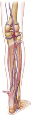
This PURL may be old news to you if you have been watching the evolution of DVT risk scoring and the role of D-dimer. This is one of those approaches for which the evidence grows over time and the adoption spreads slowly. We felt that the cumulative evidence, especially this decision analysis,1 clearly points to the most current Wells score and the D-dimer as the approach of choice for initial evaluation of suspected DVT. This more definitive evidence and the variability of practice both reported in the literature and described by our clinician reviewers led us to decide that this study is a priority update and a practice changer—even if it is a slow practice changer.
Caveats: One strategy doesn’t fit all
The authors stress that their results are most applicable to outpatients with a suspected first DVT.1
- D-dimer levels can be elevated in pregnancy, myocardial infarction, cancer, trauma, and postsurgery. These recommendations do not apply to patients with any of these conditions, patients on anticoagulation therapy, intravenous drug abusers, or patients with recurrent DVT.
- In practices where D-dimer testing is not feasible, ultrasound remains an effective approach to suspected DVT.
Challenges To Implementation: Haven’t memorized the Wells score? No problem
As simple as it seems to add up the Wells score, most of us are not likely to recall the scoring system unless we use it often enough to have memorized it. Lack of immediate access to scoring systems in the context of a hectic schedule is a barrier to adoption.
Fortunately, many handheld and Web-based electronic knowledge resources are available for easy retrieval of the Wells DVT score. Some will even dynamically calculate the score for you.
Ideally, scoring systems such as this need to be integrated into electronic health records for easy access at the point of patient care.
Which Wells score?
There are several other scoring systems for estimating DVT risk, but the Wells DVT score is the best studied. To add to the confusion, Wells and his colleagues have made continuous improvements over time, such that there are several versions of the Wells DVT score.
In a quick Google search, we found six versions in which either the criteria or the interpretation was different, not to mention multiple other systems. The one used in this meta-analysis1 and described in this PURL is the most recent, most accurate and best studied.
PURLs methodology
This study was selected and evaluated using the Family Physician Inquiries Network’s Priority Updates from the Research Literature Surveillance System (PURLs) methodology.
1. Goodacre S, Stevenson M, Wailoo A, Sampson F, Sutton AJ, Thomas S. How should we diagnose suspected deep-vein thrombosis? QJM 2006;99:377-388.
2. Fancher TL, White RH, Kravits RL. Combined use of rapid D-dimer testing and estimation of clinical probability in the diagnosis of deep vein thrombosis: systematic review. BMJ 2004;329:821.-
3. Sampson FC, Goodacre S, Kelly AM, Kerr D. How is deep vein thrombosis diagnosed and managed in UK and Australian emergency departments? Emerg Med J 2005;22:780-782.
4. Deep venous thrombosis (DVT): diagnosis. Pepid-PCP [database online]. Available at www.pepidonline.com.
5. Deep vein thrombosis (DVT). Dynamed [database online]. Updates September 24, 2006. Available at dynamicmedical.com.
6. Landaw S. Approach to the diagnosis and therapy of suspected deep vein thrombosis. UpTodate [database online]. Updated April 25, 2006. Available at uptodate.com. Accessed September 13, 2006.
7. Tapson V, Carroll B, Davidson B, et al. and the American Thoracic Society. ATS guidelines: diagnostic approach to acute venous thromboembolism. Am J Respir Crit Care Med 1999;160:1043-1066.
8. Health care guideline: venous thromboembolism. Institute for clinical Systems Improvement. Revised February 2006. Available at www.icsi.org. Accessed on September 27, 2006.
Use a combination of Wells score and D-dimer test to exclude deep vein thrombosis in low- to intermediate-risk outpatients with suggestive symptoms.1
Strength of recommendation (SOR)
A: Based on one good meta-analysis
Goodacre S, Stevenson M, Wailoo A, Sampson F, Sutton AJ, Thomas S. How should we diagnose suspected deep-vein thrombosis? QJM 2006;99:377–388.
Illustrative case
Your patient is a 47-year-old man who has had pain and swelling in his right leg since yesterday. He has no history of cancer, recent travel, surgery, or blood clots. There has been no known trauma. You observe some tenderness in the calf, and no swelling. A Homan’s sign is negative.
How would you assess for deep venous thrombosis?
Background: What are costs, benefits of different strategies?
Wouldn’t it be nice to be able to rule out deep vein thrombosis (DVT) with a simple history and blood test?
Most patients with suggestive symptoms do not have DVT, but workups for this dangerous condition “range from the accurate but expensive (contrast venography) to the cheap but unreliable (clinical assessment),” noted the researchers who conducted this extensive analysis, seeking a cost-effective strategy to put into practice throughout the United Kingdom’s National Health Service.1 The NHS Health Technology Assessment R&D Program funded the study.
Until now, there has been little clear direction from formal comparisons. Although recent studies2 suggest that combinations of simple diagnostic tests may reduce the need for expensive, definitive tests, none explicitly weigh the costs and benefits of the different strategies; despite a large amount of published data, practice varies considerably.1,3
Clinical Context: Guidelines conflict
While some clinical resources now recommend the Wells criteria and D-dimer as useful tools in the initial workup of suspected DVT,4,5 others still recommend compression ultrasound or impedance plethysmography (IPG) as the initial or confirmatory test in all patients with suspected acute DVT.6 These noninvasive tests do provide reassurance that there is no DVT, but they are costly and less convenient than the Wells score and a D-dimer.
Current guidelines give conflicting recommendations.
- The most recent American Thoracic Society guidelines, from 1999, recommend imaging with ultrasound or impedance plethysmography for all patients with suspected DVT.7
- In contrast, the Institute for Clinical Systems Improvement (ICSI) 2006 guidelines recommend first determining the clinical pretest probability of DVT using the Wells score, and then using a D-dimer test to determine which patients with a low probability test need to proceed to ultrasound. This algorithm recommends ultrasound for all patients with either a moderate or high Wells score.8
Variety of D-dimer tests, range of sensitivities
One meta-analysis of 12 studies compared a highly sensitive ELISA D-dimer assay to the less sensitive SimpliRED D-dimer assay. In studies using the highly sensitive ELISA assay, in patients with negative D-dimer and low or moderate Wells score, the 3-month incidence of DVT was 0.5%. However, using the SimpliRED assay, while the 3-month incidence of DVT was 0.5% with negative D-dimer and low Wells score, it was 3.5% with negative D-dimer and intermediate Wells score.7
Study Summary: Seeking convenience and economy
This systematic review, meta-analysis, and decision analysis sought the most practical, cost-effective strategy to detect DVT. The researchers compared the findings of 18 studies of diagnostic strategies (or algorithms) that combined Wells score (TABLE), D-dimer, ultrasound, or venography, and that followed up patients with negative results for at least 3 months. They developed a decision analysis model to compare the algorithms in a hypothetical cohort of 1000 outpatients with suspected DVT.
Applying the estimated sensitivity and specificity of each algorithm to the hypothetical population, they determined the proportions of patients with and without DVT who would receive treatment and which patients would suffer events relating to DVT or treatment, and then estimated lifetime health outcomes (quality-adjusted life years [QALYs]) and costs of testing and treatment.
Using thresholds for willingness to pay of £10,000, £20,000, and £30,000 per QALY, the study identified 2 optimal diagnostic strategies, both of which incorporated D-dimer testing and Wells score. While one strategy starts with Wells score and the other starts with D-dimer, both recommend that patients with a combination of a negative D-dimer test and an intermediate or low Wells score can be safely discharged without further testing.
One weakness of this study is that the authors made the assumption that ultrasound results are independent of Wells score or D-dimer. While it is unlikely that ultrasound results are related to D-dimer results, there is some evidence that ultrasound is more accurate in patients with higher Wells scores. However, if this true, the authors would have underestimated the cost-effectiveness of the favored strategies. Also, they did not include algorithms that involved plethysmography.
TABLE
Wells score estimates probability of deep vein thrombosis
The elements of the Wells score should be ascertained in the usual evaluation of a patient with suspected DVT.
| 1 POINT EACH FOR: | |
| Active cancer | |
| Paralysis, paresis, recent plaster immobilization of lower limb | |
| Recently bedridden for >3 days or major surgery in past 4 weeks | |
| Localized tenderness along distribution of deep venous system | |
| Entire leg swollen | |
| Calf swelling >3 cm compared to asymptomatic leg | |
| Pitting edema | |
| Collateral superficial veins | |
| –2 POINTS FOR: | |
| Alternative diagnosis as likely or more likely than that of DV T | |
| PROBABILITY: | |
| High | >3 points |
| Intermediate | 1 or 2 points |
| Low | <0 points |
What’s New?: This evidence is definitive
This PURL may be old news to you if you have been watching the evolution of DVT risk scoring and the role of D-dimer. This is one of those approaches for which the evidence grows over time and the adoption spreads slowly. We felt that the cumulative evidence, especially this decision analysis,1 clearly points to the most current Wells score and the D-dimer as the approach of choice for initial evaluation of suspected DVT. This more definitive evidence and the variability of practice both reported in the literature and described by our clinician reviewers led us to decide that this study is a priority update and a practice changer—even if it is a slow practice changer.
Caveats: One strategy doesn’t fit all
The authors stress that their results are most applicable to outpatients with a suspected first DVT.1
- D-dimer levels can be elevated in pregnancy, myocardial infarction, cancer, trauma, and postsurgery. These recommendations do not apply to patients with any of these conditions, patients on anticoagulation therapy, intravenous drug abusers, or patients with recurrent DVT.
- In practices where D-dimer testing is not feasible, ultrasound remains an effective approach to suspected DVT.
Challenges To Implementation: Haven’t memorized the Wells score? No problem
As simple as it seems to add up the Wells score, most of us are not likely to recall the scoring system unless we use it often enough to have memorized it. Lack of immediate access to scoring systems in the context of a hectic schedule is a barrier to adoption.
Fortunately, many handheld and Web-based electronic knowledge resources are available for easy retrieval of the Wells DVT score. Some will even dynamically calculate the score for you.
Ideally, scoring systems such as this need to be integrated into electronic health records for easy access at the point of patient care.
Which Wells score?
There are several other scoring systems for estimating DVT risk, but the Wells DVT score is the best studied. To add to the confusion, Wells and his colleagues have made continuous improvements over time, such that there are several versions of the Wells DVT score.
In a quick Google search, we found six versions in which either the criteria or the interpretation was different, not to mention multiple other systems. The one used in this meta-analysis1 and described in this PURL is the most recent, most accurate and best studied.
PURLs methodology
This study was selected and evaluated using the Family Physician Inquiries Network’s Priority Updates from the Research Literature Surveillance System (PURLs) methodology.
1. Goodacre S, Stevenson M, Wailoo A, Sampson F, Sutton AJ, Thomas S. How should we diagnose suspected deep-vein thrombosis? QJM 2006;99:377-388.
2. Fancher TL, White RH, Kravits RL. Combined use of rapid D-dimer testing and estimation of clinical probability in the diagnosis of deep vein thrombosis: systematic review. BMJ 2004;329:821.-
3. Sampson FC, Goodacre S, Kelly AM, Kerr D. How is deep vein thrombosis diagnosed and managed in UK and Australian emergency departments? Emerg Med J 2005;22:780-782.
4. Deep venous thrombosis (DVT): diagnosis. Pepid-PCP [database online]. Available at www.pepidonline.com.
5. Deep vein thrombosis (DVT). Dynamed [database online]. Updates September 24, 2006. Available at dynamicmedical.com.
6. Landaw S. Approach to the diagnosis and therapy of suspected deep vein thrombosis. UpTodate [database online]. Updated April 25, 2006. Available at uptodate.com. Accessed September 13, 2006.
7. Tapson V, Carroll B, Davidson B, et al. and the American Thoracic Society. ATS guidelines: diagnostic approach to acute venous thromboembolism. Am J Respir Crit Care Med 1999;160:1043-1066.
8. Health care guideline: venous thromboembolism. Institute for clinical Systems Improvement. Revised February 2006. Available at www.icsi.org. Accessed on September 27, 2006.
Copyright © 2007 The Family Physicians Inquiries Network.
All rights reserved.
Azithromycin for PID beats doxycycline on all counts
Outpatient treatment of patients with mild pelvic inflammatory disease, using 1 g of azithromycin weekly for 2 weeks, combined with 250 mg of ceftriaxone intramuscularly on the first day, is superior to the current recommended treatment with doxycycline plus ceftriaxone.1
Strength of recommendation (SOR)
A: Single well-designed RCT
Savaris RF, Teixeira LM, Torres TG, Edelweiss MI, Moncada J, Schachter J. Comparing ceftriaxone plus azithromycin or doxycycline for pelvic inflammatory disease: a randomized controlled trial. Obstet Gynecol 2007;110:53–60.
Illustrative Case
An otherwise healthy, sexually active 21-year-old woman complains of pelvic pain for a week and yellow vaginal discharge. The history and physical exam are consistent with mild, uncomplicated pelvic inflammatory disease (PID).
You believe outpatient therapy is appropriate in this case and wonder if there is a better alternative to doxycycline, particularly given the challenges of adherence to the recommended 14-day course of treatment.
Background: 2 doses or 28 doses?
In the real world, we know that adherence is better when patients have to take 2 pills than when they have to take 28 pills. For most women with mild, uncomplicated PID, outpatient treatment is appropriate2 and a shorter treatment course is related to better adherence.3 Azithromycin can be given in 2 single doses a week apart, with few side effects, and its spectrum of activity is similar to that of doxycycline,4 which requires a 14-day regimen of 2 pills daily. Earlier studies of azithromycin for PID, however, were not designed specifically for outpatient treatment, or had methodologic bias.5 Thus, the evidence has been insufficient to recommend it.
PID affects about 1 million women in the US each year, and can cause pain, scarring of the fallopian tubes, and infertility.
Current recommendations. The Centers for Disease Control and Prevention (CDC) recommends oral doxycycline 100 mg twice daily for 14 days, along with a second- or third-generation cephalosporin administered parenterally, for mild PID in ambulatory patients.5 Metronizadole can be added at the provider’s discretion. The CDC no longer routinely recommends fluoroquinolones for PID because of gonococcal resistance.6 Dynamed, PEPID PCP, and UpToDate all cite CDC guidelines. Dynamed also notes results of the article reviewed here, though treatment recommendations were not changed.7-9
A simpler approach to pelvic inflammatory disease
Patients will likely find it easier to take 1 azithromycin pill initially and 1 pill a week later, than to take a doxycycline pill twice daily for 14 days. lf so, then the advantage of azithromycin could be greater than reported in this study
Clinical context: First comparison study, first outpatient study
The study by Savaris and colleagues is the first comparison study of azithromycin and doxycycline for PID, and the first study of outpatient treatment of PID with azithromycin.
An earlier study reported that women with PID who were prescribed doxycycline took an average of 70% of the total doses, and fewer than half took it twice daily as directed.3
Azithromycin is known to be effective for treatment of Chlamydia trachomatis cervical infections,4 and single-dose azithromycin has been also been shown to have better compliance than multidose therapy for Chlamydia infection.10
We identified only 1 prior randomized controlled trial of azithromycin for treatment of PID. That trial reported that intravenous azithromycin followed by oral azithromycin with or without metronidazole is effective in the treatment of PID.11
Study summary: Azithromycin cure rate 90%, doxycycline 72%
This randomized, double-blind, controlled study evaluated the effectiveness of azithromycin plus ceftriaxone in the treatment of mild, uncomplicated PID compared to doxycycline plus ceftriaxone, in outpatients.
Patients
The study enrolled 133 women who presented to an emergency department with PID diagnosed by the following clinical criteria:
- pelvic pain for less than 30 days
- pelvic organ (adnexal or cervical) tenderness on physical examination
- cervical leukorrhea or mucopurulent cervicitis.
Method
The women were randomized into 2 groups, and both groups received 250 mg of ceftriaxone intramuscularly.
- The control group received 100 mg of doxycycline twice daily for 2 weeks.
- The study group received 1 g of azithromycin by mouth weekly for 2 weeks and a placebo twice daily for 2 weeks to maintain blinding.
Outcomes
The primary outcome was clinical cure after 2 weeks of treatment. Clinical cure was defined as an improvement in pain scale ratings by 70%. Failure was defined as worsening of pain, lack of improvement of pain, or need for additional antibiotic therapy, hospitalization, or surgery.
Of the 133 women randomized, 13 (9 from the azithromycin group and 4 from the doxycycline group) were found to have diagnoses other than PID after randomization.
Intention-to-treat analysis was performed for the remaining 120 participants. In the azithromycin group, 56/62 (90.3%; 95% confidence interval [CI], 0.80–0.96) women were classified as clinically cured, versus 42/58 (72.4%; 95% CI, 0.58–0.82) in the doxycycline group.
Adverse events. Except for oral intolerance to the first dose of medication, which was similar in both groups, adverse events were not reported.
Adherence similar in both groups
Adherence to the study protocol was similar in both groups. The study authors concluded that azithromycin was superior to doxycycline even though the adherence in the doxycycline group was good.
What’s new?: Better adherence is the probable bonus
This RCT shows that azithromycin treatment of PID in an ambulatory population is superior to doxycycline even when there is excellent compliance with taking doxycycline (unlike the reality of clinical practice). The patients in this RCT adhered well to the protocol, so it does not provide a realistic head-to-head comparison of treatment completion.
Real-world adherence
In actual practice, we speculate that taking 2 pills 1 week apart will be much easier for patients than taking 2 pills every day for 14 days. The literature on compliance would predict that to be the case. If true, then the advantage of azithromycin over doxycycline would be even greater than reported in this study.
Caveats: Apply these findings in similar cases only
This study addresses ambulatory treatment of mild, uncomplicated PID, and results should only be extrapolated to similar cases.
Azithromycin should not be prescribed to patients with an allergy to macrolide antibiotics.
One of the study authors received azithromycin donated by Pfizer for other research; however, Pfizer did not sponsor this study.
Challenges to implementation: Cost of the prescription
Prescription cost may be a consideration for patients without insurance, although azithromycin has been shown to be cost-effective in treatment of Chlamydia.12
Reminding patients to take the second dose
Some patients may have difficulty remembering to take the second dose a week after the first dose.
A follow-up visit, reminder phone call, or suggestion to “mark this on your calendar” may help enhance adherence.
PURLs methodology
This study was selected and evaluated using the Family Physician Inquiries Network’s Priority Updates from the Research Literature Surveillance System (PURLs) methodology. The criteria and findings leading to the selection of this study as a PURL can be accessed here.
1. Savaris RF, Teixeira LM, Torres TG, Edelweiss MI, Moncada J, Schachter J. Comparing ceftriaxone plus azithromycin or doxycycline for pelvic inflammatory disease: a randomized controlled trial. Obstet Gynecol 2007;110:53-60.
2. Ness RB, Soper DE, Holley RL, et al. Effectiveness of inpatient and outpatient treatment strategies for women with pelvic inflammatory disease: results from the pelvic inflammatory disease evaluation and clinical health (PEACH) randomized trial. Am J Obstet Gynecol 2002;186:929-937.
3. Dunbar-Jacob J, Sereika SM, Foley SM, Bass DC, Ness RG. Adherence to oral therapies in pelvic inflammatory disease. J Womens Health 2004;13:285-291.
4. Lau CY, Qureshi AK. Azithromycin versus doxycycline for genital chlamydial infections: a metaanalysis of randomized clinical trials. Sex Transm Dis 2002;29:497-502.
5. Workowski KA, Berman SM. Sexually transmitted diseases treatment guidelines, 2006. Centers for Disease Control and Prevention. MMWR Recomm Rep 2006;55(RR-11):1-94.Available at: www.cdc.gov/std/treatment/2006/updated-regimens.htm. Accessed on November 14, 2007.
6. Centers for Disease Control and Prevention. Update to CDC sexually transmitted diseases treatment guidelines, 2006: fluoroquinolones no longer recommended for treatment of gonococcal infections. MMWR Morb Mortal Wkly Rep 2007;56(14):332-336.Available at: www.cdc.gov/mmwr/preview/mmwrhtml/mm5614a3.htm. Accessed on November 14, 2007.
7. Pelvic inflammatory disease. In: Dynamed [online database]. Available at: www.dynamicmedical.com. Accessed on August 30, 2007.
8. Pelvic inflammatory disease. In: PEPID-PCP [online database]. Available at: www.pepidonline.com. Accessed on August 30, 2007.
9. Hynes N. Treatment of pelvic inflammatory disease. In: UptoDate [online database]. Available at: www.utdol.com. Accessed on August 30, 2007.
10. Adair CD, Gunter M, Stovall TG, McElroy G, Veille JC, Ernest JM. Chlamydia in pregnancy: a randomized trial of azithromycin and erythromycin. Obstet Gynecol 1998;91:165-168.
11. Bevan CD, Ridgway GL, Rothermel CD. Efficacy and safety of azithromycin as monotherapy or combined with metronidazole compared with two standard multidrug regimens for the treatment of acute pelvic inflammatory disease. J Int Med Res 2003;31:45-54.
12. Magid D, Douglas JM, Schwartz JS. Doxycycline compared with azithromycin for treating women with genital Chlamydia trachomatis infections: an incremental cost-effectiveness analysis. Ann Intern Med 1996;124:389-399.
Outpatient treatment of patients with mild pelvic inflammatory disease, using 1 g of azithromycin weekly for 2 weeks, combined with 250 mg of ceftriaxone intramuscularly on the first day, is superior to the current recommended treatment with doxycycline plus ceftriaxone.1
Strength of recommendation (SOR)
A: Single well-designed RCT
Savaris RF, Teixeira LM, Torres TG, Edelweiss MI, Moncada J, Schachter J. Comparing ceftriaxone plus azithromycin or doxycycline for pelvic inflammatory disease: a randomized controlled trial. Obstet Gynecol 2007;110:53–60.
Illustrative Case
An otherwise healthy, sexually active 21-year-old woman complains of pelvic pain for a week and yellow vaginal discharge. The history and physical exam are consistent with mild, uncomplicated pelvic inflammatory disease (PID).
You believe outpatient therapy is appropriate in this case and wonder if there is a better alternative to doxycycline, particularly given the challenges of adherence to the recommended 14-day course of treatment.
Background: 2 doses or 28 doses?
In the real world, we know that adherence is better when patients have to take 2 pills than when they have to take 28 pills. For most women with mild, uncomplicated PID, outpatient treatment is appropriate2 and a shorter treatment course is related to better adherence.3 Azithromycin can be given in 2 single doses a week apart, with few side effects, and its spectrum of activity is similar to that of doxycycline,4 which requires a 14-day regimen of 2 pills daily. Earlier studies of azithromycin for PID, however, were not designed specifically for outpatient treatment, or had methodologic bias.5 Thus, the evidence has been insufficient to recommend it.
PID affects about 1 million women in the US each year, and can cause pain, scarring of the fallopian tubes, and infertility.
Current recommendations. The Centers for Disease Control and Prevention (CDC) recommends oral doxycycline 100 mg twice daily for 14 days, along with a second- or third-generation cephalosporin administered parenterally, for mild PID in ambulatory patients.5 Metronizadole can be added at the provider’s discretion. The CDC no longer routinely recommends fluoroquinolones for PID because of gonococcal resistance.6 Dynamed, PEPID PCP, and UpToDate all cite CDC guidelines. Dynamed also notes results of the article reviewed here, though treatment recommendations were not changed.7-9
A simpler approach to pelvic inflammatory disease
Patients will likely find it easier to take 1 azithromycin pill initially and 1 pill a week later, than to take a doxycycline pill twice daily for 14 days. lf so, then the advantage of azithromycin could be greater than reported in this study
Clinical context: First comparison study, first outpatient study
The study by Savaris and colleagues is the first comparison study of azithromycin and doxycycline for PID, and the first study of outpatient treatment of PID with azithromycin.
An earlier study reported that women with PID who were prescribed doxycycline took an average of 70% of the total doses, and fewer than half took it twice daily as directed.3
Azithromycin is known to be effective for treatment of Chlamydia trachomatis cervical infections,4 and single-dose azithromycin has been also been shown to have better compliance than multidose therapy for Chlamydia infection.10
We identified only 1 prior randomized controlled trial of azithromycin for treatment of PID. That trial reported that intravenous azithromycin followed by oral azithromycin with or without metronidazole is effective in the treatment of PID.11
Study summary: Azithromycin cure rate 90%, doxycycline 72%
This randomized, double-blind, controlled study evaluated the effectiveness of azithromycin plus ceftriaxone in the treatment of mild, uncomplicated PID compared to doxycycline plus ceftriaxone, in outpatients.
Patients
The study enrolled 133 women who presented to an emergency department with PID diagnosed by the following clinical criteria:
- pelvic pain for less than 30 days
- pelvic organ (adnexal or cervical) tenderness on physical examination
- cervical leukorrhea or mucopurulent cervicitis.
Method
The women were randomized into 2 groups, and both groups received 250 mg of ceftriaxone intramuscularly.
- The control group received 100 mg of doxycycline twice daily for 2 weeks.
- The study group received 1 g of azithromycin by mouth weekly for 2 weeks and a placebo twice daily for 2 weeks to maintain blinding.
Outcomes
The primary outcome was clinical cure after 2 weeks of treatment. Clinical cure was defined as an improvement in pain scale ratings by 70%. Failure was defined as worsening of pain, lack of improvement of pain, or need for additional antibiotic therapy, hospitalization, or surgery.
Of the 133 women randomized, 13 (9 from the azithromycin group and 4 from the doxycycline group) were found to have diagnoses other than PID after randomization.
Intention-to-treat analysis was performed for the remaining 120 participants. In the azithromycin group, 56/62 (90.3%; 95% confidence interval [CI], 0.80–0.96) women were classified as clinically cured, versus 42/58 (72.4%; 95% CI, 0.58–0.82) in the doxycycline group.
Adverse events. Except for oral intolerance to the first dose of medication, which was similar in both groups, adverse events were not reported.
Adherence similar in both groups
Adherence to the study protocol was similar in both groups. The study authors concluded that azithromycin was superior to doxycycline even though the adherence in the doxycycline group was good.
What’s new?: Better adherence is the probable bonus
This RCT shows that azithromycin treatment of PID in an ambulatory population is superior to doxycycline even when there is excellent compliance with taking doxycycline (unlike the reality of clinical practice). The patients in this RCT adhered well to the protocol, so it does not provide a realistic head-to-head comparison of treatment completion.
Real-world adherence
In actual practice, we speculate that taking 2 pills 1 week apart will be much easier for patients than taking 2 pills every day for 14 days. The literature on compliance would predict that to be the case. If true, then the advantage of azithromycin over doxycycline would be even greater than reported in this study.
Caveats: Apply these findings in similar cases only
This study addresses ambulatory treatment of mild, uncomplicated PID, and results should only be extrapolated to similar cases.
Azithromycin should not be prescribed to patients with an allergy to macrolide antibiotics.
One of the study authors received azithromycin donated by Pfizer for other research; however, Pfizer did not sponsor this study.
Challenges to implementation: Cost of the prescription
Prescription cost may be a consideration for patients without insurance, although azithromycin has been shown to be cost-effective in treatment of Chlamydia.12
Reminding patients to take the second dose
Some patients may have difficulty remembering to take the second dose a week after the first dose.
A follow-up visit, reminder phone call, or suggestion to “mark this on your calendar” may help enhance adherence.
PURLs methodology
This study was selected and evaluated using the Family Physician Inquiries Network’s Priority Updates from the Research Literature Surveillance System (PURLs) methodology. The criteria and findings leading to the selection of this study as a PURL can be accessed here.
1. Savaris RF, Teixeira LM, Torres TG, Edelweiss MI, Moncada J, Schachter J. Comparing ceftriaxone plus azithromycin or doxycycline for pelvic inflammatory disease: a randomized controlled trial. Obstet Gynecol 2007;110:53-60.
2. Ness RB, Soper DE, Holley RL, et al. Effectiveness of inpatient and outpatient treatment strategies for women with pelvic inflammatory disease: results from the pelvic inflammatory disease evaluation and clinical health (PEACH) randomized trial. Am J Obstet Gynecol 2002;186:929-937.
3. Dunbar-Jacob J, Sereika SM, Foley SM, Bass DC, Ness RG. Adherence to oral therapies in pelvic inflammatory disease. J Womens Health 2004;13:285-291.
4. Lau CY, Qureshi AK. Azithromycin versus doxycycline for genital chlamydial infections: a metaanalysis of randomized clinical trials. Sex Transm Dis 2002;29:497-502.
5. Workowski KA, Berman SM. Sexually transmitted diseases treatment guidelines, 2006. Centers for Disease Control and Prevention. MMWR Recomm Rep 2006;55(RR-11):1-94.Available at: www.cdc.gov/std/treatment/2006/updated-regimens.htm. Accessed on November 14, 2007.
6. Centers for Disease Control and Prevention. Update to CDC sexually transmitted diseases treatment guidelines, 2006: fluoroquinolones no longer recommended for treatment of gonococcal infections. MMWR Morb Mortal Wkly Rep 2007;56(14):332-336.Available at: www.cdc.gov/mmwr/preview/mmwrhtml/mm5614a3.htm. Accessed on November 14, 2007.
7. Pelvic inflammatory disease. In: Dynamed [online database]. Available at: www.dynamicmedical.com. Accessed on August 30, 2007.
8. Pelvic inflammatory disease. In: PEPID-PCP [online database]. Available at: www.pepidonline.com. Accessed on August 30, 2007.
9. Hynes N. Treatment of pelvic inflammatory disease. In: UptoDate [online database]. Available at: www.utdol.com. Accessed on August 30, 2007.
10. Adair CD, Gunter M, Stovall TG, McElroy G, Veille JC, Ernest JM. Chlamydia in pregnancy: a randomized trial of azithromycin and erythromycin. Obstet Gynecol 1998;91:165-168.
11. Bevan CD, Ridgway GL, Rothermel CD. Efficacy and safety of azithromycin as monotherapy or combined with metronidazole compared with two standard multidrug regimens for the treatment of acute pelvic inflammatory disease. J Int Med Res 2003;31:45-54.
12. Magid D, Douglas JM, Schwartz JS. Doxycycline compared with azithromycin for treating women with genital Chlamydia trachomatis infections: an incremental cost-effectiveness analysis. Ann Intern Med 1996;124:389-399.
Copyright © 2007 The Family Physicians Inquiries Network.
All rights reserved.
Double-dose vitamin D lowers cancer risk in women over 55
Increasing the dose of vitamin D3 from the current standard of 400–600 IU per day to 1000 IU per day lowers future risk of cancer in women older than age 55 who do not get adequate vitamin D from sun exposure or diet.1
Strength of recommendation (SOR)
A: Well done randomized controlled trial2
Lappe JM, Travers-Gustafson D, Davies KM et al. Vitamin D Supplementation reduces cancer risk: results of a randomized trial. Am J Clin Nutr 2007; 85:1586–1591.
Illustrative case
A healthy 60-year-old Chicago woman who takes 1500 mg calcium and a multi vitamin daily tells you she has read that extra vitamins prevent cancer. She is particularly concerned about cancer because of her strong family history. Should you recommend that she take any additional vitamins to reduce her risk of cancer?
Background: Will this trial pass the test of time? We think so
Wouldn’t it be nice if we could recommend something as simple and safe as a daily vitamin to reduce the risk of cancer? Until now, we have had no definitive evidence to support such a recommendation. The Lappe et al trial, however, concluded that improving calcium and vitamin D nutritional status substantially reduces all-cancer risk in postmenopausal women.1 Will this single, relatively small study pass the test of time and be confirmed by future clinical trials? We think so.
- The estimated relative risk reduction was dramatic (0.232) and the 95% confidence interval was 0.09 to 0.60, meaning that the true relative risk reduction has a 95% probability of being in the range of 40% to 91%. The P value of <.005 suggests that the probability of this finding occurring by chance alone is less than 1 in 200.
- Our critical appraisal found no significant flaws in this randomized controlled trial.
- Vitamin D is known to have cancer protective effects at the cellular level.
- Prior population based studies support the association between vitamin D and cancer prevention.
For these reasons—and the fact that 1000 IU vitamin D is very safe for most patients—we find this single RCT convincing as a practice changer. For us, the potential benefit outweighs the potential harm.
United States Preventive services Task Force. A 2003 report on “routine vitamin supplementation to prevent cancer and cardiovascular disease” cited insufficient evidence to recommend the use of supplemental vitamins A, C, E, multivitamins with folic acid, or antioxidants to prevent cancer or cardiovascular disease; vitamin D is not mentioned.3
Institute of medicine. In 2005, the IOM suggested an Adequate Intake (AI) of vitamin D of 400 IU for women from 51 to 70 years of age, and 600 IU for women over 70 years of age, to maintain bone health and normal calcium metabolism in healthy women. The IOM cited epidemiologic studies showing an inverse association between either increased sun exposure or higher vitamin D levels and decreased risk of cancer, and included the caveat that it was premature to recommend taking vitamin D for cancer prevention until well-designed trials prove that vitamin D is protective against cancer.4
Electronic knowledge resources that are evidence-based and frequently updated did not recommend vitamin D for cancer prevention on the dates we searched.5-8
Clinical Context: Food, sun, supplements may not deter deficiency
Few people get enough vitamin D to match the dosage that reduced cancer incidence in this trial. In fact, inadequate vitamin D intake, even to meet current standards, is surprisingly common—even in people who are apparently conscious of their nutritional needs. A Boston hospital found that 32% of healthy students, physicians, and resident physicians were vitamin-D deficient, despite drinking a glass of milk daily, taking a daily multivitamin, and eating salmon at least once a week.9 An estimated 1 billion people worldwide have vitamin D deficiency or insufficiency.9
Many factors affect vitamin D levels (TABLE).9 Foods that contain vitamin D3 include fortified milk (100 IU per cup) and oily fish, including salmon, tuna, sardines, mackerel, and herring (200–300 IU per 3.5-oz serving). Sun exposure for 10 to 15 minutes (without sunscreen) at least twice a week to the face, arms, hands, or back is considered sufficient to provide adequate vitamin D during summer or in warm climates.
Many patients need supplements to reach the levels provided by 1000 IU daily, especially in colder climates. Most over-the-counter supplements containing vitamin D alone contain 400 to 1000 IU vitamin D3. Prescription vitamin D2 capsules contain 50,000 IU.9 Vitamin D is available as vitamin D2 and D3:
- Vitamin D2 is usually labeled vitamin D or calciferol. Vitamin D2 is only 30% as effective as vitamin D3 (doses should be adjusted accordingly).
- Vitamin D3 is labeled vitamin D3 or cholecalciferol.
TABLE
3 ways to get vitamin D: Food, sun, and supplements9
| SOURCE | AMOUNT | |
|---|---|---|
| Food | 1 cup of fortified milk | 100 IU vitamin D3 |
| One serving (3.5 oz) of oily fish (salmon, tuna, sardines, mackerel or herring) | 200 to 300 IU vitamin D | |
| Sun (ultraviolet B radiation) | Expose face, arms, hands, or back for 10 to 15 minutes (without sunscreen) at least twice a week during summer months or in warm climates | 3000 IU vitamin D3 per exposure |
| Supplements | Vitamin D3 | 1000 IU/day |
| Vitamin D2 | 50,000 IU every 2 to 4 weeks |
Study Summary: Cancer was a secondary outcome
This trial was well designed and executed, with impressive findings. The primary outcomes were related to skeletal status and calcium economy. Cancer incidence was one of the secondary outcomes.
This population-based study was randomized, double-blinded, and placebo-controlled, with concealed allocation. The researchers enrolled 1180 women older than 55 years of age, with no known cancer, and with adequate mental and physical health to allow an expected 4 years of participation in the trial. The trial was conducted in rural Nebraska. Eighty-six percent of the participants completed the study. Participants were randomly assigned to 3 groups:
- Placebo (calcium placebo plus vitamin D placebo, n=266)
- calcium-only (1400 mg calcium citrate or 1500 mg calcium carbonate plus vitamin D placebo, n=416)
- Calcium + D (1000 IU [25 mcg] vitamin D plus calcium [as above], n=403)
Every 6 months, adherence was assessed by bottle weight. Mean adherence (taking ≥80% of assigned doses) was 85.7% for vitamin D and 74.4% for calcium. Serum samples were analyzed for 25(OH)D at baseline and then yearly.1
Key results
Fifty women developed non-skin cancer during the study: 13 in the first year, and 37 during the second to fourth years. Excluding cancer diagnosed in the first year (it was assumed that these cancers were present, though undiagnosed, at entry), the relative risk reduction (RRR) for the calcium + D group was 0.232 (confidence interval [CI], 0.09–0.60; P<.005), and the RRR for the calcium-only group was 0.587 (95% CI, 0.29–1.21; P=.147) compared with the placebo group.
Number needed to treat (NNT) to prevent 1 case of cancer for the calcium + D group is 21, with an absolute risk reduction of 0.048, or approximately 5%.
Risk reduction. Using baseline 25(OH)D concentration as the predictor variable and cancer as the outcome variable in logistic regression, Lappe et al predicted a 35% reduced cancer risk for every 25 nmol/L (10 ng/mL) increase in serum 25(OH)D.1
What’s New? First RCT to show reduced cancer incidence
This is the first randomized-controlled clinical trial to show that vitamin D reduces cancer risk. (It is important to note that one prior randomized controlled trial10 found no impact on cancer incidence; however, that trial used a vitamin D3 dose of 400 IU, which is lower than the 1000 IU dose used by Lappe et al.)
Vitamin D curbs carcinogenic potential. The new findings build on prior basic research, which established the pathophysiologic process by which vitamin D may prevent cancer in humans. Vitamin D receptors are found not only in the small intestines, bones, and kidneys, but also in most other tissues, including skin, colon, prostate, breast, and brain. The interaction of 1,25(OH)2D with vitamin D receptors induces terminal differentiation and apoptosis and inhibits cellular growth, angiogenesis, and metastatic potential.10
Other studies suggest vitamin d plays a part. Previous population-based studies also suggested an association between vitamin D and reduced cancer incidence.
Lin et al, as part of the Women’s Health Study, found that higher intake of calcium and vitamin D was associated with a lower risk of breast cancer in premenopausal but not in postmenopausal women. The highest dosage quintile was >548 IU; therefore, many if not most women likely ingested an inadequate dose of vitamin D to reduce risk of cancer.11
The Health Professionals Follow-up Study, which followed a cohort of 47,800 men, from 1986 until 2000, found that low levels of vitamin D were associated with increased incidence of cancer and mortality.12
In the only other randomized controlled trial of vitamin D and cancer (also part of the Women’s Health Initiative), Wactawski-Wende et al found no difference in the risk of colorectal cancer between women taking calcium and vitamin D and women taking placebo, over an average of 7 years of follow-up. However, the vitamin D dose was only 400 IU daily, the dosage recommended for general health and bone health.13
Caveats: Consider toxicity unlikely
Although excess vitamin D intake, leading to a serum level of 25-hydroxyvitamin D (25[OH]D) >150 ng/mL, can cause toxicity, the IOM has set the tolerable upper intake level of vitamin D (a fat-soluble vitamin stored in the liver) at 2000 IU (50 mcg) for adults and children older than 1 year. Moreover, studies have shown that adults can tolerate doses as high as 10,000 IU per day.4
Symptoms of toxicity include nausea, vomiting, poor appetite, constipation, weakness, and weight loss as well as signs and symptoms of hypercalcemia, including mental status changes, renal failure, and arrhythmias.4
Diseases and drugs that affect serum levels. Patients with mild to moderate renal failure or chronic granulomatous diseases, such as sarcoidosis, are at higher risk of developing vitamin D toxicity. Patients with malabsorption syndromes, mild or moderate hepatic failure, or who take certain medications, like anticonvulsants or glucocorticoids, that increase vitamin D metabolism may need higher doses of vitamin D.9
The good sun. Exposure to sunlight never leads to vitamin D toxicity, as UV radiation destroys any excess vitamin D that is produced.10
Challenges To Implementation: A matter of time
The primary challenge is likely to be the competing demands and limited resources inherent in delivering all preventive health services in the primary care setting. By one estimate, implementing all preventive health services recommended by the US Preventive Services Task Force would require 7.4 hours per day, leaving little if any time to address the acute and chronic care needs of each individual patient.14
PURLs methodology
This study was selected and evaluated using FPIN’s Priority Updates from the Research Literature Surveillance System methodology. The criteria and findings leading to the selection of this study as a PURL can be accessed here.
1. Lappe JM, Travers-Gustafson D, Davies KM, et al. Vitamin D supplementation reduces cancer risk: results of a randomized trial. Am J Clin Nutr 2007;85:1586-15-91.
2. Ebell MH, Siwek J, Weiss BD, et al. Strength of recommendation taxonomy (SORT): A patient-centered approach to grading evidence in the medical literature. J Fam Pract 2004;53:111-120
3. US Preventive Services Task Force Web site. Routine vitamin supplementation to prevent cancer and cardiovascular disease: recommendations and rationale. June 2003. Available at: www.ahrq.gov/clinic/3rduspstf/vitamins/vitaminsrr.htm. Accessed on October 15, 2007.
4. National Institutes of Health, Office of Dietary Supplements. Dietary Supplement Fact Sheet: Vitamin D. 2005. Available at: ods.od.nih.gov/factsheets/vitamind.asp. Accessed on October 15, 2007.
5. Fletcher RH, Fairfield KM. Vitamin supplementation in disease prevention. UpToDate [database online]. Updated January 8, 2007. Available at: www.uptodateonline.com. Accessed on June 25, 2007.
6. Becker KL. Vitamin D intake and supplementation. Dynamed [database online]. Updated June 28, 2007. Available at www.dynamicmedical.com. Accessed on July 5, 2007.
7. Rosen HN. Vitamin D therapy in osteoporosis. UpToDate [database online]. Updated February 21, 2007. Available at www.uptodate.com. Accessed on July 11, 2007.
8. Vitamin D PepidPCP [database online]. Available at www.pepidonline.com. Accessed on August 6, 2007.
9. Holick MF. Vitamin D deficiency. N Engl J Med 2007;357:266-281.
10. Holick M. Vitamin D: its role in cancer prevention and treatment. Progress in Biophysics and Molecular Biology 2006;92:49-59.
11. Lin J, Manson JE, Lee I, et al. Intakes of calcium and vitamin D and breast cancer risk in women. Arch Intern Med 2007;167:1050-1059.
12. Giovannucci E, Liu Y, Rimm EB, et al. Prospective study of predictors of vitamin D status and cancer incidence and mortality in men. J Natl Cancer Inst 2006;98:451-459.
13. Wactawski-Wende J, Kotchen JM, Anderson GL, et al. Calcium plus vitamin D supplementation and the risk of colorectal cancer. N Engl J Med 2006;354:684-696.
14. Yarnall KSH, Pollak KI, Ostbye T, et al. Primary care: is there enough time for prevention? Am J Public Health 2003;93:635-641.
Increasing the dose of vitamin D3 from the current standard of 400–600 IU per day to 1000 IU per day lowers future risk of cancer in women older than age 55 who do not get adequate vitamin D from sun exposure or diet.1
Strength of recommendation (SOR)
A: Well done randomized controlled trial2
Lappe JM, Travers-Gustafson D, Davies KM et al. Vitamin D Supplementation reduces cancer risk: results of a randomized trial. Am J Clin Nutr 2007; 85:1586–1591.
Illustrative case
A healthy 60-year-old Chicago woman who takes 1500 mg calcium and a multi vitamin daily tells you she has read that extra vitamins prevent cancer. She is particularly concerned about cancer because of her strong family history. Should you recommend that she take any additional vitamins to reduce her risk of cancer?
Background: Will this trial pass the test of time? We think so
Wouldn’t it be nice if we could recommend something as simple and safe as a daily vitamin to reduce the risk of cancer? Until now, we have had no definitive evidence to support such a recommendation. The Lappe et al trial, however, concluded that improving calcium and vitamin D nutritional status substantially reduces all-cancer risk in postmenopausal women.1 Will this single, relatively small study pass the test of time and be confirmed by future clinical trials? We think so.
- The estimated relative risk reduction was dramatic (0.232) and the 95% confidence interval was 0.09 to 0.60, meaning that the true relative risk reduction has a 95% probability of being in the range of 40% to 91%. The P value of <.005 suggests that the probability of this finding occurring by chance alone is less than 1 in 200.
- Our critical appraisal found no significant flaws in this randomized controlled trial.
- Vitamin D is known to have cancer protective effects at the cellular level.
- Prior population based studies support the association between vitamin D and cancer prevention.
For these reasons—and the fact that 1000 IU vitamin D is very safe for most patients—we find this single RCT convincing as a practice changer. For us, the potential benefit outweighs the potential harm.
United States Preventive services Task Force. A 2003 report on “routine vitamin supplementation to prevent cancer and cardiovascular disease” cited insufficient evidence to recommend the use of supplemental vitamins A, C, E, multivitamins with folic acid, or antioxidants to prevent cancer or cardiovascular disease; vitamin D is not mentioned.3
Institute of medicine. In 2005, the IOM suggested an Adequate Intake (AI) of vitamin D of 400 IU for women from 51 to 70 years of age, and 600 IU for women over 70 years of age, to maintain bone health and normal calcium metabolism in healthy women. The IOM cited epidemiologic studies showing an inverse association between either increased sun exposure or higher vitamin D levels and decreased risk of cancer, and included the caveat that it was premature to recommend taking vitamin D for cancer prevention until well-designed trials prove that vitamin D is protective against cancer.4
Electronic knowledge resources that are evidence-based and frequently updated did not recommend vitamin D for cancer prevention on the dates we searched.5-8
Clinical Context: Food, sun, supplements may not deter deficiency
Few people get enough vitamin D to match the dosage that reduced cancer incidence in this trial. In fact, inadequate vitamin D intake, even to meet current standards, is surprisingly common—even in people who are apparently conscious of their nutritional needs. A Boston hospital found that 32% of healthy students, physicians, and resident physicians were vitamin-D deficient, despite drinking a glass of milk daily, taking a daily multivitamin, and eating salmon at least once a week.9 An estimated 1 billion people worldwide have vitamin D deficiency or insufficiency.9
Many factors affect vitamin D levels (TABLE).9 Foods that contain vitamin D3 include fortified milk (100 IU per cup) and oily fish, including salmon, tuna, sardines, mackerel, and herring (200–300 IU per 3.5-oz serving). Sun exposure for 10 to 15 minutes (without sunscreen) at least twice a week to the face, arms, hands, or back is considered sufficient to provide adequate vitamin D during summer or in warm climates.
Many patients need supplements to reach the levels provided by 1000 IU daily, especially in colder climates. Most over-the-counter supplements containing vitamin D alone contain 400 to 1000 IU vitamin D3. Prescription vitamin D2 capsules contain 50,000 IU.9 Vitamin D is available as vitamin D2 and D3:
- Vitamin D2 is usually labeled vitamin D or calciferol. Vitamin D2 is only 30% as effective as vitamin D3 (doses should be adjusted accordingly).
- Vitamin D3 is labeled vitamin D3 or cholecalciferol.
TABLE
3 ways to get vitamin D: Food, sun, and supplements9
| SOURCE | AMOUNT | |
|---|---|---|
| Food | 1 cup of fortified milk | 100 IU vitamin D3 |
| One serving (3.5 oz) of oily fish (salmon, tuna, sardines, mackerel or herring) | 200 to 300 IU vitamin D | |
| Sun (ultraviolet B radiation) | Expose face, arms, hands, or back for 10 to 15 minutes (without sunscreen) at least twice a week during summer months or in warm climates | 3000 IU vitamin D3 per exposure |
| Supplements | Vitamin D3 | 1000 IU/day |
| Vitamin D2 | 50,000 IU every 2 to 4 weeks |
Study Summary: Cancer was a secondary outcome
This trial was well designed and executed, with impressive findings. The primary outcomes were related to skeletal status and calcium economy. Cancer incidence was one of the secondary outcomes.
This population-based study was randomized, double-blinded, and placebo-controlled, with concealed allocation. The researchers enrolled 1180 women older than 55 years of age, with no known cancer, and with adequate mental and physical health to allow an expected 4 years of participation in the trial. The trial was conducted in rural Nebraska. Eighty-six percent of the participants completed the study. Participants were randomly assigned to 3 groups:
- Placebo (calcium placebo plus vitamin D placebo, n=266)
- calcium-only (1400 mg calcium citrate or 1500 mg calcium carbonate plus vitamin D placebo, n=416)
- Calcium + D (1000 IU [25 mcg] vitamin D plus calcium [as above], n=403)
Every 6 months, adherence was assessed by bottle weight. Mean adherence (taking ≥80% of assigned doses) was 85.7% for vitamin D and 74.4% for calcium. Serum samples were analyzed for 25(OH)D at baseline and then yearly.1
Key results
Fifty women developed non-skin cancer during the study: 13 in the first year, and 37 during the second to fourth years. Excluding cancer diagnosed in the first year (it was assumed that these cancers were present, though undiagnosed, at entry), the relative risk reduction (RRR) for the calcium + D group was 0.232 (confidence interval [CI], 0.09–0.60; P<.005), and the RRR for the calcium-only group was 0.587 (95% CI, 0.29–1.21; P=.147) compared with the placebo group.
Number needed to treat (NNT) to prevent 1 case of cancer for the calcium + D group is 21, with an absolute risk reduction of 0.048, or approximately 5%.
Risk reduction. Using baseline 25(OH)D concentration as the predictor variable and cancer as the outcome variable in logistic regression, Lappe et al predicted a 35% reduced cancer risk for every 25 nmol/L (10 ng/mL) increase in serum 25(OH)D.1
What’s New? First RCT to show reduced cancer incidence
This is the first randomized-controlled clinical trial to show that vitamin D reduces cancer risk. (It is important to note that one prior randomized controlled trial10 found no impact on cancer incidence; however, that trial used a vitamin D3 dose of 400 IU, which is lower than the 1000 IU dose used by Lappe et al.)
Vitamin D curbs carcinogenic potential. The new findings build on prior basic research, which established the pathophysiologic process by which vitamin D may prevent cancer in humans. Vitamin D receptors are found not only in the small intestines, bones, and kidneys, but also in most other tissues, including skin, colon, prostate, breast, and brain. The interaction of 1,25(OH)2D with vitamin D receptors induces terminal differentiation and apoptosis and inhibits cellular growth, angiogenesis, and metastatic potential.10
Other studies suggest vitamin d plays a part. Previous population-based studies also suggested an association between vitamin D and reduced cancer incidence.
Lin et al, as part of the Women’s Health Study, found that higher intake of calcium and vitamin D was associated with a lower risk of breast cancer in premenopausal but not in postmenopausal women. The highest dosage quintile was >548 IU; therefore, many if not most women likely ingested an inadequate dose of vitamin D to reduce risk of cancer.11
The Health Professionals Follow-up Study, which followed a cohort of 47,800 men, from 1986 until 2000, found that low levels of vitamin D were associated with increased incidence of cancer and mortality.12
In the only other randomized controlled trial of vitamin D and cancer (also part of the Women’s Health Initiative), Wactawski-Wende et al found no difference in the risk of colorectal cancer between women taking calcium and vitamin D and women taking placebo, over an average of 7 years of follow-up. However, the vitamin D dose was only 400 IU daily, the dosage recommended for general health and bone health.13
Caveats: Consider toxicity unlikely
Although excess vitamin D intake, leading to a serum level of 25-hydroxyvitamin D (25[OH]D) >150 ng/mL, can cause toxicity, the IOM has set the tolerable upper intake level of vitamin D (a fat-soluble vitamin stored in the liver) at 2000 IU (50 mcg) for adults and children older than 1 year. Moreover, studies have shown that adults can tolerate doses as high as 10,000 IU per day.4
Symptoms of toxicity include nausea, vomiting, poor appetite, constipation, weakness, and weight loss as well as signs and symptoms of hypercalcemia, including mental status changes, renal failure, and arrhythmias.4
Diseases and drugs that affect serum levels. Patients with mild to moderate renal failure or chronic granulomatous diseases, such as sarcoidosis, are at higher risk of developing vitamin D toxicity. Patients with malabsorption syndromes, mild or moderate hepatic failure, or who take certain medications, like anticonvulsants or glucocorticoids, that increase vitamin D metabolism may need higher doses of vitamin D.9
The good sun. Exposure to sunlight never leads to vitamin D toxicity, as UV radiation destroys any excess vitamin D that is produced.10
Challenges To Implementation: A matter of time
The primary challenge is likely to be the competing demands and limited resources inherent in delivering all preventive health services in the primary care setting. By one estimate, implementing all preventive health services recommended by the US Preventive Services Task Force would require 7.4 hours per day, leaving little if any time to address the acute and chronic care needs of each individual patient.14
PURLs methodology
This study was selected and evaluated using FPIN’s Priority Updates from the Research Literature Surveillance System methodology. The criteria and findings leading to the selection of this study as a PURL can be accessed here.
1. Lappe JM, Travers-Gustafson D, Davies KM, et al. Vitamin D supplementation reduces cancer risk: results of a randomized trial. Am J Clin Nutr 2007;85:1586-15-91.
2. Ebell MH, Siwek J, Weiss BD, et al. Strength of recommendation taxonomy (SORT): A patient-centered approach to grading evidence in the medical literature. J Fam Pract 2004;53:111-120
3. US Preventive Services Task Force Web site. Routine vitamin supplementation to prevent cancer and cardiovascular disease: recommendations and rationale. June 2003. Available at: www.ahrq.gov/clinic/3rduspstf/vitamins/vitaminsrr.htm. Accessed on October 15, 2007.
4. National Institutes of Health, Office of Dietary Supplements. Dietary Supplement Fact Sheet: Vitamin D. 2005. Available at: ods.od.nih.gov/factsheets/vitamind.asp. Accessed on October 15, 2007.
5. Fletcher RH, Fairfield KM. Vitamin supplementation in disease prevention. UpToDate [database online]. Updated January 8, 2007. Available at: www.uptodateonline.com. Accessed on June 25, 2007.
6. Becker KL. Vitamin D intake and supplementation. Dynamed [database online]. Updated June 28, 2007. Available at www.dynamicmedical.com. Accessed on July 5, 2007.
7. Rosen HN. Vitamin D therapy in osteoporosis. UpToDate [database online]. Updated February 21, 2007. Available at www.uptodate.com. Accessed on July 11, 2007.
8. Vitamin D PepidPCP [database online]. Available at www.pepidonline.com. Accessed on August 6, 2007.
9. Holick MF. Vitamin D deficiency. N Engl J Med 2007;357:266-281.
10. Holick M. Vitamin D: its role in cancer prevention and treatment. Progress in Biophysics and Molecular Biology 2006;92:49-59.
11. Lin J, Manson JE, Lee I, et al. Intakes of calcium and vitamin D and breast cancer risk in women. Arch Intern Med 2007;167:1050-1059.
12. Giovannucci E, Liu Y, Rimm EB, et al. Prospective study of predictors of vitamin D status and cancer incidence and mortality in men. J Natl Cancer Inst 2006;98:451-459.
13. Wactawski-Wende J, Kotchen JM, Anderson GL, et al. Calcium plus vitamin D supplementation and the risk of colorectal cancer. N Engl J Med 2006;354:684-696.
14. Yarnall KSH, Pollak KI, Ostbye T, et al. Primary care: is there enough time for prevention? Am J Public Health 2003;93:635-641.
Copyright © 2007 The Family Physicians Inquiries Network.
All rights reserved.
Stroke prevention: Age alone does not rule out warfarin
Warfarin is as safe as aspirin and more effective for stroke prevention in elders with atrial fibrillation
Strength of recommendation (SOR)
A: Well-designed randomized controlled trial of elderly patients in the primary care setting, consistent with findings from prior RCTs
Mant et al. Warfarin versus aspirin for stroke prevention in an elderly community population with atrial fibrillation (the Birmingham Atrial Fibrillation Treatment of the Aged Study [BAFTA]): a randomised controlled trial. Lancet 2007;370:493–503.1
Illustrative Case
An 85-year-old woman with hypertension and chronic atrial fibrillation has transferred her care to you. She takes an aspirin a day for cardiovascular prevention. You know that warfarin is better than aspirin for preventing stroke but worry about the increased risk of bleeding with warfarin.
Should you recommend that she stay on aspirin or switch to warfarin?
Background: BAFTA: A realistic study
We have been reluctant to use warfarin in elders with atrial fibrillation for good reason: risk of hemorrhage. Since there are few trials looking at use of warfarin among elders in primary care settings, we are uncertain about the balance of benefits and harms.
The BAFTA study1 is the first trial to compare outcomes of warfarin vs aspirin in elders specifically, in the less-than-ideal conditions of real life.
Guidelines mirror uncertainties
This uncertainty is reflected even in guidelines for anticoagulation in elderly patients with atrial fibrillation.
- The 2004 American College of Chest Physicians Seventh Conference on Antithrombotic and Thrombolytic Therapy recommends treating all patients with atrial fibrillation and high risk of stroke with warfarin. Their definition of high-risk includes any patient with 1 or more of the following risk factors: age >75 years, prior ischemic stroke, transient ischemic attack or systemic embolism, congestive heart failure, impaired left ventricular systolic function, hypertension, or diabetes mellitus.2
- In contrast, the 2006 guidelines for the management of patients with atrial fibrillation from the American College of Cardiology, American Heart Association, and European Society of Cardiology, are more conservative. They recommend that patients with more than 1 risk factor take warfarin, and patients with only 1 risk factor (for example, a patient older than 75 years of age with no other risk factors) take either warfarin or aspirin.3
Clinical context: Reasonable concerns
Fewer than half of the 10% to 12% of people older than 75 with atrial fibrillation are taking warfarin for stroke prevention. In one study, only 35% of patients 85 years and older with no known contraindication to anticoagulation received warfarin.4 Possible reasons for this low rate include:
- cost of monitoring warfarin
- concerns about compliance
- increased risk of hemorrhage
- prior studies focused on younger patients, in closely monitored settings.
These factors lead us to speculate that many physicians believe that the risks of warfarin in elderly patients in primary care settings outweigh any potential benefit.
We think this study demonstrates that we should seriously discuss and consider warfarin therapy for most of our elderly patients with atrial fibrillation.

The key finding from the BAFTA study is that advanced age alone is not a contraindication to the use of warfarin for stroke prevention in elderly patients with atrial fibrillation
Study summary: Primary care setting, elders only
This prospective randomized open-label trial was designed to test the effectiveness and safety of warfarin vs aspirin in the elderly, in a realistic primary care setting. The study compared the frequency of stroke, intracranial hemorrhage, and other significant arterial embolism in patients taking either warfarin or aspirin.
Inclusion criteria. Patients were at least 75 years old (average 81.5 years) with an ECG within the previous 2 years showing atrial fibrillation or atrial flutter. Seventy percent of the patients had been previously diagnosed with atrial fibrillation and 30% were identified because they had an irregular pulse on exam.
Exclusion criteria included rheumatic heart disease, major nontraumatic hemorrhage in the past 5 years, intracranial hemorrhage, endoscopically proven peptic ulcer disease in the past year, esophageal varices, allergy to either study drug, terminal illness, surgery in past 3 months, blood pressure greater than 180/110 mm Hg, or if the primary physician judged that a patient should either be on warfarin or not, based on risk factors.
Patient characteristics. The patients were recruited from 260 general practices in England and Wales. At baseline, 39% to 40% of the patients were already taking warfarin, 12% to 13% had had a prior stroke, 53% to 55% had hypertension, 13% to 14% had diabetes, 19% to 20% had heart failure, and 10% to 12% had a history of myocardial infarction. Patients were followed for an average of 2.7 years.
Aspirin and warfarin regimens. Patients were assigned to either aspirin at a dose of 75 mg/day or warfarin with a target international normalized ratio (INR) of 2.5 and an acceptable range of 2 to 3. Because the study aimed to reflect a realistic primary care setting, the frequency and method of INR testing was left to the discretion of participating physicians.
Patients who had been taking aspirin or warfarin prior to the study discontinued that medicine if they were assigned to the other treatment. Sixty-seven percent of the patients assigned to warfarin continued this treatment throughout the study, and 78% of those who either stopped taking warfarin or never started it were put on either aspirin or clopidogrel. Seventy-six percent of the patients assigned to aspirin took the medicine for the entire study period, while 70% of those who stopped taking aspirin or never started it were either switched to or stayed on warfarin.
INR values. Patients on warfarin had INR values between 2.0 and 3.0 for 67% of the time, below range for 19%, of the time, and above range for 14% of the time. Twenty-two percent of practices had all components of INR monitoring done at the hospital (phlebotomy, INR analysis, and warfarin dosing), 19% of the practices completed all 3 components on site, and the remaining practices had various combinations of onsite and hospital monitoring.
The primary outcomes included disabling stroke (ischemic or hemorrhagic) or clinically significant arterial embolism. There were 24 primary events (1.8% per year) in patients assigned to warfarin compared with 48 primary events (3.8% per year) in those assigned to aspirin, with a relative risk of 0.48 (95% confidence interval [CI], 0.28–0.80 (TABLE). The number needed to treat for 1 year to prevent 1 primary event was 50, when warfarin was compared to aspirin. Warfarin was superior to aspirin in all subgroup analyses, including patients over 85 years old.
Secondary outcomes. There were no significant differences between the warfarin and aspirin groups in the secondary outcomes: hospital admission or death as a result of a non-stroke vascular event (6.1% risk per year with warfarin vs 6.3% risk per year with aspirin), all-cause mortality (8.0% vs 8.4%), and major extracranial hemorrhage (1.4% vs 1.6%). Patients assigned to warfarin, including the subgroup of patients older than 85, did not have an increased risk of a major hemorrhage when compared with those assigned to aspirin (1.9% risk per year with warfarin vs 2.0% risk per year with aspirin; relative risk=0.96; 95% CI, 0.53–1.75).1
TABLE
BAFTA study: Warfarin was as safe as aspirin and more effective in preventing stroke in the elderly
| WARFARIN (488 patients) | ASPIRIN (485 patients) | ||||
|---|---|---|---|---|---|
| PRIMARY EVENTS | Total events | Risk per year | Total events | Risk per year | WARFARIN VA ASPIRIN |
| Stroke | 21 | 1.6% | 44 | 3.4% | RR=0.46 (95% CI, 0.26–0.79) P=.003 |
| Stroke, other intracranial hemorrhage, or systemic embolism | 24 | 1.8% | 48 | 3.8% | RR=0.48 (95% CI, 0.28–0.80) P=.003 |
| RR, relative risk; CI, confidence interval. | |||||
| Source: Mant J, Hobbs FD, Fletcher K et al. Warfarin versus aspirin for stroke prevention in an elderly community population with atrial fibrillation (the Birmingham Atrial Fibrillation Treatment of the Aged Study, BAFTA): a randomised controlled trial. Lancet 2007;370:493-503. | |||||
What’s new?: Age alone does not preclude warfarin
The key finding from the BAFTA study is that advanced age alone is not a contraindication to the use of warfarin for stroke prevention in elderly patients with atrial fibrillation.
This is the first randomized controlled trial of warfarin for atrial fibrillation that included only patients ages 75 and older, conducted in a primary care setting.5
Limitations of earlier studies. The most recent meta-analysis of antithrombotic therapy for stroke prevention in patients with atrial fibrillation included 29 trials with 28,044 patients. This analysis concluded that although both warfarin and aspirin are effective in reducing the risk of stroke in patients with atrial fibrillation (warfarin by 60% and aspirin by 20%), warfarin was more effective than aspirin (relative risk reduction of 39%), with very small (≤0.3% per year) absolute increases in major extracranial hemorrhage.
The average age of patients in those trials, however, was 71. The authors identified the lack of data on older patients (who are at higher risk for serious bleeding events) as a limitation of the meta-analysis. Many of these trials took place in settings with closer monitoring of INR and warfarin dosing than is customary in a primary care setting.5
Caveats: Consider the evidence on benefits and risks
Major bleeding from warfarin is a concern, especially in the elderly. A recent cohort study6 (summarized as a POEM in this journal7) reported high rates of major bleeding (13.1 per hundred person-years or 13.1%) in patients ≥80 years of age during their first year of warfarin therapy. Despite the high risk of bleeding events in this cohort study, there was considerable benefit from warfarin therapy.
None of the patients who remained on warfarin had a thrombotic stroke (personal communication with Dr Hylek by the author). The expected rate of thrombotic stroke is in the range of 5% to 6% per year in this high-risk group.
Furthermore, most of the bleeding events were gastrointestinal and did not lead to catastrophic outcomes.
Do not add warfarin to aspirin in patients >75 years
Dr Hylek also noted that 40% of the patients in their cohort study were taking both warfarin and aspirin, and, although her study did not have sufficient power to detect a difference, prior studies noted increased risk of bleeding with this combination compared to warfarin alone.8,9 For this reason we think the combination of warfarin and aspirin should be avoided in patients over 75.
Target INR <3
Our caveat is the same as the POEM author’s conclusion:7 Patients over 80 should be carefully monitored to keep the INR below 3.0 or for signs of bleeding, especially in the first 90 days of therapy when bleeding is more likely to occur.
A final point that the BAFTA authors make, which is worth repeating here, is that the prior studies showing an increased risk of bleeding complications had INR target rates of 4 to 5, whereas the target in this study was 2 to 3. Two previous studies that also compared aspirin to warfarin with an INR goal of 2 to 3 similarly showed no difference in major bleeding between the 2 groups.10,11
Challenges to Implementation: Meticulous monitoring, patient education
- Managing warfarin therapy requires meticulous care to avoid complications and optimize treatment effect.
- Patients may be reluctant to take warfarin because they may fear bleeding.
- Patients who do agree to take warfarin need education about possible medication interactions, the need for regular INR monitoring, dosage changes, and dietary issues (eg, maintaining a consistent intake of foods containing vitamin K).
Contraindications
Contraindications to the use of warfarin include hypersensitivity to warfarin, severe hepatic disease, alcoholism, recent trauma or surgery, history of falling or significant risk of falls, and active gastrointestinal, respiratory, or genitourinary bleeding.
INR testing systems
Several randomized trials support the use of monitoring systems such as a pharmacist managed anticoagulation service or decision support software, both of which can improve the percentage of patients with therapeutic INR values.12,13
Using point-of-care INR tests in the office provides immediate results which allow for more timely adjustments of warfarin dose.14
PURLs methodology
This study was selected and evaluated using the Family Physician Inquiries Network’s Priority Updates from the Research Literature Surveillance System (PURLs) methodology. The criteria and findings leading to the selection of this study as a PURL can be accessed here.
1. Mant J, Hobbs FD, Fletcher K, et al. Warfarin versus aspirin for stroke prevention in an elderly community population with atrial fibrillation (the Birmingham Atrial Fibrillation Treatment of the Aged Study, BAFTA): a randomised controlled trial. Lancet 2007;370:493-503.
2. Singer DE, Albers GW, Dalen JE, et al. Antithrombotic therapy in atrial fibrillation: The seventh ACCP (American College of Chest Physicians) conference on antithrombotic and thrombolytic therapy. Chest 2004;126:429S-456S.
3. Fuster V, Rydén LE, Cannom DS, et al. ACC/AHA/ESC 2006 guidelines for the management of patients with atrial fibrillation—executive summary: a report of the American College of Cardiology/American Heart Association Task Force on Practice Guidelines and the European Society of Cardiology Committee for Practice Guidelines (Writing Committee to Revise the 2001 Guidelines for the Management of Patients With Atrial Fibrillation). J Am Coll Cardiol 2006;48:854-906.
4. Go AS, Hylek EM, Borowsky LH, et al. Warfarin use among ambulatory patients with non-valvular atrial fibrillation: The Anticoagulation and Risk Factors in Atrial Fibrillation (ATRIA) study. Ann Intern Med 1999;131:927.-
5. Hart RG, Pearce LA, Aguilar MI. Meta-analysis: Antithrombotic therapy to prevent stroke in patients who have non-valvular atrial fibrillation. Ann Intern Med 2007;146:857-867.
6. Hylek EM, Evans-Molina C, Shea C, et al. Major hemorrhage and tolerability of warfarin in the first year of therapy among elderly patients with atrial fibrillation. Circulation 2007;115:2689-2696.
7. POEM: Bleeding risk with warfarin is high among the elderly. J Fam Pract 2007;6:709.-
8. Garcia D, Hylek E. Stroke prevention in elderly patients with atrial fibrillation. Lancet 2007;370:460-461.
9. Perez-Gomez F, Alegria E, Bejon J, et al. Comparative effects of antiplatelet, anticoagulant, or combined therapy in patients with valvular and non-valvular atrial fibrillation: a randomized multicenter study. J Am Coll Cardiol 2004;44:1557-1556.
10. Rash A, Downes T, Portner R, et al. A randomized controlled trial of warfarin versus aspirin for stroke preventions in octogenarians with atrial fibrillation (WASPO). Age Ageing 2007;36:151-156.
11. Gullov AL, Koeford BG, Petersen P, et al. Fixed minidose warfarin and aspirin alone and in combination vs adjusted-dose warfarin for stroke prevention in atrial fibrillation. Arch Intern Med 1998;158:1513-1521.
12. Witt DM, Sadler MA, Shanahan RL, et al. Effect of a centralized clinical pharmacy anticoagulation service on outcomes of anticoagulation therapy. Chest 2005;127:1515-1522.
13. Wurster M, Doran T. Anticoagulation management: a new approach. Disease Management 2006;9:201-209.
14. Dorfman DM, Goonan EM, Boutilier MK, et al. Point-of-care (POC) versus central laboratory instrumentation for monitoring oral anticoagulation. Vasc Med 2005;10:23-27.
Warfarin is as safe as aspirin and more effective for stroke prevention in elders with atrial fibrillation
Strength of recommendation (SOR)
A: Well-designed randomized controlled trial of elderly patients in the primary care setting, consistent with findings from prior RCTs
Mant et al. Warfarin versus aspirin for stroke prevention in an elderly community population with atrial fibrillation (the Birmingham Atrial Fibrillation Treatment of the Aged Study [BAFTA]): a randomised controlled trial. Lancet 2007;370:493–503.1
Illustrative Case
An 85-year-old woman with hypertension and chronic atrial fibrillation has transferred her care to you. She takes an aspirin a day for cardiovascular prevention. You know that warfarin is better than aspirin for preventing stroke but worry about the increased risk of bleeding with warfarin.
Should you recommend that she stay on aspirin or switch to warfarin?
Background: BAFTA: A realistic study
We have been reluctant to use warfarin in elders with atrial fibrillation for good reason: risk of hemorrhage. Since there are few trials looking at use of warfarin among elders in primary care settings, we are uncertain about the balance of benefits and harms.
The BAFTA study1 is the first trial to compare outcomes of warfarin vs aspirin in elders specifically, in the less-than-ideal conditions of real life.
Guidelines mirror uncertainties
This uncertainty is reflected even in guidelines for anticoagulation in elderly patients with atrial fibrillation.
- The 2004 American College of Chest Physicians Seventh Conference on Antithrombotic and Thrombolytic Therapy recommends treating all patients with atrial fibrillation and high risk of stroke with warfarin. Their definition of high-risk includes any patient with 1 or more of the following risk factors: age >75 years, prior ischemic stroke, transient ischemic attack or systemic embolism, congestive heart failure, impaired left ventricular systolic function, hypertension, or diabetes mellitus.2
- In contrast, the 2006 guidelines for the management of patients with atrial fibrillation from the American College of Cardiology, American Heart Association, and European Society of Cardiology, are more conservative. They recommend that patients with more than 1 risk factor take warfarin, and patients with only 1 risk factor (for example, a patient older than 75 years of age with no other risk factors) take either warfarin or aspirin.3
Clinical context: Reasonable concerns
Fewer than half of the 10% to 12% of people older than 75 with atrial fibrillation are taking warfarin for stroke prevention. In one study, only 35% of patients 85 years and older with no known contraindication to anticoagulation received warfarin.4 Possible reasons for this low rate include:
- cost of monitoring warfarin
- concerns about compliance
- increased risk of hemorrhage
- prior studies focused on younger patients, in closely monitored settings.
These factors lead us to speculate that many physicians believe that the risks of warfarin in elderly patients in primary care settings outweigh any potential benefit.
We think this study demonstrates that we should seriously discuss and consider warfarin therapy for most of our elderly patients with atrial fibrillation.
The key finding from the BAFTA study is that advanced age alone is not a contraindication to the use of warfarin for stroke prevention in elderly patients with atrial fibrillation
Study summary: Primary care setting, elders only
This prospective randomized open-label trial was designed to test the effectiveness and safety of warfarin vs aspirin in the elderly, in a realistic primary care setting. The study compared the frequency of stroke, intracranial hemorrhage, and other significant arterial embolism in patients taking either warfarin or aspirin.
Inclusion criteria. Patients were at least 75 years old (average 81.5 years) with an ECG within the previous 2 years showing atrial fibrillation or atrial flutter. Seventy percent of the patients had been previously diagnosed with atrial fibrillation and 30% were identified because they had an irregular pulse on exam.
Exclusion criteria included rheumatic heart disease, major nontraumatic hemorrhage in the past 5 years, intracranial hemorrhage, endoscopically proven peptic ulcer disease in the past year, esophageal varices, allergy to either study drug, terminal illness, surgery in past 3 months, blood pressure greater than 180/110 mm Hg, or if the primary physician judged that a patient should either be on warfarin or not, based on risk factors.
Patient characteristics. The patients were recruited from 260 general practices in England and Wales. At baseline, 39% to 40% of the patients were already taking warfarin, 12% to 13% had had a prior stroke, 53% to 55% had hypertension, 13% to 14% had diabetes, 19% to 20% had heart failure, and 10% to 12% had a history of myocardial infarction. Patients were followed for an average of 2.7 years.
Aspirin and warfarin regimens. Patients were assigned to either aspirin at a dose of 75 mg/day or warfarin with a target international normalized ratio (INR) of 2.5 and an acceptable range of 2 to 3. Because the study aimed to reflect a realistic primary care setting, the frequency and method of INR testing was left to the discretion of participating physicians.
Patients who had been taking aspirin or warfarin prior to the study discontinued that medicine if they were assigned to the other treatment. Sixty-seven percent of the patients assigned to warfarin continued this treatment throughout the study, and 78% of those who either stopped taking warfarin or never started it were put on either aspirin or clopidogrel. Seventy-six percent of the patients assigned to aspirin took the medicine for the entire study period, while 70% of those who stopped taking aspirin or never started it were either switched to or stayed on warfarin.
INR values. Patients on warfarin had INR values between 2.0 and 3.0 for 67% of the time, below range for 19%, of the time, and above range for 14% of the time. Twenty-two percent of practices had all components of INR monitoring done at the hospital (phlebotomy, INR analysis, and warfarin dosing), 19% of the practices completed all 3 components on site, and the remaining practices had various combinations of onsite and hospital monitoring.
The primary outcomes included disabling stroke (ischemic or hemorrhagic) or clinically significant arterial embolism. There were 24 primary events (1.8% per year) in patients assigned to warfarin compared with 48 primary events (3.8% per year) in those assigned to aspirin, with a relative risk of 0.48 (95% confidence interval [CI], 0.28–0.80 (TABLE). The number needed to treat for 1 year to prevent 1 primary event was 50, when warfarin was compared to aspirin. Warfarin was superior to aspirin in all subgroup analyses, including patients over 85 years old.
Secondary outcomes. There were no significant differences between the warfarin and aspirin groups in the secondary outcomes: hospital admission or death as a result of a non-stroke vascular event (6.1% risk per year with warfarin vs 6.3% risk per year with aspirin), all-cause mortality (8.0% vs 8.4%), and major extracranial hemorrhage (1.4% vs 1.6%). Patients assigned to warfarin, including the subgroup of patients older than 85, did not have an increased risk of a major hemorrhage when compared with those assigned to aspirin (1.9% risk per year with warfarin vs 2.0% risk per year with aspirin; relative risk=0.96; 95% CI, 0.53–1.75).1
TABLE
BAFTA study: Warfarin was as safe as aspirin and more effective in preventing stroke in the elderly
| WARFARIN (488 patients) | ASPIRIN (485 patients) | ||||
|---|---|---|---|---|---|
| PRIMARY EVENTS | Total events | Risk per year | Total events | Risk per year | WARFARIN VA ASPIRIN |
| Stroke | 21 | 1.6% | 44 | 3.4% | RR=0.46 (95% CI, 0.26–0.79) P=.003 |
| Stroke, other intracranial hemorrhage, or systemic embolism | 24 | 1.8% | 48 | 3.8% | RR=0.48 (95% CI, 0.28–0.80) P=.003 |
| RR, relative risk; CI, confidence interval. | |||||
| Source: Mant J, Hobbs FD, Fletcher K et al. Warfarin versus aspirin for stroke prevention in an elderly community population with atrial fibrillation (the Birmingham Atrial Fibrillation Treatment of the Aged Study, BAFTA): a randomised controlled trial. Lancet 2007;370:493-503. | |||||
What’s new?: Age alone does not preclude warfarin
The key finding from the BAFTA study is that advanced age alone is not a contraindication to the use of warfarin for stroke prevention in elderly patients with atrial fibrillation.
This is the first randomized controlled trial of warfarin for atrial fibrillation that included only patients ages 75 and older, conducted in a primary care setting.5
Limitations of earlier studies. The most recent meta-analysis of antithrombotic therapy for stroke prevention in patients with atrial fibrillation included 29 trials with 28,044 patients. This analysis concluded that although both warfarin and aspirin are effective in reducing the risk of stroke in patients with atrial fibrillation (warfarin by 60% and aspirin by 20%), warfarin was more effective than aspirin (relative risk reduction of 39%), with very small (≤0.3% per year) absolute increases in major extracranial hemorrhage.
The average age of patients in those trials, however, was 71. The authors identified the lack of data on older patients (who are at higher risk for serious bleeding events) as a limitation of the meta-analysis. Many of these trials took place in settings with closer monitoring of INR and warfarin dosing than is customary in a primary care setting.5
Caveats: Consider the evidence on benefits and risks
Major bleeding from warfarin is a concern, especially in the elderly. A recent cohort study6 (summarized as a POEM in this journal7) reported high rates of major bleeding (13.1 per hundred person-years or 13.1%) in patients ≥80 years of age during their first year of warfarin therapy. Despite the high risk of bleeding events in this cohort study, there was considerable benefit from warfarin therapy.
None of the patients who remained on warfarin had a thrombotic stroke (personal communication with Dr Hylek by the author). The expected rate of thrombotic stroke is in the range of 5% to 6% per year in this high-risk group.
Furthermore, most of the bleeding events were gastrointestinal and did not lead to catastrophic outcomes.
Do not add warfarin to aspirin in patients >75 years
Dr Hylek also noted that 40% of the patients in their cohort study were taking both warfarin and aspirin, and, although her study did not have sufficient power to detect a difference, prior studies noted increased risk of bleeding with this combination compared to warfarin alone.8,9 For this reason we think the combination of warfarin and aspirin should be avoided in patients over 75.
Target INR <3
Our caveat is the same as the POEM author’s conclusion:7 Patients over 80 should be carefully monitored to keep the INR below 3.0 or for signs of bleeding, especially in the first 90 days of therapy when bleeding is more likely to occur.
A final point that the BAFTA authors make, which is worth repeating here, is that the prior studies showing an increased risk of bleeding complications had INR target rates of 4 to 5, whereas the target in this study was 2 to 3. Two previous studies that also compared aspirin to warfarin with an INR goal of 2 to 3 similarly showed no difference in major bleeding between the 2 groups.10,11
Challenges to Implementation: Meticulous monitoring, patient education
- Managing warfarin therapy requires meticulous care to avoid complications and optimize treatment effect.
- Patients may be reluctant to take warfarin because they may fear bleeding.
- Patients who do agree to take warfarin need education about possible medication interactions, the need for regular INR monitoring, dosage changes, and dietary issues (eg, maintaining a consistent intake of foods containing vitamin K).
Contraindications
Contraindications to the use of warfarin include hypersensitivity to warfarin, severe hepatic disease, alcoholism, recent trauma or surgery, history of falling or significant risk of falls, and active gastrointestinal, respiratory, or genitourinary bleeding.
INR testing systems
Several randomized trials support the use of monitoring systems such as a pharmacist managed anticoagulation service or decision support software, both of which can improve the percentage of patients with therapeutic INR values.12,13
Using point-of-care INR tests in the office provides immediate results which allow for more timely adjustments of warfarin dose.14
PURLs methodology
This study was selected and evaluated using the Family Physician Inquiries Network’s Priority Updates from the Research Literature Surveillance System (PURLs) methodology. The criteria and findings leading to the selection of this study as a PURL can be accessed here.
1. Mant J, Hobbs FD, Fletcher K, et al. Warfarin versus aspirin for stroke prevention in an elderly community population with atrial fibrillation (the Birmingham Atrial Fibrillation Treatment of the Aged Study, BAFTA): a randomised controlled trial. Lancet 2007;370:493-503.
2. Singer DE, Albers GW, Dalen JE, et al. Antithrombotic therapy in atrial fibrillation: The seventh ACCP (American College of Chest Physicians) conference on antithrombotic and thrombolytic therapy. Chest 2004;126:429S-456S.
3. Fuster V, Rydén LE, Cannom DS, et al. ACC/AHA/ESC 2006 guidelines for the management of patients with atrial fibrillation—executive summary: a report of the American College of Cardiology/American Heart Association Task Force on Practice Guidelines and the European Society of Cardiology Committee for Practice Guidelines (Writing Committee to Revise the 2001 Guidelines for the Management of Patients With Atrial Fibrillation). J Am Coll Cardiol 2006;48:854-906.
4. Go AS, Hylek EM, Borowsky LH, et al. Warfarin use among ambulatory patients with non-valvular atrial fibrillation: The Anticoagulation and Risk Factors in Atrial Fibrillation (ATRIA) study. Ann Intern Med 1999;131:927.-
5. Hart RG, Pearce LA, Aguilar MI. Meta-analysis: Antithrombotic therapy to prevent stroke in patients who have non-valvular atrial fibrillation. Ann Intern Med 2007;146:857-867.
6. Hylek EM, Evans-Molina C, Shea C, et al. Major hemorrhage and tolerability of warfarin in the first year of therapy among elderly patients with atrial fibrillation. Circulation 2007;115:2689-2696.
7. POEM: Bleeding risk with warfarin is high among the elderly. J Fam Pract 2007;6:709.-
8. Garcia D, Hylek E. Stroke prevention in elderly patients with atrial fibrillation. Lancet 2007;370:460-461.
9. Perez-Gomez F, Alegria E, Bejon J, et al. Comparative effects of antiplatelet, anticoagulant, or combined therapy in patients with valvular and non-valvular atrial fibrillation: a randomized multicenter study. J Am Coll Cardiol 2004;44:1557-1556.
10. Rash A, Downes T, Portner R, et al. A randomized controlled trial of warfarin versus aspirin for stroke preventions in octogenarians with atrial fibrillation (WASPO). Age Ageing 2007;36:151-156.
11. Gullov AL, Koeford BG, Petersen P, et al. Fixed minidose warfarin and aspirin alone and in combination vs adjusted-dose warfarin for stroke prevention in atrial fibrillation. Arch Intern Med 1998;158:1513-1521.
12. Witt DM, Sadler MA, Shanahan RL, et al. Effect of a centralized clinical pharmacy anticoagulation service on outcomes of anticoagulation therapy. Chest 2005;127:1515-1522.
13. Wurster M, Doran T. Anticoagulation management: a new approach. Disease Management 2006;9:201-209.
14. Dorfman DM, Goonan EM, Boutilier MK, et al. Point-of-care (POC) versus central laboratory instrumentation for monitoring oral anticoagulation. Vasc Med 2005;10:23-27.
Copyright © 2007 The Family Physicians Inquiries Network.
All rights reserved.