User login
For MD-IQ on Family Practice News, but a regular topic for Rheumatology News
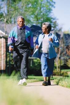
Vigorous exercise hastens knee OA progression
PARIS – Vigorous, but not moderate, physical exercise was associated with a significantly greater risk of knee osteoarthritis progression over 1 year in a longitudinal analysis of 99 patients.
Engaging in vigorous activity was associated with a 1.38-times increased risk for progression (95% confidence intervals, 1.04-1.83; P = .025), defined as an increase in either cartilage or meniscus defect scores at 1 year.
The odds ratios were not significantly increased for moderate activity (OR, 0.78; 95% CI, 0.47-1.28; P = .322) but were close to significance for walking activity (OR, 1.50; 95% CI, 1-2.25; P = .052), lead author Deepak Kumar, Ph.D., said at the World Congress on Osteoarthritis.
The analyses adjusted for age, body mass index, sex, pain, Kellgren-Lawrence (KL) score, and modified Whole Organ Magnetic Resonance Scores for cartilage and meniscus in the first block, and International Physical Activity Questionnaire (IPAQ) scores in the second block.
"We did not see that moderate activity was associated with progression, and this indicates that we need to further investigate the optimal dosage of physical activity for someone with knee osteoarthritis," said Dr. Kumar, a postdoctoral scholar, radiology department, University of California, San Francisco.
Although greater physical activity has been shown to reduce pain and improve function, the results support more recent work suggesting that certain types of activity may be associated with disease progression. Walking 10,000 steps or more per day was found to increase the risk of meniscus and cartilage lesions on MRI in people with knee OA (Ann. Rheum. Dis. 2013;72:1170-5), while high and very low levels of self-reported activity were associated with greater cartilage relaxation times on 2T MRI at 4 years in data from the Osteoarthritis Initiative (Osteoarthritis Cartilage 2013;12:1558-66).
For the current analysis, 99 participants in the ongoing, longitudinal Cartilage Loading and Unloading (CLOC) knee OA study underwent 3T MRI of the knee using a sagittal, high-resolution 3D fast spin-echo Cube sequence at baseline, which was repeated 1 year later. Cartilage and meniscus defects were graded by experienced radiologists. Participants with OA had a baseline radiographic KL score of more than 1 and were symptomatic; controls had a KL of 0 or 1 and no knee symptoms.
By 1 year, 35 participants showed progression (14 with OA and 21 controls) and 64 did not. Surprisingly, there was greater progression in the lateral compartment (11 lateral menisci, 12 lateral tibiae) and, more expectedly, in the patella in 12 persons, Dr. Kumar said at the meeting, sponsored by the Osteoarthritis Research Society International.
No significant baseline differences existed between nonprogressors and progressors with respect to age (53.2 years vs. 50.8 years), body mass index (24.4 kg/m2 vs. 24.5 kg/m2), and sex (62.5% vs. 48.5% female), he said.
Knee injury and Osteoarthritis Outcome Score subscale scores were also similar for pain (85.7 vs. 82.4) and symptoms (84.5 vs. 83.4).
Progressors had engaged, however, in significantly more metabolic equivalent-minutes per week of vigorous exercise than did nonprogressors (2,410.6 vs. 1,413.1; P = .046), Dr. Kumar said. Metabolic equivalent-minutes per week were similar for moderate activity (1,094.1 vs. 858.7; P = .396) and walking (1,646.2 vs. 1,245.1; P = .151).
During the discussion following the formal presentation, an attendee said the study is potentially very valuable because it suggests that something all clinicians want to do is "dangerous" but questioned whether some of the MRI data were "overread" given the almost 40% rate of progression in a relatively fit cohort. A Danish attendee also remarked that her group has experienced so many problems with patients filling out the IPAQ questionnaire that they no longer trust the data.
Dr. Kumar responded that the MRI readings were performed by expert radiologists and were reliable, and that more stringent definitions of progression are being explored. His group is also working on a new questionnaire to better define physical activity levels.
Data are also being analyzed from the rest of the cohort (160 participants) and over a longer, 3-year follow-up, he said in an interview.
"We are also identifying quantitative MRI and biomechanical metrics that may be more sensitive to disease progression in knee OA. These will help us understand the factors that are related to worsening of knee OA and develop therapies."
The National Institutes of Health–National Institute of Arthritis and Musculoskeletal and Skin Diseases funded the work. Dr. Kumar and his coauthors reported no conflicting interests.
AT OARSI 2014
Major finding: Vigorous activity was associated with a 1.38 times increased risk for progression (95% CI, 1.04-1.83; P = .025).
Data source: Longitudinal analysis of 99 participants in the ongoing CLOC knee OA study.
Disclosures: The National Institutes of Health–National Institute of Arthritis and Musculoskeletal and Skin Diseases funded the work. Dr. Kumar and his coauthors reported no conflicting interests.

Recent knee injuries spark rapid cascade to joint failure
PARIS – Recent knee injuries are strongly associated with accelerated knee osteoarthritis, according to an analysis from the prospective, multicenter Osteoarthritis Initiative.
"Certain injuries may initiate or coincide with an accelerated cascade towards joint failure in as little as 12 months," Jeffrey Driban, Ph.D., said at the World Congress on Osteoarthritis. "In fact, 76% of individuals with an injury and accelerated knee osteoarthritis experienced their injury in the 12 months prior to the study outcome."
The study defined accelerated knee OA as progression from a Kellgren-Lawrence grade 0 or 1 on baseline bilateral radiographs to end-stage KL grade 3 or 4 within 48 months.
Although knee OA typically has been a slowly progressive disorder, 5%-17% of patients now experience accelerated forms of OA.

"If we can better characterize this phenomenon and its potential risk factors, we can provide more insights into the nature of progression in hopes of identifying an at-risk subset," said Dr. Driban of the division of rheumatology at Tufts Medical Center, Boston.
The study by Dr. Driban and his colleagues was published in Arthritis Care & Research (2014 April 29 [doi:10.1002/acr.22359]).
A total of 1,930 participants in the Osteoarthritis Initiative, all with a KL grade of 0 or 1 on baseline bilateral radiographs, were asked at baseline and at each annual visit whether they had ever been "injured enough to limit ability to walk for at least 2 days."
On follow-up, 1,325 had no knee OA, 54 had accelerated knee OA, and 187 had typical knee OA, defined as at least one knee increased in radiographic scoring within 48 months (excluding accelerated OA).
After exclusion of 12 patients with missing data, 30% of the accelerated OA group, 28% in the typical OA group, and 35% in the no OA group had a history of knee injury before baseline. A new knee injury was reported by 32%, 13%, and 11%, respectively, with data missing from 59 persons.
In univariate analyses, participants with accelerated knee OA were significantly older than were those with typical OA or no OA (61.8 years vs. 58 years vs. 59.2 years; P = .023) and had a greater body mass index (28.9 kg/m2 vs. 27.9 kg/m2 vs. 27.1 kg/m2; P = .002), Dr. Driban said.
In multinomial logistic regression analyses that adjusted for age, sex, BMI, presence of static knee malalignment, and systolic blood pressure, there was no association between prior knee injury and accelerated OA (odds ratio, 0.84) or typical OA (OR, 0.76).
However, when the investigators looked further, participants with accelerated OA were almost 3.5 times more likely to report a recent knee injury during the observation period (OR, 3.37; 95% confidence interval, 1.82-6.25) than were those with typical OA (OR, 0.99) or no OA (reference), he said.
Moreover, if a participant experienced a knee injury 1 year before the study outcome, the risk of accelerated OA increased ninefold (OR, 9.22; CI, 4.50-18.90) versus threefold for typical OA (OR, 3.04; CI, 1.66-5.58).
Despite the focus on injuries leading to accelerated OA, the analyses can’t rule out that accelerated OA may also cause an injury or that there could be a "vicious cycle," in which an injury can cause accelerated OA, associated with joint space loss, increased symptoms, and increased risk for subsequent injury, Dr. Driban said.
This line of thought helps explain why prior injury was not associated with accelerated knee OA, but recent injury was. As patients were free of radiographic OA at baseline, those with a history of a prior injury that could cause accelerated knee OA would already have been eliminated from the study, he explained in an interview.
Secondly, if accelerated knee OA can increase the risk of injury, knee injuries from years ago would not be related to accelerated knee OA because the disorder did not exist at the time of the injury.
Finally, there also could be a recall bias, as patients often have a hard time recalling injuries that may have happened years ago.
Despite the limitations of self-reported injuries and insufficient data regarding the type, severity, status of the meniscus, mechanism, or subsequent treatment of the knee injury, the findings represent an important "starting point" in understanding the association between injuries and accelerated osteoarthritis, Dr. Driban said.
"We need to monitor older adults who report an injury, as this may initiate accelerated OA or indicate an individual experiencing accelerated OA, and we need to determine which injuries may be related to accelerated osteoarthritis," he said at the meeting, sponsored by the Osteoarthritis Research Society International.
During the discussion following the formal presentation, Dr. David Felson, professor of medicine and epidemiology at Boston University, said, "I think what you are saying is exactly right," but suggested that the investigators exclude patients with spontaneous osteonecrosis of the knee and osteochondritis dissecans, as both conditions are more common than anticipated and can drive very rapid development of OA. Conversely, inclusion of patients with osteophyte-only knee OA would increase the number likely identified with accelerated OA, he said.
Dr. Driban reported no conflicting interests.
PARIS – Recent knee injuries are strongly associated with accelerated knee osteoarthritis, according to an analysis from the prospective, multicenter Osteoarthritis Initiative.
"Certain injuries may initiate or coincide with an accelerated cascade towards joint failure in as little as 12 months," Jeffrey Driban, Ph.D., said at the World Congress on Osteoarthritis. "In fact, 76% of individuals with an injury and accelerated knee osteoarthritis experienced their injury in the 12 months prior to the study outcome."
The study defined accelerated knee OA as progression from a Kellgren-Lawrence grade 0 or 1 on baseline bilateral radiographs to end-stage KL grade 3 or 4 within 48 months.
Although knee OA typically has been a slowly progressive disorder, 5%-17% of patients now experience accelerated forms of OA.
"If we can better characterize this phenomenon and its potential risk factors, we can provide more insights into the nature of progression in hopes of identifying an at-risk subset," said Dr. Driban of the division of rheumatology at Tufts Medical Center, Boston.
The study by Dr. Driban and his colleagues was published in Arthritis Care & Research (2014 April 29 [doi:10.1002/acr.22359]).
A total of 1,930 participants in the Osteoarthritis Initiative, all with a KL grade of 0 or 1 on baseline bilateral radiographs, were asked at baseline and at each annual visit whether they had ever been "injured enough to limit ability to walk for at least 2 days."
On follow-up, 1,325 had no knee OA, 54 had accelerated knee OA, and 187 had typical knee OA, defined as at least one knee increased in radiographic scoring within 48 months (excluding accelerated OA).
After exclusion of 12 patients with missing data, 30% of the accelerated OA group, 28% in the typical OA group, and 35% in the no OA group had a history of knee injury before baseline. A new knee injury was reported by 32%, 13%, and 11%, respectively, with data missing from 59 persons.
In univariate analyses, participants with accelerated knee OA were significantly older than were those with typical OA or no OA (61.8 years vs. 58 years vs. 59.2 years; P = .023) and had a greater body mass index (28.9 kg/m2 vs. 27.9 kg/m2 vs. 27.1 kg/m2; P = .002), Dr. Driban said.
In multinomial logistic regression analyses that adjusted for age, sex, BMI, presence of static knee malalignment, and systolic blood pressure, there was no association between prior knee injury and accelerated OA (odds ratio, 0.84) or typical OA (OR, 0.76).
However, when the investigators looked further, participants with accelerated OA were almost 3.5 times more likely to report a recent knee injury during the observation period (OR, 3.37; 95% confidence interval, 1.82-6.25) than were those with typical OA (OR, 0.99) or no OA (reference), he said.
Moreover, if a participant experienced a knee injury 1 year before the study outcome, the risk of accelerated OA increased ninefold (OR, 9.22; CI, 4.50-18.90) versus threefold for typical OA (OR, 3.04; CI, 1.66-5.58).
Despite the focus on injuries leading to accelerated OA, the analyses can’t rule out that accelerated OA may also cause an injury or that there could be a "vicious cycle," in which an injury can cause accelerated OA, associated with joint space loss, increased symptoms, and increased risk for subsequent injury, Dr. Driban said.
This line of thought helps explain why prior injury was not associated with accelerated knee OA, but recent injury was. As patients were free of radiographic OA at baseline, those with a history of a prior injury that could cause accelerated knee OA would already have been eliminated from the study, he explained in an interview.
Secondly, if accelerated knee OA can increase the risk of injury, knee injuries from years ago would not be related to accelerated knee OA because the disorder did not exist at the time of the injury.
Finally, there also could be a recall bias, as patients often have a hard time recalling injuries that may have happened years ago.
Despite the limitations of self-reported injuries and insufficient data regarding the type, severity, status of the meniscus, mechanism, or subsequent treatment of the knee injury, the findings represent an important "starting point" in understanding the association between injuries and accelerated osteoarthritis, Dr. Driban said.
"We need to monitor older adults who report an injury, as this may initiate accelerated OA or indicate an individual experiencing accelerated OA, and we need to determine which injuries may be related to accelerated osteoarthritis," he said at the meeting, sponsored by the Osteoarthritis Research Society International.
During the discussion following the formal presentation, Dr. David Felson, professor of medicine and epidemiology at Boston University, said, "I think what you are saying is exactly right," but suggested that the investigators exclude patients with spontaneous osteonecrosis of the knee and osteochondritis dissecans, as both conditions are more common than anticipated and can drive very rapid development of OA. Conversely, inclusion of patients with osteophyte-only knee OA would increase the number likely identified with accelerated OA, he said.
Dr. Driban reported no conflicting interests.
AT OARSI 2014
Key clinical point: Older adults who report a knee injury should be monitored for accelerated knee OA.
Major finding: Knee injury within 1 year of the study outcome increased the odds of accelerated OA ninefold (OR, 9.22; CI, 4.50-18.90).
Data source: Person-based analyses of 1,930 participants in the Osteoarthritis Initiative.
Disclosures: Dr. Driban reported no conflicting interests.

Childhood sports knee injuries carry heavy burden
PARIS – Knee trauma from a childhood sports injury can have serious consequences in young adults, judging from preliminary results from a historical cohort.
When assessed 3-10 years after the sports injury, at an average age of 22 years, young adults appear to be at higher risk of abnormalities visible on MRI that are consistent with future arthropathy and have poorer knee-related quality of life and more knee symptoms.
They are at higher risk of having structural asymmetry of the vastus medialis muscle and being categorized as being overweight or obese.
There also appears to be some trends for increased percent body fat and reduced participation in physical activity, Jackie L. Whittaker, Ph.D., said at the World Congress on Osteoarthritis.
She stressed that these are preliminary findings and that the group hopes to quadruple the size of the cohort this year in order to better understand the relationships they’ve identified.
"With that being said, if we are seeing what we think we’re seeing, there may be some clinical, structural, physiologic, and behavioral markers that can be used to identify individuals who are at risk and target them with secondary prevention strategies at an earlier age," said Dr. Whittaker, a postdoctoral fellow and physiotherapist, University of Calgary Sport Injury Prevention Research Centre, Alberta, Canada.
The historical cohort study involved 25 patients recruited from previous sports injury epidemiology studies who had an intra-articular knee injury in ice hockey (male and female), soccer, basketball, or other sports that required medical attention and time off play, and 25 uninjured controls matched on age, sex, and sport. The median age at injury was 15 years (range 9-18 years). Eight patients in the injured group had contralateral knee surgeries, in addition to surgery on the primary knee. The median age in both groups was 22 years.
Using the Knee injury and Osteoarthritis Outcome Scores (KOOS) outcome, injured participants scored significantly lower, indicating worse function, on all five, 100-point subscales: pain (mean 93 vs. 97), symptoms (mean 82.9 vs. 92.4), activities of daily living (mean 96.3 vs. 99.3), sport/recreation (mean 90.6 vs. 97.4), and knee-related quality of life (mean 89.7 vs. 97.7), Dr. Whittaker said.
Further, in a subsample of 10 matched pairs, injured participants were three times more likely to have an MRI Osteoarthritis Score (MOAKS) of one than were matched controls, according to the study, led by her colleague Carolyn Emery, Ph.D.
Although there was no difference in quadriceps strength between groups, injured participants were 3.8 times more likely to have a difference in the cross-sectional area of the vastus medialis muscle greater than 15%.
"We think that this may be clinically relevant, as there has been some recent prospective work (Arthritis Rheum. 2012;64:3917-25) that has shown a temporal relationship between a decrease in size of the vastus medialis muscle and progression or loss of tibial joint cartilage," she said.
Within 3-10 years after their knee trauma, injured participants have a significantly higher mean body mass index (BMI) than do uninjured participants (25.4 kg/m2 vs. 23.2 kg/m2) and a trend for more body fat (20.8% vs. 18.7%).
Not surprisingly, these troubling findings were coupled with a trend for injured participants to spend less time each week participating in moderate to strenuous activity (97.8 minutes vs. 101.4 minutes) and also fewer participated in sports in the previous year (4% vs. 16%).
"If we dichotomize BMI, what we see is that those individuals in the injured group are two times more likely to have a BMI that is [classified as] overweight or obese," Dr. Whittaker said at the meeting, sponsored by the Osteoarthritis Research Society International.
During a discussion of the results, the suggestion was made to stratify future data by age at injury, as puberty has an influence on response to injury.
Dr. Whittaker reported funding from the Alberta Children’s Hospital Foundation, Alberta Innovates Health Solutions Alberta Team Osteoarthritis, Canadian Institutes of Health Research, and the University of Calgary Sport Injury Prevention Research Centre.
AT OARSI 2014
Major finding: Preliminary evidence suggests that young adults with history of a sports-related knee injury differ in symptomatology, physiology, knee muscle morphology, and joint structure from uninjured controls 3-10 years post injury.
Data source: Historical cohort of 25 sport-related knee injured and 25 uninjured young adults.
Disclosures: Dr. Whittaker reported funding from the Alberta Children’s Hospital Foundation, Alberta Innovates Health Solutions Alberta Team Osteoarthritis, Canadian Institutes of Health Research, and the University of Calgary Sport Injury Prevention Research Centre.
PCPs all over the map in conservative OA therapy use
PARIS – Use of conservative, nonpharmacologic osteoarthritis therapies was relatively low and varied considerably across 10 primary care clinics within the same U.S. health care system.
In seven of the clinics, for example, less than half of patients had ever received physical therapy for knee osteoarthritis, with a range of 24%-61%.
Physical therapy for hip OA was even less common, ranging from 0%-71%, Kelli D. Allen, Ph.D., reported at the World Congress on Osteoarthritis.
In contrast, 70%-88% of patients were currently using pain medications such as opioids or NSAIDs to treat their symptomatic hip and/or knee OA, according to the cross-sectional analysis.
Patients received care at 1 of 10 family and internal medicine Duke Primary Care clinics in North Carolina. All were overweight and not meeting physical activity recommendations. Patients had OA symptoms for an average of 10 years, their average WOMAC (Western Ontario and McMaster Osteoarthritis Index) score was 39, and average age was 63 years.
Four of the clinics were in a rural setting (population less than 20,000), one in a medium-size town (20,000-99,999), four in small cities (100,000-250,000), and one in a large city (more than 250,000). The clinics had an average of 6.4 physicians and one nurse practitioner or physician’s assistant.
Knee brace use of any type ranged from 40%-64% across clinics, Dr. Allen reported. Use of knee braces with metal supports was even lower at 0%-18%.
The relatively low use of PT and knee braces with metal supports "may signal a need for more specific treatment recommendations or guidance for consistent application," suggested Dr. Allen, a health services researcher and exercise physiologist, Duke University Medical Center and Durham VA Medical Center, both in Durham, N.C.
Knee injections were reported by 43%-70% of patients.
Intraclass correlation coefficients (ICCs) were calculated to measure between-clinic variation in treatment use, with an ICC of more than .01 indicating considerable variation.
ICCs were 0.01 for the proportion using any pain medications, 0 for opioids, NSAIDS, knee injections and knee braces, 0.02 for metal knee braces, 0.07 for knee PT, and 0.08 for hip PT, according to Dr. Allen.
Rural clinics were typically below the median for nonpharmacologic use, possibly because of fewer resources in these areas, according to the poster presentation.
Clinics with fewer patients reporting "fair" or "poor" general health, however, tended to have higher use across treatment categories.
"These OA therapies may be more highly utilized when patients have fewer competing health problems," she suggested at the meeting, sponsored by the Osteoarthritis Research Society International.
Overall, 20% of patients in the study reported having fair or poor health, but rates ranged from a low of 10% to a high of 35%.
The study was supported by the National Institute of Arthritis and Musculoskeletal and Skin Diseases. Dr. Allen reported no conflicts of interest.
In seven of the clinics, for example, less than half of patients had ever received physical therapy for knee osteoarthritis, with a range of 24%-61%.
Physical therapy for hip OA was even less common, ranging from 0%-71%, Kelli D. Allen, Ph.D., reported at the World Congress on Osteoarthritis.
In contrast, 70%-88% of patients were curre
PARIS – Use of conservative, nonpharmacologic osteoarthritis therapies was relatively low and varied considerably across 10 primary care clinics within the same U.S. health care system.
In seven of the clinics, for example, less than half of patients had ever received physical therapy for knee osteoarthritis, with a range of 24%-61%.
Physical therapy for hip OA was even less common, ranging from 0%-71%, Kelli D. Allen, Ph.D., reported at the World Congress on Osteoarthritis.
In contrast, 70%-88% of patients were currently using pain medications such as opioids or NSAIDs to treat their symptomatic hip and/or knee OA, according to the cross-sectional analysis.
Patients received care at 1 of 10 family and internal medicine Duke Primary Care clinics in North Carolina. All were overweight and not meeting physical activity recommendations. Patients had OA symptoms for an average of 10 years, their average WOMAC (Western Ontario and McMaster Osteoarthritis Index) score was 39, and average age was 63 years.
Four of the clinics were in a rural setting (population less than 20,000), one in a medium-size town (20,000-99,999), four in small cities (100,000-250,000), and one in a large city (more than 250,000). The clinics had an average of 6.4 physicians and one nurse practitioner or physician’s assistant.
Knee brace use of any type ranged from 40%-64% across clinics, Dr. Allen reported. Use of knee braces with metal supports was even lower at 0%-18%.
The relatively low use of PT and knee braces with metal supports "may signal a need for more specific treatment recommendations or guidance for consistent application," suggested Dr. Allen, a health services researcher and exercise physiologist, Duke University Medical Center and Durham VA Medical Center, both in Durham, N.C.
Knee injections were reported by 43%-70% of patients.
Intraclass correlation coefficients (ICCs) were calculated to measure between-clinic variation in treatment use, with an ICC of more than .01 indicating considerable variation.
ICCs were 0.01 for the proportion using any pain medications, 0 for opioids, NSAIDS, knee injections and knee braces, 0.02 for metal knee braces, 0.07 for knee PT, and 0.08 for hip PT, according to Dr. Allen.
Rural clinics were typically below the median for nonpharmacologic use, possibly because of fewer resources in these areas, according to the poster presentation.
Clinics with fewer patients reporting "fair" or "poor" general health, however, tended to have higher use across treatment categories.
"These OA therapies may be more highly utilized when patients have fewer competing health problems," she suggested at the meeting, sponsored by the Osteoarthritis Research Society International.
Overall, 20% of patients in the study reported having fair or poor health, but rates ranged from a low of 10% to a high of 35%.
The study was supported by the National Institute of Arthritis and Musculoskeletal and Skin Diseases. Dr. Allen reported no conflicts of interest.
In seven of the clinics, for example, less than half of patients had ever received physical therapy for knee osteoarthritis, with a range of 24%-61%.
Physical therapy for hip OA was even less common, ranging from 0%-71%, Kelli D. Allen, Ph.D., reported at the World Congress on Osteoarthritis.
In contrast, 70%-88% of patients were curre
AT OARSI 2014
Data source: Cross-sectional analysis of 537 OA patients at 10 PCP clinics.
Disclosures: The study was supported by the National Institute of Arthritis and Musculoskeletal and Skin Diseases. Dr. Allen reported no conflicting interests.
Negative moods tied to OA pain flares
PARIS – Patients with knee osteoarthritis are significantly more likely to experience a flare if they report a higher negative mood in the previous 10 days.
Passive coping strategies such as "I restrict my social activities" or "I focus on the location and intensity of pain" were also significantly associated with an increased risk of flares, while more active coping strategies were protective in a Web-based, case-crossover study.

"We really need to elaborate on this, replicate it, and elucidate this connection because psychosocial factors of mood and pain coping are modifiable and ultimately may improve our patients experience of this disease, osteoarthritis," Dr. David J. Hunter said at the World Congress on Osteoarthritis.
The investigators recruited 298 patients with symptomatic knee osteoarthritis (OA) and asked them to log on to the study website if they experienced a knee pain exacerbation over 3 months of follow-up.
Daily mood (negative/positive) in the previous 10 days was assessed using the Profile of Mood States (POMS) questionnaire. The Pain Coping Inventory (PCI) was used to assess daily pain coping in the previous 30 days.
Pain exacerbation during follow-up was defined as a 2-unit increase in the patient’s visual analog knee pain score (10-point scale) from his or her mildest pain score reported at the baseline visit, said Dr. Hunter, the Florance and Cope Chair of Rheumatology, Northern Clinical School, University of Sydney, Australia.
Most of the patients were female (61%) and white (92%), and their average body mass index was 29.4 kg/m2. Their average baseline Knee injury and Osteoarthritis Outcome Score (KOOS) symptoms subscale score was 44.5 and KOOS pain subscale score 55.6.
The average POMS positive mood score was 33.9 and average negative mood (for example, distressed, irritable, nervous) score 16.5, both on a 50-point scale. The average PCI passive coping score was 8.1 on a 24-point scale, and average PCI active coping score 12.2 on a 20-point scale.
In conditional logistic regression analyses, increasing negative affect scores were significantly associated with having a pain exacerbation (P less than .001) such that patients with a score of 13-17 were nearly three times more likely to experience a flare (odds ratio, 2.77) and those with a score of 18 or more were 6.5 times more likely to do so (OR, 6.5), Dr. Hunter said.
Patients with higher PCI passive coping scores were also significantly more likely to have a pain exacerbation (OR, 1.26; P = .01), while those with more active coping strategies such as "I stay busy or active," or "I clear my mind of bothersome thoughts" were less likely to have a pain flare (OR, 0.81; P = .03).
During a discussion of the results, Dr. Hunter said they have yet to look at trajectories to see whether patients returned to their prior pain level.
Session comoderator Aileen Davis, Ph.D., senior scientist with the Toronto Western Research Institute, said the general literature recognizes there is a psychosocial component to OA pain, but that this is the first study to make the direct link with OA pain flares.
If the results can be replicated, "It means there’s a lot more we can do in terms of coping strategies and in recognizing this a lot more often," she said in an interview at the meeting, sponsored by the Osteoarthritis Research Society International.
Since many OA patients are cared for by primary care physicians, Dr. Davis suggested they may want to have a psychosocial profile of their OA patients and push management strategies or referrals to psychologists or social workers skilled in coping strategies to patients who are "fluctuators," so they can "proactively change how they’re doing things."
Dr. Hunter reported support from the Australian National Health and Medical Research Council.
Passive coping strategies, pain, active coping strategies, psychosocial factors, mood, pain, Dr. David J. Hunter, World Congress on Osteoarthritis, symptomatic knee osteoarthritis, knee pain exacerbation, Profile of Mood States questionnaire, The Pain Coping Inventory, Knee injury and Osteoarthritis Outcome Score symptoms subscale score,
PARIS – Patients with knee osteoarthritis are significantly more likely to experience a flare if they report a higher negative mood in the previous 10 days.
Passive coping strategies such as "I restrict my social activities" or "I focus on the location and intensity of pain" were also significantly associated with an increased risk of flares, while more active coping strategies were protective in a Web-based, case-crossover study.
"We really need to elaborate on this, replicate it, and elucidate this connection because psychosocial factors of mood and pain coping are modifiable and ultimately may improve our patients experience of this disease, osteoarthritis," Dr. David J. Hunter said at the World Congress on Osteoarthritis.
The investigators recruited 298 patients with symptomatic knee osteoarthritis (OA) and asked them to log on to the study website if they experienced a knee pain exacerbation over 3 months of follow-up.
Daily mood (negative/positive) in the previous 10 days was assessed using the Profile of Mood States (POMS) questionnaire. The Pain Coping Inventory (PCI) was used to assess daily pain coping in the previous 30 days.
Pain exacerbation during follow-up was defined as a 2-unit increase in the patient’s visual analog knee pain score (10-point scale) from his or her mildest pain score reported at the baseline visit, said Dr. Hunter, the Florance and Cope Chair of Rheumatology, Northern Clinical School, University of Sydney, Australia.
Most of the patients were female (61%) and white (92%), and their average body mass index was 29.4 kg/m2. Their average baseline Knee injury and Osteoarthritis Outcome Score (KOOS) symptoms subscale score was 44.5 and KOOS pain subscale score 55.6.
The average POMS positive mood score was 33.9 and average negative mood (for example, distressed, irritable, nervous) score 16.5, both on a 50-point scale. The average PCI passive coping score was 8.1 on a 24-point scale, and average PCI active coping score 12.2 on a 20-point scale.
In conditional logistic regression analyses, increasing negative affect scores were significantly associated with having a pain exacerbation (P less than .001) such that patients with a score of 13-17 were nearly three times more likely to experience a flare (odds ratio, 2.77) and those with a score of 18 or more were 6.5 times more likely to do so (OR, 6.5), Dr. Hunter said.
Patients with higher PCI passive coping scores were also significantly more likely to have a pain exacerbation (OR, 1.26; P = .01), while those with more active coping strategies such as "I stay busy or active," or "I clear my mind of bothersome thoughts" were less likely to have a pain flare (OR, 0.81; P = .03).
During a discussion of the results, Dr. Hunter said they have yet to look at trajectories to see whether patients returned to their prior pain level.
Session comoderator Aileen Davis, Ph.D., senior scientist with the Toronto Western Research Institute, said the general literature recognizes there is a psychosocial component to OA pain, but that this is the first study to make the direct link with OA pain flares.
If the results can be replicated, "It means there’s a lot more we can do in terms of coping strategies and in recognizing this a lot more often," she said in an interview at the meeting, sponsored by the Osteoarthritis Research Society International.
Since many OA patients are cared for by primary care physicians, Dr. Davis suggested they may want to have a psychosocial profile of their OA patients and push management strategies or referrals to psychologists or social workers skilled in coping strategies to patients who are "fluctuators," so they can "proactively change how they’re doing things."
Dr. Hunter reported support from the Australian National Health and Medical Research Council.
Passive coping strategies, pain, active coping strategies, psychosocial factors, mood, pain, Dr. David J. Hunter, World Congress on Osteoarthritis, symptomatic knee osteoarthritis, knee pain exacerbation, Profile of Mood States questionnaire, The Pain Coping Inventory, Knee injury and Osteoarthritis Outcome Score symptoms subscale score,
Passive coping strategies, pain, active coping strategies, psychosocial factors, mood, pain, Dr. David J. Hunter, World Congress on Osteoarthritis, symptomatic knee osteoarthritis, knee pain exacerbation, Profile of Mood States questionnaire, The Pain Coping Inventory, Knee injury and Osteoarthritis Outcome Score symptoms subscale score,
AT OARSI 2014
Key clinical point: Clinicians may want to help OA patients with coping strategies by requesting a psychosocial profile and tailoring management strategies to it or referral to psychologists or social workers skilled in coping strategies.
Major finding: Patients with a negative affect score of 18 or more were 6.5 times more likely to experience an OA pain flare (OR, 6.5).
Data source: A case-crossover study in 298 patients with knee OA.
Disclosures: Dr. Hunter reported support from the Australian National Health and Medical Research Council.

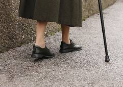
Walking disability raises red flag in diabetics with OA
PARIS – Walking disability from hip or knee osteoarthritis puts patients with comorbid diabetes at increased risk for serious diabetes complications, according to a retrospective cohort study.
After a median follow-up of 6.6 years among 437 patients with moderately severe symptomatic osteoarthritis (OA) and type 1 or type 2 diabetes, 37% experienced the composite diabetes-specific primary outcome of hospitalization for hypo- or hyperglycemia, soft tissue infection, amputation, or initiation of chronic dialysis.
Specifically, there were 51 hospitalizations for hypoglycemia, 11 for hyperglycemia, 127 for soft tissue infections, 10 amputations, and 4 patients who went on chronic dialysis.
After adjusting for age, sex, and preexisting cardiovascular disease, baseline walking disability was a significant independent predictor of risk for a non-CVD, diabetes-specific complication (Hazard ratio per unit increase in Health Assessment Questionnaire [HAQ] walking score, 1.26; P = .02), Dr. Gillian Hawker reported at the World Congress on Osteoarthritis.
In sensitivity analyses, the results were unchanged after additional adjustment for post-baseline receipt of a primary, elective hip or knee joint replacement or when retinopathy was included as an outcome.

"In people with diabetes, OA functional limitations may impede their ability to carry out diabetes self-management activities, increasing their risk for diabetes complications," noted Dr. Hawker, professor of medicine at the University of Toronto and physician-in-chief of medicine at Women’s College Hospital, Toronto.
This is particularly worrisome since the coprevalence of diabetes with OA is high, with as many as 50%-60% of patients with longstanding diabetes also having clinically evident hand, hip, or knee OA.
In an oral presentation at the same meeting, Dr. Hawker stressed the need for early identification of walking disability after reporting that greater walking disability was an independent predictor of all-cause death and major cardiovascular events in the overall cohort of patients with symptomatic hip or knee OA alone.
The retrospective cohort study linked provincial health administrative databases with surveys from a population cohort of 2,156 patients with at least moderately severe symptomatic hip or knee OA who were recruited from 1996 to 1998 through a screening survey in Ontario.
At baseline, 457 patients self-reported diabetes or met criteria for inclusion in the Ontario Diabetes Database (sensitivity 86%, specificity 97%) and were without preexisting retinopathy or renal failure. After censoring patients who died, emigrated, or had insufficient data, 434 patients were included in the current analysis. The study could not differentiate type 1 from type 2 diabetes.
Their mean age was 71.6 years (all were 55 years or older), 41% were obese, and 53.2% had preexisting CVD. Their median HAQ walking disability score was 2 on a 3-point scale, median HAQ grip score was 1 on a 3-point scale, and mean Western Ontario and McMaster Osteoarthritis Index (WOMAC) summary score was 43.4. Overall, 55.5% used a walking aid, and 39% reported using NSAIDs. (A walking disability score of 2 corresponds with walking outdoors on flat ground with much difficulty, whereas a score of 3 means the person is unable to do it. A grip score of 1 corresponds with some difficulty in just one of the following tasks: opening car doors, opening previously open jars, or turning faucets on and off.)
In multivariable analyses, no effect was found for baseline grip strength (adjusted HR, 1.16; P = .07), Dr. Hawker reported at the meeting, sponsored by the Osteoarthritis Research Society International.
However, in sensitivity analyses that further adjusted for receipt of a diabetes drug prescription or NSAID in patients at least 66 years old at baseline and thus eligible for drug benefits, both HAQ walking and grip scores were significant predictors of risk for a diabetes complication, she reported. Specific data were not shown in the poster presentation, but the adjusted hazard ratios per unit increase in HAQ were 1.36 (P = .003) and 1.26 (P = .01), respectively, according to the abstract.
"Controlling for confounders, reduced grip strength and increased walking disability were identified as potentially modifiable risk factors for serious diabetes complications in people with both OA and diabetes," the authors concluded. "Additional studies are warranted to confirm or refute our findings, and if confirmed, to elucidate potential mechanisms."
Dr. Hawker reported having no financial disclosures.
PARIS – Walking disability from hip or knee osteoarthritis puts patients with comorbid diabetes at increased risk for serious diabetes complications, according to a retrospective cohort study.
After a median follow-up of 6.6 years among 437 patients with moderately severe symptomatic osteoarthritis (OA) and type 1 or type 2 diabetes, 37% experienced the composite diabetes-specific primary outcome of hospitalization for hypo- or hyperglycemia, soft tissue infection, amputation, or initiation of chronic dialysis.
Specifically, there were 51 hospitalizations for hypoglycemia, 11 for hyperglycemia, 127 for soft tissue infections, 10 amputations, and 4 patients who went on chronic dialysis.
After adjusting for age, sex, and preexisting cardiovascular disease, baseline walking disability was a significant independent predictor of risk for a non-CVD, diabetes-specific complication (Hazard ratio per unit increase in Health Assessment Questionnaire [HAQ] walking score, 1.26; P = .02), Dr. Gillian Hawker reported at the World Congress on Osteoarthritis.
In sensitivity analyses, the results were unchanged after additional adjustment for post-baseline receipt of a primary, elective hip or knee joint replacement or when retinopathy was included as an outcome.
"In people with diabetes, OA functional limitations may impede their ability to carry out diabetes self-management activities, increasing their risk for diabetes complications," noted Dr. Hawker, professor of medicine at the University of Toronto and physician-in-chief of medicine at Women’s College Hospital, Toronto.
This is particularly worrisome since the coprevalence of diabetes with OA is high, with as many as 50%-60% of patients with longstanding diabetes also having clinically evident hand, hip, or knee OA.
In an oral presentation at the same meeting, Dr. Hawker stressed the need for early identification of walking disability after reporting that greater walking disability was an independent predictor of all-cause death and major cardiovascular events in the overall cohort of patients with symptomatic hip or knee OA alone.
The retrospective cohort study linked provincial health administrative databases with surveys from a population cohort of 2,156 patients with at least moderately severe symptomatic hip or knee OA who were recruited from 1996 to 1998 through a screening survey in Ontario.
At baseline, 457 patients self-reported diabetes or met criteria for inclusion in the Ontario Diabetes Database (sensitivity 86%, specificity 97%) and were without preexisting retinopathy or renal failure. After censoring patients who died, emigrated, or had insufficient data, 434 patients were included in the current analysis. The study could not differentiate type 1 from type 2 diabetes.
Their mean age was 71.6 years (all were 55 years or older), 41% were obese, and 53.2% had preexisting CVD. Their median HAQ walking disability score was 2 on a 3-point scale, median HAQ grip score was 1 on a 3-point scale, and mean Western Ontario and McMaster Osteoarthritis Index (WOMAC) summary score was 43.4. Overall, 55.5% used a walking aid, and 39% reported using NSAIDs. (A walking disability score of 2 corresponds with walking outdoors on flat ground with much difficulty, whereas a score of 3 means the person is unable to do it. A grip score of 1 corresponds with some difficulty in just one of the following tasks: opening car doors, opening previously open jars, or turning faucets on and off.)
In multivariable analyses, no effect was found for baseline grip strength (adjusted HR, 1.16; P = .07), Dr. Hawker reported at the meeting, sponsored by the Osteoarthritis Research Society International.
However, in sensitivity analyses that further adjusted for receipt of a diabetes drug prescription or NSAID in patients at least 66 years old at baseline and thus eligible for drug benefits, both HAQ walking and grip scores were significant predictors of risk for a diabetes complication, she reported. Specific data were not shown in the poster presentation, but the adjusted hazard ratios per unit increase in HAQ were 1.36 (P = .003) and 1.26 (P = .01), respectively, according to the abstract.
"Controlling for confounders, reduced grip strength and increased walking disability were identified as potentially modifiable risk factors for serious diabetes complications in people with both OA and diabetes," the authors concluded. "Additional studies are warranted to confirm or refute our findings, and if confirmed, to elucidate potential mechanisms."
Dr. Hawker reported having no financial disclosures.
AT OARSI 2014
Key clinical point: Early identification of walking disability is important in patients with OA and diabetes to help them to carry out diabetes self-management activities.
Major finding: Baseline walking disability was associated with an increased risk for a non-CVD, diabetes-specific complication (adjusted HR, 1.26; P = .02).
Data source: A retrospective cohort study in 437 patients with OA and diabetes.
Disclosures: Dr. Hawker reported having no financial disclosures.
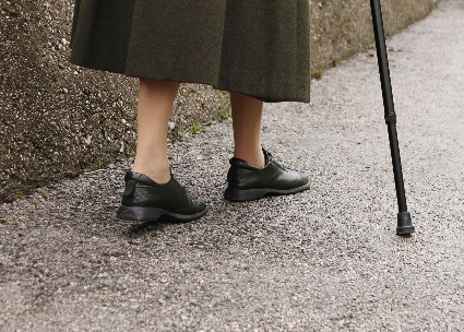
OA-related walking disability on par with diabetes as CVD risk factor
PARIS – Greater walking disability is an independent predictor of all-cause death and major cardiovascular events in patients with symptomatic hip or knee osteoarthritis, according to a population-based study that confirms previous research.
For the composite cardiovascular disease (CVD) outcome of hospitalization for angina, acute myocardial infarction, coronary revascularization, congestive heart failure, stroke, or transient ischemic attack, the risk associated with a Health Assessment Questionnaire (HAQ) walking disability score of 2 was the same as that of diabetes and pre-existing CVD.

Total joint replacement reduced those risks by about 40%, Dr. Gillian A. Hawker said at the World Congress on Osteoarthritis (OA).
"I think the key piece here is that these data at least suggest that walking disability is a cardiovascular risk factor similar to diabetes," she said. "When we’re talking about risk assessment for cardiovascular outcomes, which everyone does pretty routinely in medicine, walking disability, which is really osteoarthritis, is one of the risk factors that they should be assessing ... If we can just start asking, hopefully it will get us thinking about, ‘Wow, this person has OA,’ and we need to start assessing it."
Session comoderator Ana M. Valdes, Ph.D., of the University of Nottingham (England) commented that the results are fascinating because this is not the first study to identify these associations. In 2011, British investigators reported that all-cause mortality, particularly due to CVD causes, was significantly related to baseline walking disability in patients with knee or hip OA (Br. Med. J. 2011;342:d1165).
"One of the things that happens often with OA is that it’s seen as just pain and if we can give them plenty of relief, they’ll be OK, but it’s actually life-threatening because if we do not address their disability, they are at risk of dying," Dr. Valdes said in an interview. "I think this should be put on the front pages of the weekly medical journals, so that people are aware of the dangers of not treating osteoarthritis properly."
Comorbidity is a barrier to OA care, and 90% of those aged 65 years and older with OA have at least one other chronic condition, such as diabetes and heart disease, observed Dr. Hawker, professor of medicine at the University of Toronto and physician-in-chief of medicine, Women’s College Hospital in Toronto.
Her group reported in a separate presentation at the meeting that walking disability also raises the risk for diabetes complications in those with both OA and diabetes.
For the current population-based study, the investigators linked provincial health administrative databases to baseline surveys from a population cohort of 2,156 patients with symptomatic moderate to severe OA who were recruited in 1996-1998 through a screening survey in Ontario.
Their mean age was 71 years (all were 55 years or older), 72% were female, 34% obese, 20% had diabetes, and 40% had prebaseline CVD. Their average HAQ walking disability score was 2 on a 3-point scale, and they had a mean WOMAC (Western Ontario and McMaster Universities Osteoarthritis Index) summary score of 41. Overall, 44% used a walking aid. (A walking disability score of 2 corresponds with walking outdoors on flat ground with much difficulty, whereas a score of 3 means the person is unable to do it.)
After a median follow-up period of 13.2 years, 57% of patients had died, and 38% experienced the composite CVD outcome with a median of 9.2 years follow-up.
In multivariable analysis, there was a very clear dose-response, where increased walking disability was associated with increasing risk of all-cause death, Dr. Hawker said. The adjusted hazard ratio per unit increase in HAQ walking score was 1.30 (P less than .001), after controlling for confounders including age, sex, preexisting CVD, diabetes, hypertension, body mass index, and smoking status.
A HAQ walking score of 2 was associated with an adjusted hazard ratio of 1.69, which was actually greater than that associated with preexisting cardiovascular disease or diabetes, she said.
For the composite CVD outcome, there was also a significant association with HAQ walking score (adjusted HR, 1.17; P = .001).
Sensitivity analyses performed in 402 patients (18%) receiving postbaseline total joint arthroplasty (TJA ) showed that controlling for TJA did not change the effect of baseline disability but was protective for both all-cause death (HR, 0.62; P less than .001) and CVD events (HR, 0.66; P less than .001), Dr. Hawker said at the meeting sponsored by the Osteoarthritis Research Society International.
Although the data were not dichotomized, she noted that there was "beautiful discrimination" when they looked at the outcomes based on whether or not patients used a walking aid.
"Obviously, it’s nice to see a dose-response, but simply knowing that someone’s using a walking aid and getting them to the point where they’ve got improved walking ability, could go a long way," she said.
Though the study was not designed to address potential mechanisms, Dr. Hawker said she’s long argued that physical activity and mobility are a key player of downstream effects on fitness, blood pressure, glucose control, and ability to participate in self-management activities and physician visits. The potential role for pain, mood, and stress also can not be excluded.
One audience member asked whether walking disability may be a "cheap and dirty version" of the stress test.
Dr. Hawker responded that walking disability is a proxy for a lot of things and is definitely OA-related, adding, "Yes, I think it is a very cheap and dirty stress test. It’s a lovely thing that if we focused more explicitly on it in randomized trials, for instance as a primary outcome, or in rehabilitation strategies, it would have global benefit."
The authors reported no competing interests.
PARIS – Greater walking disability is an independent predictor of all-cause death and major cardiovascular events in patients with symptomatic hip or knee osteoarthritis, according to a population-based study that confirms previous research.
For the composite cardiovascular disease (CVD) outcome of hospitalization for angina, acute myocardial infarction, coronary revascularization, congestive heart failure, stroke, or transient ischemic attack, the risk associated with a Health Assessment Questionnaire (HAQ) walking disability score of 2 was the same as that of diabetes and pre-existing CVD.
Total joint replacement reduced those risks by about 40%, Dr. Gillian A. Hawker said at the World Congress on Osteoarthritis (OA).
"I think the key piece here is that these data at least suggest that walking disability is a cardiovascular risk factor similar to diabetes," she said. "When we’re talking about risk assessment for cardiovascular outcomes, which everyone does pretty routinely in medicine, walking disability, which is really osteoarthritis, is one of the risk factors that they should be assessing ... If we can just start asking, hopefully it will get us thinking about, ‘Wow, this person has OA,’ and we need to start assessing it."
Session comoderator Ana M. Valdes, Ph.D., of the University of Nottingham (England) commented that the results are fascinating because this is not the first study to identify these associations. In 2011, British investigators reported that all-cause mortality, particularly due to CVD causes, was significantly related to baseline walking disability in patients with knee or hip OA (Br. Med. J. 2011;342:d1165).
"One of the things that happens often with OA is that it’s seen as just pain and if we can give them plenty of relief, they’ll be OK, but it’s actually life-threatening because if we do not address their disability, they are at risk of dying," Dr. Valdes said in an interview. "I think this should be put on the front pages of the weekly medical journals, so that people are aware of the dangers of not treating osteoarthritis properly."
Comorbidity is a barrier to OA care, and 90% of those aged 65 years and older with OA have at least one other chronic condition, such as diabetes and heart disease, observed Dr. Hawker, professor of medicine at the University of Toronto and physician-in-chief of medicine, Women’s College Hospital in Toronto.
Her group reported in a separate presentation at the meeting that walking disability also raises the risk for diabetes complications in those with both OA and diabetes.
For the current population-based study, the investigators linked provincial health administrative databases to baseline surveys from a population cohort of 2,156 patients with symptomatic moderate to severe OA who were recruited in 1996-1998 through a screening survey in Ontario.
Their mean age was 71 years (all were 55 years or older), 72% were female, 34% obese, 20% had diabetes, and 40% had prebaseline CVD. Their average HAQ walking disability score was 2 on a 3-point scale, and they had a mean WOMAC (Western Ontario and McMaster Universities Osteoarthritis Index) summary score of 41. Overall, 44% used a walking aid. (A walking disability score of 2 corresponds with walking outdoors on flat ground with much difficulty, whereas a score of 3 means the person is unable to do it.)
After a median follow-up period of 13.2 years, 57% of patients had died, and 38% experienced the composite CVD outcome with a median of 9.2 years follow-up.
In multivariable analysis, there was a very clear dose-response, where increased walking disability was associated with increasing risk of all-cause death, Dr. Hawker said. The adjusted hazard ratio per unit increase in HAQ walking score was 1.30 (P less than .001), after controlling for confounders including age, sex, preexisting CVD, diabetes, hypertension, body mass index, and smoking status.
A HAQ walking score of 2 was associated with an adjusted hazard ratio of 1.69, which was actually greater than that associated with preexisting cardiovascular disease or diabetes, she said.
For the composite CVD outcome, there was also a significant association with HAQ walking score (adjusted HR, 1.17; P = .001).
Sensitivity analyses performed in 402 patients (18%) receiving postbaseline total joint arthroplasty (TJA ) showed that controlling for TJA did not change the effect of baseline disability but was protective for both all-cause death (HR, 0.62; P less than .001) and CVD events (HR, 0.66; P less than .001), Dr. Hawker said at the meeting sponsored by the Osteoarthritis Research Society International.
Although the data were not dichotomized, she noted that there was "beautiful discrimination" when they looked at the outcomes based on whether or not patients used a walking aid.
"Obviously, it’s nice to see a dose-response, but simply knowing that someone’s using a walking aid and getting them to the point where they’ve got improved walking ability, could go a long way," she said.
Though the study was not designed to address potential mechanisms, Dr. Hawker said she’s long argued that physical activity and mobility are a key player of downstream effects on fitness, blood pressure, glucose control, and ability to participate in self-management activities and physician visits. The potential role for pain, mood, and stress also can not be excluded.
One audience member asked whether walking disability may be a "cheap and dirty version" of the stress test.
Dr. Hawker responded that walking disability is a proxy for a lot of things and is definitely OA-related, adding, "Yes, I think it is a very cheap and dirty stress test. It’s a lovely thing that if we focused more explicitly on it in randomized trials, for instance as a primary outcome, or in rehabilitation strategies, it would have global benefit."
The authors reported no competing interests.
AT OARSI 2014
Key clinical point: A study confirming previous research suggests that a high level of walking disability in OA patients should be considered a strong risk factor for CVD.
Major finding: The adjusted hazard ratio per unit increase in HAQ walking score was 1.30 (P less than .001) for all-cause mortality and 1.17 (P = .001) for composite CVD events.
Data source: A population-based, cohort study in 2,156 hip and knee OA patients.
Disclosures: The authors reported no competing interests.

Intraoperative wound anesthetic cut chronic pain after hip replacement
PARIS – Adding local anesthetic to the wound during total hip replacement surgery for osteoarthritis reduced chronic pain, according to a double-blind, randomized controlled trial.
At 12 months postoperative, the number of patients with severe pain was 8.6% with standard care, compared with 1.4% among those who also received 60 mL of 0.25% bupivacaine with lidocaine (Xylocaine) 1% with adrenaline 100 mcg/20 mL injected directly into the wound prior to closure.

The difference was statistically significant (P = .004; odds ratio, 10.19), but the confidence intervals were wide (95% C.I., 2.10-49.55) because of the small number of patients with severe pain, Vikki Wylde, Ph.D., said at the World Congress on Osteoarthritis.
She noted that 7%-23% of patients report moderate to severe chronic pain in the long term after total hip replacement, according to a recent systematic review (BMJ Open 2012:2:e000435), and that the severity of postoperative pain is a known risk factor for chronic postsurgical pain.
Local anesthetic wound infiltration has been shown to be effective at reducing the severity of acute postoperative pain after total hip replacement (J. Am Coll. Surg. 2006:203:914-32), but this is the first double-blind trial to assess its long-term effects.
Investigators at high-volume orthopedic centers in the United Kingdom randomly assigned 322 patients undergoing total hip replacement for osteoarthritis to standard care (spinal anesthesia with or without general anesthesia) alone or with local anesthetic infiltration. The primary outcome was WOMAC (Western Ontario and McMaster Universities Osteoarthritis Index) pain scale scores at 12 months postoperatively.
Use of in-patient analgesia or length of hospitalization did not differ between the groups, said Dr. Wylde, a research associate with the University of Bristol, England. Acute postoperative pain severity was also similar, except that patients in the intervention group reported less severe pain on postoperative night 2 (P = .008). This was unexpected, but the study was not powered to detect differences in these secondary measures, and no conclusions can be drawn from these data, she said at the meeting, sponsored by the Osteoarthritis Research Society International.
There were no differences in other secondary measures including the WOMAC function or stiffness scales or in ICOAP (Intermittent and Constant Osteoarthritis Pain) scores.
Local anesthetic infiltration, however, significantly reduced neuropathic pain at 12 months, as assessed using the PainDETECT questionnaire, Dr. Wylde said.
"Our study suggests that local anesthetic infiltration is unlikely to change long-term pain outcomes for the majority of patients, but potentially can improve pain relief for a small number of patients who may otherwise go on to develop severe long-term pain after surgery," she concluded.
In a separate interview, Dr. Jeffrey Katz, codirector, Brigham Spine Center, Brigham & Women’s Hospital and professor at Harvard Medical School, both in Boston, said the study was very interesting mechanistically and potentially interesting from a public health standpoint because there are about 400,000 hips done a year in the United States and thus 4,000 or so of these patients are having chronic pain. Moreover, the problem of chronic pain after surgery is even more common in the knee than in the hip.
"The difference in severe pain of 8% vs. 1% or 2% is a striking difference, but it’s a small incidence, and so it does bear replication," he added. "I thought it was a very exciting study and think it’s important information for folks to begin to work with and might change practice."
Dr. Wylde reported funding from the National Institute for Health Research, London.
PARIS – Adding local anesthetic to the wound during total hip replacement surgery for osteoarthritis reduced chronic pain, according to a double-blind, randomized controlled trial.
At 12 months postoperative, the number of patients with severe pain was 8.6% with standard care, compared with 1.4% among those who also received 60 mL of 0.25% bupivacaine with lidocaine (Xylocaine) 1% with adrenaline 100 mcg/20 mL injected directly into the wound prior to closure.
The difference was statistically significant (P = .004; odds ratio, 10.19), but the confidence intervals were wide (95% C.I., 2.10-49.55) because of the small number of patients with severe pain, Vikki Wylde, Ph.D., said at the World Congress on Osteoarthritis.
She noted that 7%-23% of patients report moderate to severe chronic pain in the long term after total hip replacement, according to a recent systematic review (BMJ Open 2012:2:e000435), and that the severity of postoperative pain is a known risk factor for chronic postsurgical pain.
Local anesthetic wound infiltration has been shown to be effective at reducing the severity of acute postoperative pain after total hip replacement (J. Am Coll. Surg. 2006:203:914-32), but this is the first double-blind trial to assess its long-term effects.
Investigators at high-volume orthopedic centers in the United Kingdom randomly assigned 322 patients undergoing total hip replacement for osteoarthritis to standard care (spinal anesthesia with or without general anesthesia) alone or with local anesthetic infiltration. The primary outcome was WOMAC (Western Ontario and McMaster Universities Osteoarthritis Index) pain scale scores at 12 months postoperatively.
Use of in-patient analgesia or length of hospitalization did not differ between the groups, said Dr. Wylde, a research associate with the University of Bristol, England. Acute postoperative pain severity was also similar, except that patients in the intervention group reported less severe pain on postoperative night 2 (P = .008). This was unexpected, but the study was not powered to detect differences in these secondary measures, and no conclusions can be drawn from these data, she said at the meeting, sponsored by the Osteoarthritis Research Society International.
There were no differences in other secondary measures including the WOMAC function or stiffness scales or in ICOAP (Intermittent and Constant Osteoarthritis Pain) scores.
Local anesthetic infiltration, however, significantly reduced neuropathic pain at 12 months, as assessed using the PainDETECT questionnaire, Dr. Wylde said.
"Our study suggests that local anesthetic infiltration is unlikely to change long-term pain outcomes for the majority of patients, but potentially can improve pain relief for a small number of patients who may otherwise go on to develop severe long-term pain after surgery," she concluded.
In a separate interview, Dr. Jeffrey Katz, codirector, Brigham Spine Center, Brigham & Women’s Hospital and professor at Harvard Medical School, both in Boston, said the study was very interesting mechanistically and potentially interesting from a public health standpoint because there are about 400,000 hips done a year in the United States and thus 4,000 or so of these patients are having chronic pain. Moreover, the problem of chronic pain after surgery is even more common in the knee than in the hip.
"The difference in severe pain of 8% vs. 1% or 2% is a striking difference, but it’s a small incidence, and so it does bear replication," he added. "I thought it was a very exciting study and think it’s important information for folks to begin to work with and might change practice."
Dr. Wylde reported funding from the National Institute for Health Research, London.
AT OARSI 2014
Major finding: Severe pain at 12 months was reduced from 8.6% with standard care to 1.4% with the addition of local anesthetic wound infiltration (P = .004).
Data source: Double-blind, randomized controlled trial in 322 patients undergoing total hip replacement for osteoarthritis.
Disclosures: Dr. Wylde reported funding from the National Institute for Health Research, London.

PCPs waffle in referrals for total joint replacement
PARIS – Primary care physicians are only moderately confident about which patients to refer for total hip or knee replacement, according to cross-sectional survey of 218 physicians.
One-quarter (27.4%) were unsure about the risks of total joint arthroplasty (TJA), and 44.3% reported they were discouraged from referring patients for TJA because of a lack of clarity about surgical indications.
When asked how confident they were in deciding which patients should be referred to orthopedic surgery for consideration of hip or knee replacement, the average response was 6.9 on a 10-point scale from "not at all" to "highly confident," indicating a moderate level of confidence, Esther* Waugh, Ph.D., reported at the World Congress on Osteoarthritis.
"The results suggest the need for increased guidance for primary care physicians regarding which patient, and when, total joint arthroplasty should be considered," she concluded. "While lack of confidence does not necessarily equate with decreased competence, additional clarity may result in enhanced physician confidence, improved decision making, and ultimately better surgical outcomes through better patient selection of appropriate candidates."
A total of 376 primary care physicians (PCPs) were recruited from among those providing care to participants of an established community cohort with symptomatic hip or knee osteoarthritis (OA) in Ontario, Canada, with 218 (58%) completing the standardized questionnaire by mail or online.
One-third reported seeing more than 10 moderate to severe hip/knee OA patients in the previous 2 weeks.
Most had been in practice for more than 15 years (77%), were in group practice (63%), and were older than age 50 years (65%). One-third practiced in a rural location, and 31% had more than 10 TJA referrals per year.
Interestingly, total hip and total knee arthroplasty was perceived as "very effective" by 53% and 40% of physicians, respectively, according to Dr. Waugh of the physical therapy department at the University of Toronto.
Still, TJA was viewed as carrying high risks of death by 11.3%, of complications by 35%, and need for revision surgery by 12.3%.
Lower confidence in referring patients for TJA was independently associated with lack of clarity about surgical indications (P less than .0001; parameter estimate, –1.02) and female sex (P = .02; parameter estimate, –0.54), in multiple linear regression analysis.
Both factors were associated with fewer years of practice, she noted. Female physicians were significantly more likely to report fewer years of practice (P less than .0001), fewer OA patients (P = .0004), practicing in a group practice (P = .003), urban practice location (P = .003), and being unsure about risks (P = .02).
Confidence was not associated with uncertainty of TJA risks, volume of OA patients in the prior 2 weeks, group practice, years of practice, or rural practice setting, Dr. Waugh reported at the meeting, sponsored by the Osteoarthritis Research Society International.
Prior research by the University of Toronto team demonstrated underuse of total joint arthroplasty, but concerns have recently been raised regarding possible overuse, particularly in younger patients. In Canada, PCPs are responsible for referrals to orthopedic surgery for TJA. Thus, "inappropriate under- or overuse of TJA may be a consequence of uncertainty among PCPs about indications for TJA," the authors noted in the poster presentation
Conflict of interest disclosures were not available at press time.
*Correction, 5/8/2014: An earlier version of this story misspelled Dr. Esther Waugh's name.
AT OARSI 2014
Major finding: Lack of clarity about indications thwarted 44.3% of primary care physicians from referring patients for arthroplasty.
Data source: A cross-sectional survey of 218 primary care physicians.
Disclosures: Conflict of interest disclosures were not available at press time.
Men have more major complications after hip, knee replacement
PARIS – After total hip or knee replacement, men are at higher risk than women for heart attack, infection, and revision surgery, according to a large database analysis.
The elevated risk was particularly apparent following total knee arthroplasty (TKA), Dr. Gillian Hawker said at the World Congress on Osteoarthritis.

Prior research suggests that younger patients and males who have joint replacement are at higher risk of certain complications such as early revision, and that joint replacement is underutilized in women.
Based on the current study, underuse of total joint arthroplasty among women versus men does not appear to be explained by higher surgical risk, she said.
The investigators used administrative databases to analyze 97,445 patients who underwent primary, elective total joint arthroplasty in Ontario, Canada, between 2002 and 2009. In all, 59,564 patients (39% male) underwent TKA and 37,881 patients (46% male) had total hip arthroplasty.
Knee arthroplasty
Men had more acute myocardial infarctions within 90 days of undergoing TKA than did women (1.1% vs. 0.8%), more infection within 2 years of surgery (1.1% vs. 0.7%), and more revision TKA within 2 years (1.5% vs. 1%), said Dr. Hawker, professor of medicine at the University of Toronto and physician-in-chief of medicine, Women’s College Hospital, Toronto.
At baseline, the men were similar in age to women, but less likely to be frail (4% vs. 6.7%) and more likely to have a Charlson Comorbidity Index score of 2 or more (5.7% vs. 3.4%).
In Cox regression analysis, the risk for acute MI was significantly increased (hazard ratio, 1.79; P less than .0001) after adjustment for age, sex, income quartile, rurality, frailty, Charlson score, and hospital volume, she said.
Men were also at significantly increased risk for infection (HR, 1.67; P less than .0001) and revision TKA within 2 years (HR, 1.49; P less than .0001), after further adjustment for surgeon volume.
The investigators, led by colleague and orthopedic surgery resident Bheeshma Ravi, Ph.D., postulate that the increased risk of infection and revision among men is due to the high-impact activities that men may engage in after their prosthesis.
No sex differences were found for venous thromboembolism within 90 days or periprosthetic fracture within 2 years.
Hip arthroplasty
Men undergoing total hip replacement had higher rates of early acute MI than women (0.9% vs. 0.7%), but lower rates of periprosthetic fracture within 2 years (0.3% vs. 0.5%), Dr. Hawker said.
At baseline, male patients were younger than their female counterparts (65 years vs. 70 years) and less likely to be frail (3.5% vs. 6.6%), but more likely to have a Charlson score of 2 or more (5.3% vs. 3.7%).
After full adjustment, men were at significantly increased risk for acute MI (HR, 1.64; P less than .0001) and reduced risk for periprosthetic fracture (HR, 0.52; P = .0005), Dr. Hawker said at the meeting, which was sponsored by the Osteoarthritis Research Society International.
"We think the potential explanations for acute MI after both hip and knee replacement may be additional cardiovascular risk factors," she said. "We did not control for preexisting cardiovascular risk; that is something we are doing now."
The study excluded patients who had a pre–joint replacement fragility fracture, but because of the quality of the data postoperatively and the availability of drug benefit data, "we don’t feel we have adequate control for the presence of osteoporosis," Dr. Hawker observed.
No differences were found between sexes in the hip replacement cohort for infection, death, venous thromboembolism, dislocation, or revision.
Session comoderator Dr. Martin Englund, of Lund (Sweden) University, commented that the study was well conducted and the findings very interesting, but he cautioned against generalizing the results too broadly.
"We have seen before, that results from Sweden are not necessarily the same as in North America," he said in an interview. "I’m sure these results are probably very generalizable in Canada and in that type of health care setting, but may not be the same elsewhere. ... These are also things that might change over time. So we just need to keep monitoring outcomes and repeating these studies and adjust our treatment to what we find."
Dr. Ravi reported fellowship support from the University of Toronto, Canadian Institutes of Health Research, and Women’s College Research Institute.
PARIS – After total hip or knee replacement, men are at higher risk than women for heart attack, infection, and revision surgery, according to a large database analysis.
The elevated risk was particularly apparent following total knee arthroplasty (TKA), Dr. Gillian Hawker said at the World Congress on Osteoarthritis.
Prior research suggests that younger patients and males who have joint replacement are at higher risk of certain complications such as early revision, and that joint replacement is underutilized in women.
Based on the current study, underuse of total joint arthroplasty among women versus men does not appear to be explained by higher surgical risk, she said.
The investigators used administrative databases to analyze 97,445 patients who underwent primary, elective total joint arthroplasty in Ontario, Canada, between 2002 and 2009. In all, 59,564 patients (39% male) underwent TKA and 37,881 patients (46% male) had total hip arthroplasty.
Knee arthroplasty
Men had more acute myocardial infarctions within 90 days of undergoing TKA than did women (1.1% vs. 0.8%), more infection within 2 years of surgery (1.1% vs. 0.7%), and more revision TKA within 2 years (1.5% vs. 1%), said Dr. Hawker, professor of medicine at the University of Toronto and physician-in-chief of medicine, Women’s College Hospital, Toronto.
At baseline, the men were similar in age to women, but less likely to be frail (4% vs. 6.7%) and more likely to have a Charlson Comorbidity Index score of 2 or more (5.7% vs. 3.4%).
In Cox regression analysis, the risk for acute MI was significantly increased (hazard ratio, 1.79; P less than .0001) after adjustment for age, sex, income quartile, rurality, frailty, Charlson score, and hospital volume, she said.
Men were also at significantly increased risk for infection (HR, 1.67; P less than .0001) and revision TKA within 2 years (HR, 1.49; P less than .0001), after further adjustment for surgeon volume.
The investigators, led by colleague and orthopedic surgery resident Bheeshma Ravi, Ph.D., postulate that the increased risk of infection and revision among men is due to the high-impact activities that men may engage in after their prosthesis.
No sex differences were found for venous thromboembolism within 90 days or periprosthetic fracture within 2 years.
Hip arthroplasty
Men undergoing total hip replacement had higher rates of early acute MI than women (0.9% vs. 0.7%), but lower rates of periprosthetic fracture within 2 years (0.3% vs. 0.5%), Dr. Hawker said.
At baseline, male patients were younger than their female counterparts (65 years vs. 70 years) and less likely to be frail (3.5% vs. 6.6%), but more likely to have a Charlson score of 2 or more (5.3% vs. 3.7%).
After full adjustment, men were at significantly increased risk for acute MI (HR, 1.64; P less than .0001) and reduced risk for periprosthetic fracture (HR, 0.52; P = .0005), Dr. Hawker said at the meeting, which was sponsored by the Osteoarthritis Research Society International.
"We think the potential explanations for acute MI after both hip and knee replacement may be additional cardiovascular risk factors," she said. "We did not control for preexisting cardiovascular risk; that is something we are doing now."
The study excluded patients who had a pre–joint replacement fragility fracture, but because of the quality of the data postoperatively and the availability of drug benefit data, "we don’t feel we have adequate control for the presence of osteoporosis," Dr. Hawker observed.
No differences were found between sexes in the hip replacement cohort for infection, death, venous thromboembolism, dislocation, or revision.
Session comoderator Dr. Martin Englund, of Lund (Sweden) University, commented that the study was well conducted and the findings very interesting, but he cautioned against generalizing the results too broadly.
"We have seen before, that results from Sweden are not necessarily the same as in North America," he said in an interview. "I’m sure these results are probably very generalizable in Canada and in that type of health care setting, but may not be the same elsewhere. ... These are also things that might change over time. So we just need to keep monitoring outcomes and repeating these studies and adjust our treatment to what we find."
Dr. Ravi reported fellowship support from the University of Toronto, Canadian Institutes of Health Research, and Women’s College Research Institute.
AT OARSI 2014
Major finding: Men had more acute MIs within 90 days after total knee arthroplasty than did women (1.1% vs. 0.8%), more infection within 2 years of surgery (1.1% vs. 0.7%), and more revision surgery within 2 years (1.5% vs. 1%). After total hip replacement, men had a higher rate of early acute MI (0.9% vs. 0.7%), but less periprosthetic fracture within 2 years (0.3% vs. 0.5%).
Data source: Database analysis of 97,445 patients who received primary, elective hip or knee arthroplasty.
Disclosures: Dr. Ravi reported fellowship support from the University of Toronto, Canadian Institutes of Health Research, and Women’s College Research Institute.