Article

Reflectance Confocal Microscopy as a Diagnostic Aid in Allergic Contact Dermatitis to Mango Sap
The mango tree (Mangifera indica) produces nutrient-dense fruit that is consumed across the world. Interestingly, despite widespread...
Article
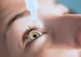
Does allergic conjunctivitis always require prescription eyedrops?
We reserve prescription drops for patients with persistent bothersome symptoms despite using over-the-counter drops.
Article
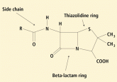
Penicillin allergy: A practical guide for clinicians
Clinical presentation is key in classifying reactions as either medicated by immunoglobulin E or not.