User login
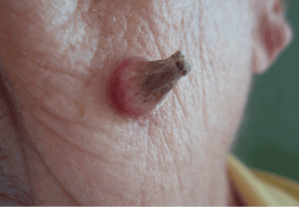
A facial cutaneous horn
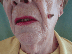
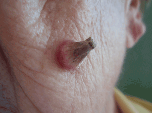
The patient says she has had intense sunlight exposure during her life, due to agricultural work.
Q: What is the most likely diagnosis?
- Basal cell carcinoma
- Cutaneous leishmaniasis
- Squamous cell carcinoma
- Viral wart
- Pyoderma gangrenosum
A: Squamous cell carcinoma is the most likely diagnosis. It is the second most common form of skin cancer (after basal cell carcinoma) that arises on the sun-exposed skin of elderly individuals.
Basal cell carcinoma usually presents as a slow-growing, pearly nodule with telangiectasias over the surface, but without a keratotic component. Cutaneous leishmaniasis is caused by a protozoan and is transmitted by the bite of a sandfly. It presents as a crusted papule or ulcer at the site of the bite, only rarely presenting as a cutaneous horn. Viral warts may present as horns (filiform warts), but the horn is slender and is usually around the lips, eyelids, or nares. Although warts can occur at any age, they are more frequently seen among school-aged children, and they peak between ages 12 and 16. Pyoderma gangrenosum is considered a reactive dermatosis sometimes secondary to a systemic disease such as inflammatory bowel disease. It usually presents as a small, red papule or pustule that progresses to a larger ulcerative lesion.
SQUAMOUS CELL CARCINOMA: CAUSES, RISK FACTORS
Exposure to ultraviolet radiation is the most common cause of squamous cell carcinoma. Other risk factors include exposure to ionizing radiation, human papilloma virus, thermal injury, burns, immunosuppression, exposure to chemical carcinogens, and diseases such as xeroderma pigmentosum, oculocutaneous albinism, and junctional epidermolysis bullosa.1,2 Organ transplantation is another significant risk factor, and cutaneous squamous cell carcinoma is the most common malignancy in renal transplant recipients.3
People born in areas that receive high amounts of ultraviolet radiation from the sun have a risk of squamous cell carcinoma three times higher than people who move to those areas in adulthood.1 Occupational exposure to ultraviolet radiation is also implicated in this type of cancer.
CLINICAL APPEARANCE
Clinically, squamous cell carcinoma of the skin may manifest as a variety of primary morphologies with or without associated symptoms. The characteristic invasive squamous cell carcinoma is a raised, firm, pink or flesh-colored keratotic papule or plaque arising on sun-exposed skin. Approximately 70% of all squamous cell carcinomas occur on the head and neck, with an additional 15% found on the upper extremities. Surface changes may include scaling, ulceration, crusting, or the presence of a cutaneous horn. Squamous cell carcinoma can also present as an eczematous patch that does not resolve with emollient or topical steroids. Less commonly, it may manifest as a pink cutaneous nodule with no overlying surface changes. Often, the patient has a background of severely sun-damaged skin, including solar elastosis, mottled dyspigmentation, telangiectasia, and multiple actinic keratoses.
MAKING THE DIAGNOSIS
Skin biopsy usually confirms the clinical diagnosis. Histologic examination shows neoplastic proliferation of atypical keratinocytes extending into the dermis, with large hyperchromatic and pleomorphic nuclei.
Imaging is not routinely indicated for diagnosing cutaneous squamous cell carcinoma. However, radiologic imaging should be obtained in patients with regional lymphadenopathy or neurologic symptoms suggestive of perineural involvement.
Other lesions found at the base of a cutaneous horn include actinic keratosis, keratoacanthoma, basal cell carcinoma, and benign lesions such as seborrheic keratosis and verruca vulgaris.4,5
MANAGEMENT
Most cutaneous squamous cell carcinomas are easily treated, and the rate of cure is high. Treatment options include topical chemotherapy, cryosurgery, electrodesiccation and curettage, surgical excision, Mohs micrographic surgery, radiotherapy, and photodynamic therapy. Fractionated radiation treatment may be preferred for patients who are unable to tolerate surgery or who have inoperable tumors, and it may provide favorable functional and cosmetic results.1
Photodynamic therapy and topical chemotherapy with imiquimod are currently not recommended for invasive squamous cell carcinoma.
Low-risk tumors are usually cured with surgical therapy. However, patients who develop one squamous cell carcinoma have a 40% risk of developing additional squamous cell carcinomas within the next 2 years. This risk is likely even greater as more time elapses. Thus, patients with a history of this cancer should be evaluated with a complete skin examination every 6 to 12 months.
OUR PATIENT’S TREATMENT
Our patient underwent total surgical excision of the lesion with a safety margin of 0.5 cm. Histologic results confirmed the diagnosis of squamous cell carcinoma with tumor-free margins. No clinical relapse was observed after 1 year of follow-up.
- Alam M, Ratner D. Cutaneous squamous-cell carcinoma. N Engl J Med 2001; 344:975–983.
- Mallipeddi R, Keane FM, Mcgrath JA, Mayou BJ, Eady RA. Increased risk of squamous cell carcinoma in junctional epidermolysis bullosa. J Eur Acad Dermatol Venereol 2004; 18:521–526.
- Rubel JR, Milford EL, Abdi R. Cutaneous neoplasms in renal transplant recipients. Eur J Dermatol 2002; 12:532–535.
- Aydogan K, Ozbek S, Balaban Adim S, Tokgöz N. Irritated seborrhoeic keratosis presenting as a cutaneous horn. J Eur Acad Dermatol Venereol 2006; 20:626–628.
- Vañó-Galván S, Sanchez-Olaso A. Images in clinical medicine. Squamous-cell carcinoma manifesting as a cutaneous horn. N Engl J Med 2008; 359:e10.
The patient says she has had intense sunlight exposure during her life, due to agricultural work.
Q: What is the most likely diagnosis?
- Basal cell carcinoma
- Cutaneous leishmaniasis
- Squamous cell carcinoma
- Viral wart
- Pyoderma gangrenosum
A: Squamous cell carcinoma is the most likely diagnosis. It is the second most common form of skin cancer (after basal cell carcinoma) that arises on the sun-exposed skin of elderly individuals.
Basal cell carcinoma usually presents as a slow-growing, pearly nodule with telangiectasias over the surface, but without a keratotic component. Cutaneous leishmaniasis is caused by a protozoan and is transmitted by the bite of a sandfly. It presents as a crusted papule or ulcer at the site of the bite, only rarely presenting as a cutaneous horn. Viral warts may present as horns (filiform warts), but the horn is slender and is usually around the lips, eyelids, or nares. Although warts can occur at any age, they are more frequently seen among school-aged children, and they peak between ages 12 and 16. Pyoderma gangrenosum is considered a reactive dermatosis sometimes secondary to a systemic disease such as inflammatory bowel disease. It usually presents as a small, red papule or pustule that progresses to a larger ulcerative lesion.
SQUAMOUS CELL CARCINOMA: CAUSES, RISK FACTORS
Exposure to ultraviolet radiation is the most common cause of squamous cell carcinoma. Other risk factors include exposure to ionizing radiation, human papilloma virus, thermal injury, burns, immunosuppression, exposure to chemical carcinogens, and diseases such as xeroderma pigmentosum, oculocutaneous albinism, and junctional epidermolysis bullosa.1,2 Organ transplantation is another significant risk factor, and cutaneous squamous cell carcinoma is the most common malignancy in renal transplant recipients.3
People born in areas that receive high amounts of ultraviolet radiation from the sun have a risk of squamous cell carcinoma three times higher than people who move to those areas in adulthood.1 Occupational exposure to ultraviolet radiation is also implicated in this type of cancer.
CLINICAL APPEARANCE
Clinically, squamous cell carcinoma of the skin may manifest as a variety of primary morphologies with or without associated symptoms. The characteristic invasive squamous cell carcinoma is a raised, firm, pink or flesh-colored keratotic papule or plaque arising on sun-exposed skin. Approximately 70% of all squamous cell carcinomas occur on the head and neck, with an additional 15% found on the upper extremities. Surface changes may include scaling, ulceration, crusting, or the presence of a cutaneous horn. Squamous cell carcinoma can also present as an eczematous patch that does not resolve with emollient or topical steroids. Less commonly, it may manifest as a pink cutaneous nodule with no overlying surface changes. Often, the patient has a background of severely sun-damaged skin, including solar elastosis, mottled dyspigmentation, telangiectasia, and multiple actinic keratoses.
MAKING THE DIAGNOSIS
Skin biopsy usually confirms the clinical diagnosis. Histologic examination shows neoplastic proliferation of atypical keratinocytes extending into the dermis, with large hyperchromatic and pleomorphic nuclei.
Imaging is not routinely indicated for diagnosing cutaneous squamous cell carcinoma. However, radiologic imaging should be obtained in patients with regional lymphadenopathy or neurologic symptoms suggestive of perineural involvement.
Other lesions found at the base of a cutaneous horn include actinic keratosis, keratoacanthoma, basal cell carcinoma, and benign lesions such as seborrheic keratosis and verruca vulgaris.4,5
MANAGEMENT
Most cutaneous squamous cell carcinomas are easily treated, and the rate of cure is high. Treatment options include topical chemotherapy, cryosurgery, electrodesiccation and curettage, surgical excision, Mohs micrographic surgery, radiotherapy, and photodynamic therapy. Fractionated radiation treatment may be preferred for patients who are unable to tolerate surgery or who have inoperable tumors, and it may provide favorable functional and cosmetic results.1
Photodynamic therapy and topical chemotherapy with imiquimod are currently not recommended for invasive squamous cell carcinoma.
Low-risk tumors are usually cured with surgical therapy. However, patients who develop one squamous cell carcinoma have a 40% risk of developing additional squamous cell carcinomas within the next 2 years. This risk is likely even greater as more time elapses. Thus, patients with a history of this cancer should be evaluated with a complete skin examination every 6 to 12 months.
OUR PATIENT’S TREATMENT
Our patient underwent total surgical excision of the lesion with a safety margin of 0.5 cm. Histologic results confirmed the diagnosis of squamous cell carcinoma with tumor-free margins. No clinical relapse was observed after 1 year of follow-up.
- Alam M, Ratner D. Cutaneous squamous-cell carcinoma. N Engl J Med 2001; 344:975–983.
- Mallipeddi R, Keane FM, Mcgrath JA, Mayou BJ, Eady RA. Increased risk of squamous cell carcinoma in junctional epidermolysis bullosa. J Eur Acad Dermatol Venereol 2004; 18:521–526.
- Rubel JR, Milford EL, Abdi R. Cutaneous neoplasms in renal transplant recipients. Eur J Dermatol 2002; 12:532–535.
- Aydogan K, Ozbek S, Balaban Adim S, Tokgöz N. Irritated seborrhoeic keratosis presenting as a cutaneous horn. J Eur Acad Dermatol Venereol 2006; 20:626–628.
- Vañó-Galván S, Sanchez-Olaso A. Images in clinical medicine. Squamous-cell carcinoma manifesting as a cutaneous horn. N Engl J Med 2008; 359:e10.