Article
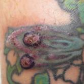
Pseudoepitheliomatous Hyperplasia Arising From Purple Tattoo Pigment
Pseudoepitheliomatous hyperplasia is a rare benign condition that can arise in response to multiple underlying triggers such as tattoo pigment.
Article
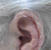
Trichodysplasia Spinulosa in the Setting of Colon Cancer
Trichodysplasia spinulosa (TS) is a rare skin condition seen in immunosuppressed patients that is characterized by folliculocentric papules with...