Article
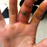
5 Points on Pyogenic Flexor Tenosynovitis of the Hand
Pyogenic flexor tenosynovitis (PFT) is a common closed space infection of the flexor tendon sheaths of the hand and remains one of the most...
Article
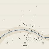
Prospective Evaluation of Opioid Consumption After Distal Radius Fracture Repair Surgery
Pain management and opioid consumption after distal radius fracture (DRF) open reduction and internal fixation (ORIF) are highly variable and...