Article
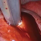
Hysterectomy in patients with history of prior cesarean delivery: A reverse dissection technique for vesicouterine adhesions
Vesicouterine adhesions resulting from prior CDs or other surgeries can distort the pelvic anatomy and present challenges during...
Article

A patient with severe adenomyosis requests uterine-sparing surgery
Combined laparoscopy and, when necessary, minilaparotomy is the authors’ preferred technique. It can relieve many symptoms of...
News
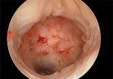
Cesarean scar defect: What is it and how should it be treated?
Hysteroscopic resection and laparoscopic repair can reduce a woman’s symptoms arising from cesarean scar defect. The technique of choice depends...