Article
Partial Flexor Tendon Laceration Assessment: Interobserver and Intraobserver Reliability
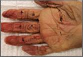
Accurate assessment of partial-thickness flexor tendon lacerations in the hand is difficult owing to the subjectivity of evaluation. In this...
Accurate assessment of partial-thickness flexor tendon lacerations in the hand is difficult owing to the subjectivity of evaluation. In this...