Article

What’s Eating You? Bedbugs
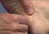
Bedbugs are an increasing problem in the workplace. Employees must be educated on the signs of a bedbug infestation and take preventive measures...
Article
Skin Cancer in Military Pilots: A Special Population With Special Risk Factors
Military pilots may be at greater risk for skin cancer, particularly melanoma. Military-specific studies are limited, but skin cancer rates in...
Article
How to Teach the Potassium Hydroxide Preparation: A Disappearing Clinical Art Form
Using potassium hydroxide (KOH) preparations in the diagnosis of superficial fungal infections is a technique that has been handed down from...