News
A Step-by-Step Guide for Diagnosing Cushing Syndrome
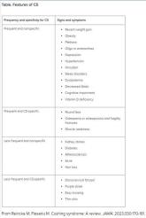
CS is defined as prolonged cortisol increase, due to exogenous steroid use or excess endogenous cortisol production.
News
PTSD Needs a New Name, Experts Say — Here’s Why
Because of stigma, many people with PTSD — especially those in the military — don’t get help.
News
‘Alarming’ Rise in Mental Health Hospital Admissions Involving Methamphetamine
“The data suggest that there are synergistic effects with methamphetamine use and mental health disorder, highlighting this patient group’s unique...
News
Atogepant May Prevent Rebound Headache From Medication Overuse in Chronic Migraine
“Treatment with atogepant may potentially decrease the risk of developing rebound headache by reducing the use of pain medications.”
News
Is Anxiety a Prodromal Feature of Parkinson’s Disease?
The risk for Parkinson’s disease was double in those with anxiety, compared with the non-anxiety group.
News
Two-Drug Combo Promising for Methamphetamine Use Disorder
Extended-release injectable naltrexone combined with extended-release oral bupropion was linked to reduced use of the drug.
News
New Clues on How Blast Exposure May Lead to Alzheimer’s Disease
A higher index of suspicion for dementia or Alzheimer’s disease may be warranted in patients with a history of blast exposure or subconcussive...
News

Early-Life Excess Weight Tied to Subsequent Stroke Risk
“Being overweight may have long-term health effects, even if the excess weight is temporary.”
News
Sharp Rise in US Pediatric ADHD Diagnoses
The prevalence estimates can be used by clinicians, policymakers, and others “to plan for the needs of children with ADHD, such as by ensuring...
News
Early Memory Problems Linked to Increased Tau
“Understanding the earliest signs of Alzheimer’s disease is even more important now that new disease-modifying drugs are becoming available.”
News
Recently Incarcerated Account for Nearly 20% of US Suicides
"Suicide prevention efforts should focus on people who have spent at least 1 night in jail in the past year."
News
Migraine Disability Nearly Doubled in US Between 2005 and 2018
Although prevalence remained roughly the same during the past 30 years, the proportion of people with moderate to severe MIDAS disability has...
News
New Expert Guidance on Antiseizure Medication Use During Pregnancy
The small risk of pregnancy-related problems is partly due to seizures and partly due to the effects of antiseizure medications.
News
Do Antipsychotic Overprescribing Warning Letters Work?
The intervention reduced quetiapine use among all patients with dementia.
News
Mental Health Worsens in Trans, Gender-Nonconforming Adults
“Mental health and primary care providers must be prepared to address the unique psychosocial needs of gender minority adults.”