Article
Biomechanical Evaluation of Two Arthroscopic Biceps Tenodesis Techniques: Proximal Interference Screw and Modified Percutaneous Intra-Articular Transtendon
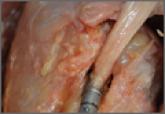
The percutaneous intra-articular transtendon (PITT) technique has recently been shown to have results comparable to those of more accepted...