User login
Researchers Puzzled by Reduced IVF Outcomes in Minority Patients
SANTA BARBARA, CALIF. – Although there are hints, researchers remain largely baffled about why ethnic minority patients have lower pregnancy and live birth rates than do whites when they undergo in vitro fertilization, especially as natural conception rates do not appear to be disparate.
Many potential explanations have been hypothesized, including differences in obesity rates, leiomyomata prevalence, inflammatory processes, and estrogen metabolism, Dr. Marcelle I. Cedars said at a meeting on in vitro fertilization and embryo transfer, which was sponsored by the University of California, Los Angeles.
None seem to fully account for diminished IVF outcomes in Asian Americans, blacks, and Hispanics. Pregnancy loss rates associated with assisted reproductive techniques also are higher for blacks and Hispanics, she noted.
Economic disparities and unequal access to IVF have been suggested as playing a role, but military studies, in which all patients have equal access to care, confirm reduced outcomes numerically – although the numbers did not quite reach statistical significance.
Dr. Cedars’ group at the University of California, San Francisco (UCSF), has focused on IVF outcomes in Asian Americans, who represent approximately a third of their patient population. Differences came under scrutiny beginning about 6 years ago.
"If we looked at live birth rate overall in any age group, we saw a significantly decreased rate in the Asian population," said Dr. Cedars, director of the in vitro fertilization program and director of the division of reproductive endocrinology and infertility at UCSF.
The clinical pregnancy rate among Asian patients also was lower than among white patients in their clinic, but the number of eggs, number of embryos, and number of high-quality embryos were similar.
"We don’t think there is something intrinsically different in terms of their ovarian reserve," she said.
Further investigation revealed higher estradiol levels during stimulation in Asian patients, even among those on a strictly controlled regimen, when "everyone gets the same dose," she noted.
Asian oocyte donors, like IVF patients, had peak serum estradiol rates 23% higher than those of white donors, but their implantation, clinical pregnancy, and live birth rates were not statistically different.
Environmental or lifestyle factors, such as higher rates of mercury exposure through fish consumption, have been hypothesized as contributing to poorer IVF outcomes, said Dr. Cedars, who also is a professor of obstetrics, gynecology, and reproductive sciences at UCSF.
But such a factor would influence spontaneous conception and pregnancy as well as IVF.
"When we looked at spontaneous pregnancies and time to pregnancy, there were absolutely no decrements in the Asian population," she said.
Indeed, Dr. Cedars believes more attention should be focused on possible differences in the way patients metabolize drugs used during ovarian stimulation. "I think we really need to think about our stimulation protocols, which were not developed with [proportional representation] of these minorities."
In the meantime, "much still remains to be explained, and this is an active area for investigation," she concluded.
Dr. Cedars reported no financial relationships relevant to her talk.
EXPERT ANALYSIS FROM A MEETING ON IN VITRO FERTILIZATION AND EMBRYO TRANSFER

Ovarian Cortex Autografts in Cancer Survivors Yield Live Births
SANTA BARBARA, CALIF. – Ovarian cortex orthotransplantation has resulted in the live births of at least 21 babies to cancer survivors in Europe, where the technique was pioneered and is being refined, Dr. Antonio Pellicer reported at a meeting on in vitro fertilization and embryo transfer.
Unlike the freezing of oocytes or embryos to preserve potential fertility, which requires ovarian stimulation, the ovarian cortex can be harvested from a cancer patient without delay, permitting immediate initiation of chemotherapy and/or radiation therapy. The tissue is cryopreserved until the patient is in remission.
If cancer treatment results in premature ovarian failure and the patient wishes to become pregnant, her autologous ovarian cortex can then be reintroduced.
Ovarian function generally resumes within 3-4 months, said Dr. Pellicer, professor of obstetrics and gynecology and dean of the medical school at the University of Valencia (Spain). Follicle stimulating hormone rates do not reach normal levels, but are sufficient in many cases for resumption of menses and pregnancy, either naturally or through assisted reproductive techniques.
The technique is currently believed to be safe for breast cancer patients and those with Hodgkin’s and non-Hodgkin’s lymphoma, based on histologic and immunologic evaluations of harvested ovarian tissue, Dr. Pellicer said at the meeting, which was sponsored by the University of California, Los Angeles.
It is considered unsafe for patients with leukemia, as metastatic cells might well circulate through the bloodstream to the ovaries. Because of its highly metastatic potential, Ewing’s sarcoma is also considered a contraindication for the procedure, according to Dr. Pellicer.
The technique offers hope, potentially, for prepubertal girls and adolescents with other types of cancer, as well as adult cancer patients, although much remains unknown about the viability and usefulness of the treatment, explained Dr. Pellicer.
At the Valencia Program of Fertility Preservation, more than 600 cancer patients from across Spain have undergone removal of the ovarian cortex around the time of diagnosis, said Dr. Pellicer.
He reported on results in 583 of those patients who were treated since 2005, 55% of whom had been diagnosed with breast cancer.
Regular menses and fertility were restored in some patients who received ovarian autografts, said Dr. Pellicer. In all, 16 pregnancies and 3 live births have occurred, some following in vitro fertilization and some following natural conception.
Those results, along with published studies from programs in France, Germany, Denmark, Belgium, and other countries, indicate that at least 21 and perhaps 23 or more live births have resulted from the technique.
The problem, as Dr. Pellicer sees it, is a lack of cohesive follow-up or evidence that would put those births into perspective.
"We don’t know the number of failed attempts," he said. "There are no registries. There are no real data. Are we doing something which is really helpful? Or are the unsuccessful cases more [typical] than the successful cases?"
"This is a concern to me," he said.
Responding to a question from an audience member, Dr. Pellicer acknowledged that the removal of one ovarian cortex prior to cancer treatment might diminish fertility potential rather than enhance it, because some cancer patients conceive naturally following remission.
Dr. Pellicer reported that he had no relevant financial relationships to disclose.
AT A MEETING ON IN VITRO FERTILIZATION AND EMBRYO TRANSFER
Major Finding: Sixteen pregnancies and three live births have occurred, some following in vitro fertilization and some following natural conception.
Data Source: This was a study of 583 patients who received ovarian autografts since 2005, 55% of whom had been diagnosed with breast cancer.
Disclosures: Dr. Pellicer reported that he had no relevant financial relationships to disclose.

Chromosomal Screening May Make Single Embryo Transfer Realistic
SANTA BARBARA, CALIF. – Routine comprehensive chromosomal screening so improves implantation rates that it makes single embryo transfer a realistic and highly cost-efficient procedure for couples undergoing in vitro fertilization, according to Dr. Richard T. Scott Jr.
In a randomized clinical trial, comprehensive chromosome screening led to successful implantation in 54 of 71 cycles (a 76.1% implantation rate), compared with 42 of 81 cycles (51.8%) in those treated in a standard fashion at Reproductive Medicine Associates of New Jersey, Morristown, the clinical center serving the division of reproductive endocrinology at the Robert Wood Johnson Medical School. Dr. Scott serves as professor and director of the division, and as clinical and scientific director of the medical group.
A 4-hour polymerase chain reaction (PCR) screening assay chromosome screen resulted in a delivery rate of 87.2% (34 of 39 patients), compared with a control rate of 68.3% (28 of 41) in a randomized trial conducted at his center, reported Dr. Scott at the conference on in vitro fertilization and embryo transfer, which was sponsored by the University of California, Los Angeles.
By upping the odds of a successful implantation and delivery, routine chromosome screening may one day outpace aneuploidy screening as the method of choice for selecting prime candidates for transfer, Dr. Scott predicted.
Indeed, his group has challenged the accuracy of aneuploidy screening, suggesting that it results in a high rate of false aneuploid results, thus lowering the transfer rate of embryos that might result in healthy infants. Along the way, the temptation builds to implant more than one embryo, despite the risks inherent in twin or higher-order multiple pregnancies, he said.
"It’s mathematically impossible for one to be as good as two," he said. "The gain approaches 25%. The bottom line is, it’s always tempting."
Dr. Scott said that the results from using comprehensive chromosomal screening for embryo selection are so superior that they might override that temptation, resulting in healthier singleton pregnancies at a reasonable cost to patients.
"I think it’s safe to say one screened embryo does as well or better than two unscreened embryos," he said. "The question is, is it cost effective?"
Answering his own question, Dr. Scott asserted, "It’s overwhelmingly and amazingly cost effective."
The cost of a singleton pregnancy, delivery, and newborn care following comprehensive chromosomal screening and IVF vs. aneuploidy screening and IVF followed by a twin pregnancy saves $1.8 million per 100 patients, or $18,700 per patient, he estimated.
The cost of infertility treatment in such an equation basically costs out at zero for couples who decide to transfer only one chromosomally selected embryo, said Dr. Scott.
The chance of abnormal gestations following comprehensive chromosomal screening is "not zero, but awfully low," he reported.
Specifically, Dr. Scott’s clinic’s clinical error rate was 0.2% in over 3,500 screened euploid embryos transferred, in more than 2,400 clinical implantations.
When products of conception were subjected to DNA fingerprinting to establish embryo origins, five abnormal gestations were traced to the procedure, including one diagnostic error (a tetraploid embryo), one trisomy 13, one Turner’s syndrome, one trisomy 21, and one healthy baby born with the opposite gender than what was predicted, through a lab error.
Dr. Scott reported that he receives grant research support and is on the speakers bureau and the scientific advisory board for EMD Serono and Ferring Pharmaceuticals. He also receives grant research support and is on the speakers bureau for Merck.
AT A CONFERENCE ON IN VITRO FERTILIZATION AND EMBRYO TRANSFER
ART Risks to Mother/Baby Likely Small, but Remain Uncertain
SANTA BARBARA, CALIF. – To err on the side of caution, couples who are concerned about the risks to mother and baby from in vitro fertilization might be advised to avoid intracytoplasmic sperm injection, to transfer frozen embryos rather than fresh, and to ask their fertility specialists to restrict the estrogen level during stimulation to less than 3,450 pg/mL on the day of HCG administration, according to Dr. Joseph C. Gambone.
The problem is, none of those guidelines is clear-cut, he said at a conference on in vitro fertilization and embryo transfer, sponsored by the University of California, Los Angeles.
Flaws in scientific methodology permeate studies that hint at elevated risks of maternal and childhood cancer, perinatal complications, and birth defects, making it difficult to advise patients about which risks are truly elevated because of assisted reproductive technology (ART).
"It’s just so enticing to get into these big databases and start fishing," said Dr. Gambone, who is in private practice in Durango, Colo. "There certainly are people who violate [valid scientific method] more than others."
Common scientific errors include inappropriate data mining, the failure to include subfertile women as controls, and the failure to consider the potential contribution of male infertility or advanced paternal age to genetic abnormalities.
Pointing to one example, Dr. Gambone reviewed a recent report from an autism conference implying an increased rate of autism among infants born through ART. However, fertile women in the general population were used as the control group, rather than subfertile women who did not undergo ART treatment prior to becoming pregnant.
Other studies failed to control for maternal age, although in vitro fertilization (IVF) mothers are generally far older than women who become pregnant on their own, and would be at higher risk for some negative outcomes based on their age alone.
That said, Dr. Gambone updated the audience on the new studies – albeit with limitations – that might give providers and prospective parents some perspective on the following IVF risks:
• Birth defects. A study based on a large Australian registry found significant differences in the rate of birth defects in ART babies, compared with the rate in babies of a control group comprising fertile and subfertile women (8.3% vs. 5.8%); the highest rate of birth defects (9.9%) was in babies conceived via intracytoplasmic sperm injection (N. Engl. J. Med. 2012;366:1803-13). No increase in risk was seen when frozen embryo transfer was employed, noted Dr. Gambone.
Accumulating data suggest that frozen embryo transfer "may be the way for us to head with this technology," he said.
• Maternal breast cancer. No overall increase in breast cancer risk was seen in women who underwent IVF in a large, population-based cohort study that was also drawn from Australian hospital and registry data. However, a 59% increase in risk was seen among younger women, defined in this study as those who started IVF at age 24 years (Fertil. Steril. 2012;98:334-40).
• Preeclampsia and SGA risks. Like many studies, a new report from the Massachusetts General Hospital in Boston found an increased risk of small-for-gestational-age infants in singleton pregnancies among women who underwent IVF (Fertil. Steril. 2012;97:1374-9). Preeclampsia rates were also higher in pregnancies associated with elevated peak serum estradiol levels during controlled ovarian hyperstimulation, but "this was interesting, and actually gave us a threshold," said Dr. Gambone. "It’s a novel and interesting way of looking at it."
Higher risk for both events was associated with elevated peak serum estradiol levels (greater than 3,450 pg/mL) on the day of HCG administration.
Dr. Gambone reported that he has no relevant financial disclosures.
AT A CONFERENCE ON IN VITRO FERTILIZATION AND EMBRYO TRANSFER SPONSORED BY UCLA
Look to the Past for Cheaper IVF Alternatives
SANTA BARBARA, CALIF. – Given the state of the economy, lower cost alternatives to in vitro fertilization appear to be gaining favor, spurring a look back into history at the potential for superovulation and intrauterine insemination to achieve pregnancy in infertile couples.
At a conference on in vitro fertilization and embryo transfer, an audience response survey found that 58% of the audience of more than 100 specialists reported that they currently performed more superovulation/intrauterine insemination (IUI) cycles than IVF cycles.
"Last year, it was a third" of the audience, said Dr. Bill Yee, medical director of Reproductive Partners Medical Group in Westminster, Calif., and a presenter at the meeting, which was sponsored by the University of California, Los Angeles.
Dr. Yee reviewed early papers on superovulation/IUI, including a series documenting a pregnancy rate of 16%/cycle in 85 patients (Fertil. Steril. 1987;48:441-5).
"That was damned good in the mid-’80s," and rivaled the results of early IVF, said Dr. Yee.
A renewed interest in the technique led Dr. Yee to investigate recent results using gonadotropin/IUI in more than 7,000 cycles among participants in IntegraMed America’s group of fertility centers that includes Dr. Yee’s offices.
In 2009, the group’s overall success rate averaged 10%/cycle in patients aged 41-42 years; 13.6% in those aged 38-40 years; 15.5% in 35- to 37-year-olds, and 20.3% in those younger than 35. Dr. Yee’s own practice’s results during 2009 and 2010 were 7.8%, 10.7%, 13.5%, and 24.3% in the same age categories, respectively.
"The overall success rate per cycle is not too bad," he concluded, noting that each cycle costs far less than an IVF cycle.
The use of clomiphene followed by IUI was less successful in Dr. Yee’s practice, achieving pregnancy rates of 6%-9% per cycle in patients younger than 40, and no pregnancies in the group older than 40.
Higher-order pregnancy – a risk of hyperstimulation and IUI – was the result in 2%-3% of pregnancies in the large IntegraMed series.
As Dr. Yee was collecting the data, a pertinent overview appeared "just by chance" on the occasion of the quarter-centennial of the technique’s use in treating infertility (Fertil. Steril. 2012;97:802-9). The overview authors compared higher-order multiple rates in 10 studies that employed high doses (150 IU or more) of gonadotropins vs. 7 studies using 75 IU or less and determined that the risk of triplets or above was "practically zero," when the lower dose was used.
Indeed, although twins resulted from as many as 29% of successful inseminations in the low-dose studies, no higher-order multiples were reported in 1,123 cycles. When higher doses of gonadotropins were used, as many as 9.3% of pregnancies involved higher-order multiples.
"Maybe the take-home message is that despite the fact the patient is 45 [years old,] maybe we should stick to 75 IU or less just to prevent the risk of higher-order pregnancies," said Dr. Yee.
Dr. Yee also discussed natural or "minimal" stimulation using clomiphene during IVF and noted that the approach can achieve a 10% pregnancy rate/cycle in so-called "poor responders" as a final alternative to using donor eggs.
"You have to individualize," he said, but such an approach would cost a couple approximately $5,000/cycle vs. $16,000-$17,000, primarily by reducing visits, procedures, and medications.
Dr. Yee made the following general recommendations for budget-conscious patients:
• In patients younger than 40 years with unexplained infertility or mild "male factor" infertility, try one cycle of clomiphene/IUI followed by one cycle of low-dose FSH/IUI.
• In poor responders who require IVF, try natural or minimal stimulation IVF with clomiphene alone as a "viable alternative" to standard IVF.
• For clomiphene IVF cycles, freezing embryos with subsequent frozen embryo transfer appears to lead to higher success rates.
Dr. Yee reported no financial disclosures relevant to his talk.
AT A CONFERENCE ON IN VITRO FERTILIZATION AND EMBRYO TRANSFER
Noninvasive Embryo Selection Forecast for IVF
SANTA BARBARA, CALIF. – Unique protein signatures in the media surrounding embryos may soon provide a noninvasive means of identifying viability and aneuploidy, Dr. William B. Schoolcraft predicted at a conference on in vitro fertilization and embryo transfer sponsored by the University of California, Los Angeles.
Biopsies performed during preimplantation genetic diagnosis or comprehensive chromosome screening are "very invasive procedures" that are comparable to surgery or a forceps delivery, said Dr. Schoolcraft.
Removal of the embryo from an incubator, exposure to the heat of a laser, and traumatic manipulation may cause subtle harm, resulting in short- or long-term complications, he added.
But fascinating developments in the laboratory have confirmed dynamic, day-by-day evolutions in the pattern of proteins that are taken up – and secreted – by embryos in culture, providing evidence of distinctive signatures indicating viability, gene expression, and prospects for implantation, Dr. Schoolcraft noted.
To date, his group has identified more than 250 proteins in spent media from embryos, 74 of which are uniquely expressed in that environment.
"Some are excreted only by early embryos, some by embryos throughout preimplantation development, and most interestingly, some proteins are just excreted by embryos on day 3 to day 5, suggesting they might be markers for viability," said Dr. Schoolcraft, medical director of the Colorado Center for Reproductive Medicine, Lone Tree.
Indeed, 14 biomarkers are differentially expressed in culture on day 5, which holds great promise for selection of the embryos that are most likely to be successfully implanted.
Nine specific proteins have been identified as candidate biomarkers, including lipocalin-1, which has proved to demonstrate increased expression in aneuploid blastocyst secretome, the material secreted from the embryo.
Significant differences in pregnancy rates have also been found based on reactive oxygen species in the spent media of day 3 embryos, highlighting "another potential marker" for embryo viability and health, said Dr. Schoolcraft.
"The concept would be that in addition to morphology – certainly not in place of morphology – we would be able to look at spent media and identify proteins" that suggest high viability, and select only those embryos that "don’t possess a signature of aneuploidy," he said.
Dr. Schoolcraft reported that he had no relevant financial disclosures.
AT A CONFERENCE ON IN VITRO FERTILIZATION AND EMBRYO TRANSFER
Cesarean-Induced Isthmoceles Eyed as Secondary Infertility Cause
SANTA BARBARA, CALIF. – Surgical hysteroscopy may be the most efficient way to restore fertility in women with cesarean-induced isthmoceles, but no randomized trials exist to guide clinicians concerning what may loom as a notable contributor to secondary infertility.
Reservoirlike pouch defects develop on the anterior wall of the uterine isthmus at the site of a cesarean section scar in an unknown percentage of women, explained Dr. Mousa Shamonki at an annual conference on in vitro fertilization and embryo transfer sponsored by the University of California, Los Angeles.
"This may be a cause of fluid accumulation in the uterine cavity, or be associated with abnormal uterine bleeding," said Dr. Shamonki, director of in vitro fertilization and assisted reproduction at the university.
Surgical correction, he added, "may correct bleeding and fluid accumulation, and potentially restore fertility."
"You’ll notice I’m being very careful in my verbiage here," he emphasized, noting that the lack of epidemiologic data or randomized trials makes the scope of the problem and its treatment unclear.
With cesarean deliveries’ constituting nearly a third of live births in the United States, any common complication is likely to powerfully impact gynecologic and obstetrical practice.
In a recent review, Dr. Pasquale Florio and his associates at the University of Siena (Italy) suggested that cesarean section–induced isthmoceles may interfere with passage of menstrual blood through the cervix, and the accumulated fluid may lead to suprapubic pelvic pain and postmenstrual abnormal uterine bleeding (Curr. Opin. Obstet. Gynecol. 2012;24:180-6).
"Moreover," the authors wrote, "persistence of the menstrual blood after menstruation in the cervix may negatively influence the mucus quality and sperm quality, obstruct sperm transport through the cervical canal, [and] interfere with embryo implantation, leading to secondary infertility."
The diagnosis of cesarean-induced isthmocele can be reliably made by transvaginal ultrasound, they stated.
In the largest treatment series to date, the same group reported in 2011 on 41 consecutive patients in whom other causes of infertility had been ruled out, and whose cesarean-induced isthmoceles were hysteroscopically excised (J. Minim. Invasive Gynecol. 2011;18:234-7).
"Believe it or not, all patients spontaneously conceived 12-24 months after isthmoplasty," said Dr. Shamonki.
Also, isthmoplasty resolved the postmenstrual abnormal uterine bleeding and suprapubic pelvic pain in all patients.
"It appears promising," he acknowledged. "But again, this is not a randomized trial. It’s very important that we get more information."
The talk generated considerable interest at the meeting, which draws reproductive medicine specialists from around the world.
In an interview, Dr. Ivan Valencia from Quito, Ecuador, estimated that in his reproductive endocrinology practice, 60%-70% of infertile patients with a history of cesarean section have evidence of an isthmocele. Deep suturing or an exaggerated healing response is likely to blame, he noted.
In such patients, he creates a small hole through the stenotic tissue on hysteroscopy that is performed without anesthesia during patients’ initial office visit. "Many patients get pregnant the next month and you don’t have to do IVF."
In others, the procedure eases the embryo transfer procedure, said Dr. Valencia.
Dr. Valencia hypothesized that isthmocele is very common following cesarean deliveries. "In some cases, this scar is not affecting them," he surmised. "They don’t come to see a specialist."
Dr. Shamonki reported that he had no relevant financial disclosures.
AT THE ANNUAL IN-VITRO FERTILIZATION AND EMBRYO TRANSFER CONFERENCE
Oncologists Favor Psychosocial Care, But Give It Short Shrift
MIAMI – Oncologists endorse the idea of connecting cancer patients to psychosocial care at the conclusion of active treatment. But practice doesn’t align with beliefs, perhaps because they are unfamiliar with where to refer their patients for care.
Among 57 oncologists who responded to a survey in the southeastern United States, 35, or 61%, considered psychosocial care to be beneficial. A majority thought it was "important" following cancer treatment, reported Laurie Freeman-Gibb at the annual conference of the American Psychosocial Oncology Society (APOS).
But the oncologists said they spent just 4.2 minutes, on average, discussing psychosocial care during consultations, according to Ms. Freeman-Gibb, a lecturer in the department of nursing at the University of Windsor in Ontario, and her colleague Dr. Andrew Hatchett, Ph.D., of the University of Louisiana at Lafayette’s department of kinesiology.
And since only about 1 in 6 oncologists responded to the survey – it was sent to 350 practitioners – the findings may present an overly optimistic picture of what happens in real-life practice when a patient leaves active treatment and returns to the community for care.
"I think it’s sometimes a time constraint," said Ms. Freeman-Gibb. "If you only have 20 minutes to see this person and you open the floodgates to what’s really going on, you might never get out the door ... especially if you don’t know whom to tell the patient to call."
Dr. Hatchett said the impetus for the study was a series of conversations he had with survivors, in which they seemed to indicate a "disconnect" in support after their active treatment ended. "It seemed as though after treatment the survivor was left to their own devices to acquire any additional help," he said.
Many oncologists told the researchers that they would like to refer survivors for follow-up psychosocial care, but they don’t know what’s available, the investigators said.
No comprehensive registry exists that would outline the locations and qualifications of therapists, exercise and rehabilitation specialists, and support agencies that specialize in the psychosocial needs of cancer survivors. In Ireland, a national registry does just that, detailing not only the services available but also their cost, said Ms. Freeman-Gibb.
The organization that sponsored the meeting, APOS, offers a free helpline intended to connect cancer patients and survivors with community counseling services and other sources of support. However, the oncologists in the survey were unaware of that resource, the investigators noted.
Development of a "network of resources" remains a goal of the researchers, who plan to conduct an expanded online survey of a larger pool of oncologists to build on the findings of their pilot questionnaire.
Having a better sense of available resources might make oncologists more comfortable bringing up survivors’ psychosocial adjustment, added Ms. Freeman-Gibb: "Their attitude is great. They say they would love to refer patients. But they don’t."
No outside funding was used to conduct the study.
FROM THE ANNUAL CONFERENCE OF THE AMERICAN PSYCHOSOCIAL ONCOLOGY SOCIETY
New Trial Proposed for Asymptomatic Stenosis
SAN DIEGO – Citing vastly improved management of carotid stenosis, the principal investigator of the Carotid Revascularization Endarterectomy vs. Stenting Trial has called for yet another trial, this one to clarify risks and benefits of carotid revascularization with either endarterectomy or stenting vs. aggressive medical management to prevent stroke in asymptomatic patients.
Every contemporary intervention to prevent strokes – endarterectomy (CEA), carotid stenting (CAS), and aggressive medical management of risk factors – is becoming safer and more efficacious, said Dr. Thomas G. Brott of the Mayo Clinic, Jacksonville, Fla., at the annual meeting of the American Neurological Association. The dilemma, according to Dr. Brott: "We don’t know how they stack up."
Current clinical practice was shaped by results of the ACAS (Asymptomatic Carotid Atherosclerosis Study) and the ACST (Asymptomatic Carotid Surgery Trial), in which CEA trumped medical management for prevention of stroke.
CREST (Carotid Revascularization Endarterectomy vs. Stenting Trial), which began enrolling only symptomatic patients, added asymptomatic subjects after publication of ACST results in 2004. In the end, it concluded that perioperative stroke and death rates were "low and similar" for CAS (2.5%) and CEA (1.4%).
Revascularization with CAS has remained somewhat controversial, with the current body of evidence suggesting the need for better control of rare, but real, complications and mortality, particularly in Medicare-aged patients.
Meanwhile, recent epidemiologic studies demonstrate profoundly lowered stroke rates without surgery or stenting, via intensive medical therapy to control risk factors such as hypertension, hyperlipidemia, and insulin resistance. For example, a population-based study in the United Kingdom found that the rate of ipsilateral stroke in medically treated patients who had carotid stenosis of 50% or greater was 0.3%.
Two new, randomized trials suggest that intensive medical therapy can indeed produce far more impressive results than anticipated in a prospective study of patients with asymptomatic carotid stenosis (Arch. Neurol. 2010;67:180-6), and even in patients with severe intracranial artery stenosis in the SAMMPRIS (Stenting vs. Aggressive Medical Management for Preventing Recurrent Stroke in Intracranial Stenosis) trial (N. Engl. J. Med. 2011;365:993-1003).
Various groups, including CMS, the American Heart Association, and the American Stroke Association, have weighed in with opinions on the management of asymptomatic patients in the hopes of preventing an estimated 5%-10% of all strokes.
The problem, Dr. Brott said, is not a lack of guidance for treatment decisions, but the lack of a direct comparison of carotid revascularization (either CEA or CAS) vs. contemporary medical therapy.
"We have evolving opinion without evolving data," he asserted.
Much is at stake, with CEA selected for 80,000-90,000 asymptomatic patients each year in the United States, and CAS for another 40,000 patients.
Dr. Brott proposed a trial to enroll 950 patients at 70 centers, with the CREST team providing the interventional arm (either endarterectomy or stenting) and the SAMMPRIS team providing the medical management arm in asymptomatic patients exhibiting at least 70% carotid stenosis by angiography or ultrasound.
The effect size of 1.2% would be equal to the absolute difference in the primary end point (periprocedural stroke and death or subsequent ipsilateral stroke) in the ACAS trial, a difference substantial enough to alter clinical practice.
Dr. Brot said he had no disclosures.
There is building consensus among vascular surgeons and other providers who manage patients with asymptomatic carotid stenosis that the results from the medical arm of ACAS are obsolete considering current medical therapy. For instance, since ACAS was published, statin use has become very prevalent. I agree that a modern trial comparing carotid intervention with medical therapy is necessary but the design of such a trial should be carefully considered.
It would be a huge mistake to fail to stratify for high-grade(around 80%) vs. moderate (around 50%-79%) stenosis with enough power in each group to draw separate conclusions. Similarly, it would be a mistake not to include morphological studies of plaque composition (calcification, hemorrhage, fibrous cap thickness, necrotic core size) which are now possible with sophisticated MRI protocols. Whether these important study design elements can be satisfied with a trial of 950 patients is unclear from this report of Dr Brott’s comments. In particular, a trial that fails to discriminate between high- and moderate-risk patients by lumping them all together will have little credibility upon completion and is likely to be money wasted.
Dr. Larry W. Kraiss is chief of Vascular Surgery at the University of Utah, Salt Lake City. and an associate medical editor of Vascular Specialist.
SAN DIEGO – Citing vastly improved management of carotid stenosis, the principal investigator of the Carotid Revascularization Endarterectomy vs. Stenting Trial has called for yet another trial, this one to clarify risks and benefits of carotid revascularization with either endarterectomy or stenting vs. aggressive medical management to prevent stroke in asymptomatic patients.
Every contemporary intervention to prevent strokes – endarterectomy (CEA), carotid stenting (CAS), and aggressive medical management of risk factors – is becoming safer and more efficacious, said Dr. Thomas G. Brott of the Mayo Clinic, Jacksonville, Fla., at the annual meeting of the American Neurological Association. The dilemma, according to Dr. Brott: "We don’t know how they stack up."
Current clinical practice was shaped by results of the ACAS (Asymptomatic Carotid Atherosclerosis Study) and the ACST (Asymptomatic Carotid Surgery Trial), in which CEA trumped medical management for prevention of stroke.
CREST (Carotid Revascularization Endarterectomy vs. Stenting Trial), which began enrolling only symptomatic patients, added asymptomatic subjects after publication of ACST results in 2004. In the end, it concluded that perioperative stroke and death rates were "low and similar" for CAS (2.5%) and CEA (1.4%).
Revascularization with CAS has remained somewhat controversial, with the current body of evidence suggesting the need for better control of rare, but real, complications and mortality, particularly in Medicare-aged patients.
Meanwhile, recent epidemiologic studies demonstrate profoundly lowered stroke rates without surgery or stenting, via intensive medical therapy to control risk factors such as hypertension, hyperlipidemia, and insulin resistance. For example, a population-based study in the United Kingdom found that the rate of ipsilateral stroke in medically treated patients who had carotid stenosis of 50% or greater was 0.3%.
Two new, randomized trials suggest that intensive medical therapy can indeed produce far more impressive results than anticipated in a prospective study of patients with asymptomatic carotid stenosis (Arch. Neurol. 2010;67:180-6), and even in patients with severe intracranial artery stenosis in the SAMMPRIS (Stenting vs. Aggressive Medical Management for Preventing Recurrent Stroke in Intracranial Stenosis) trial (N. Engl. J. Med. 2011;365:993-1003).
Various groups, including CMS, the American Heart Association, and the American Stroke Association, have weighed in with opinions on the management of asymptomatic patients in the hopes of preventing an estimated 5%-10% of all strokes.
The problem, Dr. Brott said, is not a lack of guidance for treatment decisions, but the lack of a direct comparison of carotid revascularization (either CEA or CAS) vs. contemporary medical therapy.
"We have evolving opinion without evolving data," he asserted.
Much is at stake, with CEA selected for 80,000-90,000 asymptomatic patients each year in the United States, and CAS for another 40,000 patients.
Dr. Brott proposed a trial to enroll 950 patients at 70 centers, with the CREST team providing the interventional arm (either endarterectomy or stenting) and the SAMMPRIS team providing the medical management arm in asymptomatic patients exhibiting at least 70% carotid stenosis by angiography or ultrasound.
The effect size of 1.2% would be equal to the absolute difference in the primary end point (periprocedural stroke and death or subsequent ipsilateral stroke) in the ACAS trial, a difference substantial enough to alter clinical practice.
Dr. Brot said he had no disclosures.
Major Finding:
Data Source:
Disclosures:
Chronic Inflammation Implies Perineural Invasion
SAN DIEGO – Evidence of chronic inflammation noted during Mohs surgery is a telltale sign of perineural invasion.
"If you see chronic inflammatory infiltrate within or proximal to a neurovascular bundle, look for perineural tumor," said Dr. Alexander Miller, a dermatologic surgeon in private practice in Yorba Linda, Calif. Histologically, that means abundant lymphocytes and perhaps histiocytes.
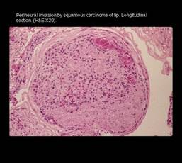
An abundance of neutrophils, however, is likely indicative of an acute inflammatory response. Perhaps even a response resulting from electrocautery during a Mohs procedure.
Neutrophils might also be present if a keratinizing tumor has ruptured into stroma, generating a microabscess. "But then it’s pretty darned obvious what you have," said Dr. Miller at a meeting sponsored by the American Society for Mohs Surgery.
Finding perineural tumor cells within a cluster of inflammatory cells can be like hunting for the proverbial needle in a haystack. A low-power view might miss them, he said. On medium or higher power views, tumor cells may appear as minute dots, a tiny stripe, or a sliver along one edge of a nerve.
To demonstrate, Dr. Miller displayed a slide depicting voluminous chronic inflammation surrounding an artery, vessel, and nerve.
"No tumor," he said.
"But if one looks carefully, two sections down, same slide, same patient, lo and behold there’s the tumor. Complacency should not be had here. One has to ensure that particularly when there’s inflammation, one needs to look very carefully at all sections of the slide."
Dr. Miller reported having no financial disclosures relevant to his talk.
SAN DIEGO – Evidence of chronic inflammation noted during Mohs surgery is a telltale sign of perineural invasion.
"If you see chronic inflammatory infiltrate within or proximal to a neurovascular bundle, look for perineural tumor," said Dr. Alexander Miller, a dermatologic surgeon in private practice in Yorba Linda, Calif. Histologically, that means abundant lymphocytes and perhaps histiocytes.
An abundance of neutrophils, however, is likely indicative of an acute inflammatory response. Perhaps even a response resulting from electrocautery during a Mohs procedure.
Neutrophils might also be present if a keratinizing tumor has ruptured into stroma, generating a microabscess. "But then it’s pretty darned obvious what you have," said Dr. Miller at a meeting sponsored by the American Society for Mohs Surgery.
Finding perineural tumor cells within a cluster of inflammatory cells can be like hunting for the proverbial needle in a haystack. A low-power view might miss them, he said. On medium or higher power views, tumor cells may appear as minute dots, a tiny stripe, or a sliver along one edge of a nerve.
To demonstrate, Dr. Miller displayed a slide depicting voluminous chronic inflammation surrounding an artery, vessel, and nerve.
"No tumor," he said.
"But if one looks carefully, two sections down, same slide, same patient, lo and behold there’s the tumor. Complacency should not be had here. One has to ensure that particularly when there’s inflammation, one needs to look very carefully at all sections of the slide."
Dr. Miller reported having no financial disclosures relevant to his talk.
EXPERT ANALYSIS FROM A MEETING SPONSORED BY THE AMERICAN SOCIETY FOR MOHS SURGERY