Article

Punked By the Punctum: Domestically Acquired Cutaneous Myiasis
Cutaneous myiasis is a skin infestation with dipterous larvae that feed on the host’s tissue and cause a wide range of manifestations depending on...
Article

Pityriasis Rosea Associated With COVID-19 Vaccination: A Common Rash Following Administration of a Novel Vaccine
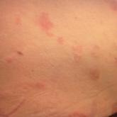
We report a clinically typical case of pityriasis rosea that developed following COVID-19 vaccination.