Article
Bullous Systemic Lupus Erythematosus Successfully Treated With Rituximab
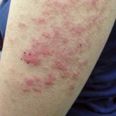
Bullous systemic lupus erythematosus can present with a waxing and waning course punctuated by flares. This case illustrates the role of rituximab...
Bullous systemic lupus erythematosus can present with a waxing and waning course punctuated by flares. This case illustrates the role of rituximab...