Article

Lung cancer screening: New evidence, updated guidance
Emerging evidence supports lower thresholds for age and smoking history when screening for lung cancer. Here’s how the USPSTF and others have...
Article

Plantar Fasciitis: How Best to Treat?
In addition to stretching exercises and orthotics, consider steroid injections as part of your first-line treatment options. For recalcitrant pain...
Article
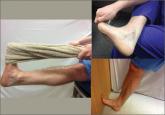
Plantar fasciitis: How best to treat?
In addition to stretching exercises and orthotics, consider steroid injections as part of your first-line treatment options. For recalcitrant pain...