Affiliations
Bridgeport Hospital and Yale University School of Medicine, Bridgeport, Connecticut
Given name(s)
Cristina
Family name
Gheorghe
Degrees
MD

Rapid Bedside Diagnosis of Shock

Article Type
Changed
Mon, 01/02/2017 - 19:34
Display Headline
Accuracy of bedside physical examination in distinguishing categories of shock

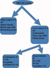
Shock has been defined as failure to deliver and/or utilize adequate amounts of oxygen1 and is a common cause of critical illness. Few studies have examined the predictive utility of bedside clinical examination in predicting the category of shock. Scholars have suggested a bedside approach that uses simple examination techniques and applied physiology to rapidly identify a patients' circulation as high vs. low cardiac output. Those with a high‐output examination are designated as high‐output, most often septic shock. Low‐output patients are further categorized as heart full or heart empty to distinguish cardiogenic from hypovolemic categories of shock, respectively.2 The predictive characteristics of this simple algorithm have not been studied. In this study, we examine the operating characteristics of selected elements of this algorithm when administered at the bedside by trainees in Internal Medicine.

Methods

This study was performed after approval of the Institutional Review Board; informed consent was waived. Patients with nonsurgical problems who present to the hospital or who develop sustained hypotension are managed by medical house officers on the intensive care and/or rapid response team with the supervision of patients' attending physicians. All house officers were asked to document explicitly in their assessment notes the following examination findings: finger capillary refill (same/quicker vs. slower than examiner's), hand skin temperature (same/warmer vs. cooler than examiner's) and pulse pressure (ie, same/wider vs. thinner than examiner's), presence or absence of crackles >1/3 from base on bilateral lung examination and jugular venous pressure (JVP) vs. <8 cmH2O. The documented examinations of either the rapid response team (PGY2; n = 14) or intensive care unit (ICU) resident (PGY3; n = 14) for patients evaluated between September 2008 and February 2009 were used for this study. Resuscitation was administered entirely by house officers, occasionally guided in person, but always supervised by attending physicians.

In May 2009, clinical data, including electrocardiograms/echocardiograms and laboratory (eg, cardiac enzymes, culture) results were abstracted from medical records of subjects. These were presented to a blinded senior clinician (DK) to review and apply evidence‐based or consensus criteria,36 whenever possible, to categorize the type of shock (septic vs. cardiogenic vs. hypovolemic) based on data acquired after the onset of shock. For example, patients with microbiologic and/or radiologic evidence of infection were classified as septic shock,1, 3, 4 those with acute left or right ventricular dysfunction on echocardiogram were classified as cardiogenic shock,1, 6 and those with clinical evidence of acute hemorrhage with hypovolemic shock.1, 5 While some of the patients were examined by DK as part of clinical care, he was blinded to the identity of patients and their algorithm‐related physical examination findings when he reviewed the abstracted data (>2 months after study closure) to adjudicate the final diagnosis of shock. These diagnoses were considered the reference standard for this study. The operating characteristics (sensitivity = true positive/true positive + false negative; specificity = true negative/true negative + false positive; negative predictive value (NPV) = true negative/all negatives; positive predictive value (PPV) = true positive/all positives; accuracy = true results/all results) were calculated for combinations of physical examination findings and correct final diagnosis (Figure 1).

Figure 1
Bedside algorithm for defining various categories of shock (ie, high output, low output heart empty [hypovolemic], low output heart full [cardiogenic]).

Results

A total of 68 patients, averaging 71 16 years, were studied; 57% were male, and 66% were White, and 20% were Black. Table 1 lists characteristics of patients. A total of 37 patients were diagnosed as having septic shock, 11 had cardiogenic shock and 10 hypovolemic shock. Operating characteristics of the bedside examination techniques for predicting mechanism of shock are listed in Table 2. Capillary refill and skin temperature were 100% concordant yielding sensitivity of 89% (95% confidence interval [CI], 75‐97%), specificity of 68% (95% CI, 46‐83%), PPV of 77% (95% CI, 61‐88%), NPV of 84% (95% CI, 64‐96%) and overall accuracy of 79% (95% CI, 68‐88%) for diagnosis of high output (ie, septic shock). JVP 8 cmH2O was more accurate than crackles for predicting cardiogenic shock in low‐output patients with sensitivity of 82% (95% CI, 48‐98%), specificity of 79% (95% CI, 41‐95%), PPV of 75% (95% CI, 43‐95%), NPV of 85% (95% CI, 55‐98%), and overall accuracy of 80% (95%CI, 59‐93%). Using just skin temperature and JVP, the bedside approach misdiagnosed 19 of 75 cases (overall accuracy, 75%; 95% CI, 16‐37%).

Characteristics of Patients and Their Final Diagnoses
n Total
  • Abbreviations: AMI, acute myocardial infarction; SIRS, systemic inflammatory response syndrome.

Gender, n (%) n = 68
Male 39 (57)
Age, years 71 16
Race, n (%)
White 45 (66)
Black 15 (22)
Hispanic 7 (10)
Other 1 (2)
High output, n (%) n = 37
Sepsis
Pneumonia 10 (27)
Urinary tract 17 (46)
Skin 3 (8)
Gastrointestinal 5 (14)
Non‐infectious SIRS 2 (5)
Low output heart full, n (%) n = 18
Pulmonary embolism 3 (16)
AMI 7 (40)
Cardiomyopathy 5 (28)
Rhythm disturbance 3 (16)
Low output heart empty, n (%) n = 13
Hemorrhagic 9 (70)
NPO 1 (7)
Diarrhea 2 (14)
Adrenal crisis 1 (7)
Predictive Characteristics of Bedside Examination for SIRS and Cardiogenic (vs. Hypovolemic) Shock
Prediction of SIRS Capillary Refill Same/Faster (%) Skin Same/ Warm (%) Bounding Pulses (%)
  • Abbreviations: JVP, jugular venous pressure; SIRS, systemic inflammatory response syndrome.

Sensitivity 89 89 65
Specificity 68 68 74
Accuracy 79 79 69
Prediction of SIRS Capillary Refill Same/Faster + Warm Skin + Bounding Pulse (%) Capillary Refill Same/Faster + Warm Skin (%) Any Other Combination of 2 (%)
Sensitivity 62 89 62
Specificity 74 68 74
Accuracy 67 79 67
Prediction of Cardiogenic JVP (%) Crackles (%) JVP + Crackles (%)
Sensitivity 82 55 55
Specificity 79 71 100
Accuracy 80 64 80

Discussion

This is the first study to examine the predictive characteristics of simple bedside physical examination techniques in correctly predicting the category/mechanism of shock. Overall, the algorithm performed well, and accurately predicted the category of shock in three‐quarters of patients. It also has the benefit of being very rapid, taking only seconds to complete, using bedside techniques that even inexperienced clinicians can apply.

Very few studies have examined the accuracy of examination techniques specifically for diagnosis of shock. In humans injected with endotoxin, body temperature and cardiac output increased, but skin temperature and capillary refill times are not well described.79 Schriger and Baraff10 reported that capillary refill >2 seconds was only 59% sensitive for diagnosing hypovolemia in patients with hypovolemic shock or orthostatic changes in blood pressure. Sensitivity was 77% in 13 patients with hypovolemic shock.10 However, some studies have demonstrated that age, sex, external temperature11 and fever12 can affect capillary refill times. Otieno et al.13 demonstrated a kappa statistic value of 0.49 for capillary refill 4 seconds, suggesting that reproducibility of this technique could be a major drawback. McGee et al.14 reviewed examination techniques for diagnosing hypovolemic states and concluded that postural changes in heart rate and blood pressure were the most accurate; capillary refill was not recommended. Stevenson and Perloff15 demonstrated that crackles and elevated JVP were absent in 18 of 43 patients with pulmonary capillary wedge pressures >22 mmHg. Butman et al.16 showed that elevated JVP was 82% accurate for predicting a wedge pressure >18 mmHg. Connors et al.17 demonstrated that clinicians' predictions of heart filling pressures and cardiac output were accurate (relative to pulmonary artery catheter measurements) in less than 50% of cases, though the examination techniques used were not qualified or quantified. No previous study has combined simple, semiobjective physical examination techniques for the purpose of distinguishing categories of shock.

Since identification of the pathogenesis of shock has important treatment/prognostic implications (eg, fluid and vasopressor therapies, early search for drainable focus of infection in sepsis, reestablishing vessel patency in myocardial infarction and pulmonary embolus), we believe that this simple, rapidly administered algorithm will prove useful in clinical medicine. In some clinical situations, the approach can lead to timely identification of the causative mechanism, allowing prompt definitive treatment. For example, a patient presenting with high‐output hypotension is so often sepsis/septic shock that treatment with antibiotics is justified (since success is time‐sensitive) even when the exact site/microbe has not yet been identified. Acute right heart overfilled low‐output hypotension should be considered pulmonary embolism (which also requires time‐sensitive therapies) until proven otherwise. Yet, a sizeable number of cases do not fit neatly into a single category. For example, 11% of patients with septic shock presented with cool extremities in the early phases of illness. In clinical decision‐making, 2 diagnostic‐therapeutic paradigms are common. In the first, the diagnosis is relatively certain and narrowly‐directed, mechanism‐specific treatment is appropriate. The second paradigm is 1 of significant uncertainty, when clinicians must treat empirically the most likely causes until more data become available to permit safe narrowing of therapies. For example, a patient presenting with hypotension, cool extremities, leukocytosis and apparent pneumonia should be treated empirically for septic shock while exploring explanations for the incongruous low‐output state (eg, profound hypovolemia, adrenal insufficiency, concurrent or precedent myocardial dysfunction). Patients often have several mechanisms contributing to hypotension. Since patients are not ideal forms, there can be no perfect decision‐tool; clinicians would be fool‐hardy to prematurely close decision‐making prior to definitive diagnosis. In the case of shock, such diagnostic arrogance would delay time‐sensitive therapies and thus contribute to morbidity and mortality. Nonetheless, this physical examination algorithmunderstanding its operating characteristics and limitationsmay add to the bedside clinician's diagnostic armamentarium.

Our study has several notable limitations. First, bedside examinations were performed by multiple observers who had limited (1 electronic mail) instruction on how to perform and document the data gathered for this study. So these results should be generalized cautiously until reproduced at other centers with greater numbers of observers (than the 28 of this study). The central supposition, that skin cooler, capillary refill longer, and pulse pressure more narrow than theirs, presupposes reasonable homogeneity of the normal state which is not necessarily true.11 Interobserver variability of physical examination further compromises the fidelity of findings recorded for this study.13 Since we conducted a retrospective review, and because of the emergency nature of the clinical problem, it would be difficult to conduct a study in which multiple examiners performed the same physical examinations to quantify interobserver variability. Irrespective, we would expect interobserver variability to systematically reduce accuracy; it is all‐the‐more impressive that trainees' examination results correctly diagnosed mechanism of shock in three‐quarters of cases. Also, examiners were not blinded to clinical history, so results of their examination could have been biased by their pre‐examination hypotheses of pathogenesis. Of course, they were not aware of the expert's final categorization of mechanism performed much later in time. Since there is no absolute reference standard for classification of the pathogenesis of shock, we depended upon careful review of selected data (same parameters for each patient) by a single senior investigatoralbeit armed with evidence‐based or consensus‐based standards of diagnosing shock. Finally, it can be argued that all forms of shock are mixed (with hypovolemia) early in the course; sepsis requires refilling of a leaky and dilated vasculature and the noncompliant ischemic ventricle often requires a higher filling pressure than normal to empty. To complicate even more, patients may have preexistent conditions (eg, chronic congestive heart failure, cirrhosis) that limit cardiovascular responses to acute shock. Our diagnostic approach was to identify the principal cause of the acute decompensation, assuming that many patients will have more than 1 single mechanism accounting for hypotension.

In conclusion, this is the first study to examine the utility of this simple physical examination algorithm to diagnose the mechanism of shock. Some have discounted or underemphasized examination techniques in favor of more time‐intensive and labor‐intensive diagnostic modalities, such as bedside echocardiography, which may waste precious time and resources. The simple physical examination algorithm assessed in this study has favorable operating characteristics and can be performed readily by even novice clinicians. If replicated at other centers and by greater numbers of observers, this approach could assist clinicians and teachers who train clinicians to rapidly diagnose and manage patients with shock.

References
  1. Antonelli M,Levy M,Andrews PJD, et al.Hemodynamic monitoring in shock and implications for management. International consensus conference. Paris, France, 27–28 April 2006.Intensive Care Med.2007;33:575590.
  2. Wood LDH.The pathophysiology of the circulation in critical illness. In:Principles of Critical Care.New York:McGraw Hill;2005.
  3. Levy MM,Fink MP,Marshall JC, et al.2001 SCCM/ESICM/ACCP/ATS/SIS. International Sepsis Definitions Conference.Crit Care Med.2003;31:12501256.
  4. Bone RC,Balk RA,Cerra FB, et al.Definitions for sepsis and organ failure and guidelines for the use of innovative therapies in sepsis.Chest.1992;101:16441655;
  5. Shoemaker WC,Peitzman AB,Bellamy R, et al.Resuscitation from severe hemorrhage.Crit Care Med.1996;24(2 Suppl):S12S23.
  6. Reynolds HR,Hochman JS.Cardiogenic shock: current concepts and improving outcomes circulation.Circulation.2008;117:686697.
  7. Suffredini AF,Fromm RE,Parker MM, et al.The cardiovascular response of normal humans to the administration of endotoxin.N Engl J Med.1989;321:280287.
  8. van Deventer SJ,Buller HR,ten Cate JW,Aarden LA,Hack CE,Sturk A.Experimental endotoxemia in humans: análysis of cytokine release and coagulation, fibrinolytic, an complement pathways.Blood.1990;76:25202526.
  9. Feemster J,Idezuki Y,Bloch J,Lillehei R,Dietzman RH.Peripheral resistance changes during shock in man.Angiology.1968;19:268276.
  10. Schriger DL,Baraff LJ.Capillary refill—is it a useful predictor of hypovolemic states?Ann Emerg Med.1991;20:601605.
  11. Schriger DL,Baraff L.Defining normal capillary refill: variation with age, sex, and temperature.Ann Emerg Med.1988;17:113116.
  12. Gorelick MH,Shaw KN,Murphy KO,Baker MD.Effect of fever on capillary refill time.Pediatr Emerg Care.1997;13:305307.
  13. Otieno H,Were E,Ahmed I,Charo E,Brent A,Maitland K.Are bedside features of shock reproducible between different observers?Arch Dis Child.2004;89:977979.
  14. McGee S,Abernethy WB,Simel DL.Is this patient hypovolemic?JAMA.1999;281:10221029.
  15. Stevenson LW,Perloff JK.The limited reliability of physical signs for estimating hemodynamics in chronic heart failure.JAMA.1989;261:884888.
  16. Butman SM,Ewy GA,Standen JR,Kern KB,Hahn E.Bedside cardiovascular examination in patients with severe chronic heart failure: importance of rest or inducible jugular venous distension.J Am Coll Cardiol.1993;22:968974.
  17. Connors AF,McCaffree DR,Gray BA.Evaluation of right‐heart catheterization in the critically ill patients without acute myocardial infarction.N Engl J Med.1983;308(5):263267.
Article PDF
Issue
Journal of Hospital Medicine - 5(8)
Publications
Page Number
471-474
Legacy Keywords
cardiogenic shock, hemorrhage, hypovolemic shock, physical examination, sepsis, septic shock, shock
Sections
Article PDF
Article PDF

Shock has been defined as failure to deliver and/or utilize adequate amounts of oxygen1 and is a common cause of critical illness. Few studies have examined the predictive utility of bedside clinical examination in predicting the category of shock. Scholars have suggested a bedside approach that uses simple examination techniques and applied physiology to rapidly identify a patients' circulation as high vs. low cardiac output. Those with a high‐output examination are designated as high‐output, most often septic shock. Low‐output patients are further categorized as heart full or heart empty to distinguish cardiogenic from hypovolemic categories of shock, respectively.2 The predictive characteristics of this simple algorithm have not been studied. In this study, we examine the operating characteristics of selected elements of this algorithm when administered at the bedside by trainees in Internal Medicine.

Methods

This study was performed after approval of the Institutional Review Board; informed consent was waived. Patients with nonsurgical problems who present to the hospital or who develop sustained hypotension are managed by medical house officers on the intensive care and/or rapid response team with the supervision of patients' attending physicians. All house officers were asked to document explicitly in their assessment notes the following examination findings: finger capillary refill (same/quicker vs. slower than examiner's), hand skin temperature (same/warmer vs. cooler than examiner's) and pulse pressure (ie, same/wider vs. thinner than examiner's), presence or absence of crackles >1/3 from base on bilateral lung examination and jugular venous pressure (JVP) vs. <8 cmH2O. The documented examinations of either the rapid response team (PGY2; n = 14) or intensive care unit (ICU) resident (PGY3; n = 14) for patients evaluated between September 2008 and February 2009 were used for this study. Resuscitation was administered entirely by house officers, occasionally guided in person, but always supervised by attending physicians.

In May 2009, clinical data, including electrocardiograms/echocardiograms and laboratory (eg, cardiac enzymes, culture) results were abstracted from medical records of subjects. These were presented to a blinded senior clinician (DK) to review and apply evidence‐based or consensus criteria,36 whenever possible, to categorize the type of shock (septic vs. cardiogenic vs. hypovolemic) based on data acquired after the onset of shock. For example, patients with microbiologic and/or radiologic evidence of infection were classified as septic shock,1, 3, 4 those with acute left or right ventricular dysfunction on echocardiogram were classified as cardiogenic shock,1, 6 and those with clinical evidence of acute hemorrhage with hypovolemic shock.1, 5 While some of the patients were examined by DK as part of clinical care, he was blinded to the identity of patients and their algorithm‐related physical examination findings when he reviewed the abstracted data (>2 months after study closure) to adjudicate the final diagnosis of shock. These diagnoses were considered the reference standard for this study. The operating characteristics (sensitivity = true positive/true positive + false negative; specificity = true negative/true negative + false positive; negative predictive value (NPV) = true negative/all negatives; positive predictive value (PPV) = true positive/all positives; accuracy = true results/all results) were calculated for combinations of physical examination findings and correct final diagnosis (Figure 1).

Figure 1
Bedside algorithm for defining various categories of shock (ie, high output, low output heart empty [hypovolemic], low output heart full [cardiogenic]).

Results

A total of 68 patients, averaging 71 16 years, were studied; 57% were male, and 66% were White, and 20% were Black. Table 1 lists characteristics of patients. A total of 37 patients were diagnosed as having septic shock, 11 had cardiogenic shock and 10 hypovolemic shock. Operating characteristics of the bedside examination techniques for predicting mechanism of shock are listed in Table 2. Capillary refill and skin temperature were 100% concordant yielding sensitivity of 89% (95% confidence interval [CI], 75‐97%), specificity of 68% (95% CI, 46‐83%), PPV of 77% (95% CI, 61‐88%), NPV of 84% (95% CI, 64‐96%) and overall accuracy of 79% (95% CI, 68‐88%) for diagnosis of high output (ie, septic shock). JVP 8 cmH2O was more accurate than crackles for predicting cardiogenic shock in low‐output patients with sensitivity of 82% (95% CI, 48‐98%), specificity of 79% (95% CI, 41‐95%), PPV of 75% (95% CI, 43‐95%), NPV of 85% (95% CI, 55‐98%), and overall accuracy of 80% (95%CI, 59‐93%). Using just skin temperature and JVP, the bedside approach misdiagnosed 19 of 75 cases (overall accuracy, 75%; 95% CI, 16‐37%).

Characteristics of Patients and Their Final Diagnoses
n Total
  • Abbreviations: AMI, acute myocardial infarction; SIRS, systemic inflammatory response syndrome.

Gender, n (%) n = 68
Male 39 (57)
Age, years 71 16
Race, n (%)
White 45 (66)
Black 15 (22)
Hispanic 7 (10)
Other 1 (2)
High output, n (%) n = 37
Sepsis
Pneumonia 10 (27)
Urinary tract 17 (46)
Skin 3 (8)
Gastrointestinal 5 (14)
Non‐infectious SIRS 2 (5)
Low output heart full, n (%) n = 18
Pulmonary embolism 3 (16)
AMI 7 (40)
Cardiomyopathy 5 (28)
Rhythm disturbance 3 (16)
Low output heart empty, n (%) n = 13
Hemorrhagic 9 (70)
NPO 1 (7)
Diarrhea 2 (14)
Adrenal crisis 1 (7)
Predictive Characteristics of Bedside Examination for SIRS and Cardiogenic (vs. Hypovolemic) Shock
Prediction of SIRS Capillary Refill Same/Faster (%) Skin Same/ Warm (%) Bounding Pulses (%)
  • Abbreviations: JVP, jugular venous pressure; SIRS, systemic inflammatory response syndrome.

Sensitivity 89 89 65
Specificity 68 68 74
Accuracy 79 79 69
Prediction of SIRS Capillary Refill Same/Faster + Warm Skin + Bounding Pulse (%) Capillary Refill Same/Faster + Warm Skin (%) Any Other Combination of 2 (%)
Sensitivity 62 89 62
Specificity 74 68 74
Accuracy 67 79 67
Prediction of Cardiogenic JVP (%) Crackles (%) JVP + Crackles (%)
Sensitivity 82 55 55
Specificity 79 71 100
Accuracy 80 64 80

Discussion

This is the first study to examine the predictive characteristics of simple bedside physical examination techniques in correctly predicting the category/mechanism of shock. Overall, the algorithm performed well, and accurately predicted the category of shock in three‐quarters of patients. It also has the benefit of being very rapid, taking only seconds to complete, using bedside techniques that even inexperienced clinicians can apply.

Very few studies have examined the accuracy of examination techniques specifically for diagnosis of shock. In humans injected with endotoxin, body temperature and cardiac output increased, but skin temperature and capillary refill times are not well described.79 Schriger and Baraff10 reported that capillary refill >2 seconds was only 59% sensitive for diagnosing hypovolemia in patients with hypovolemic shock or orthostatic changes in blood pressure. Sensitivity was 77% in 13 patients with hypovolemic shock.10 However, some studies have demonstrated that age, sex, external temperature11 and fever12 can affect capillary refill times. Otieno et al.13 demonstrated a kappa statistic value of 0.49 for capillary refill 4 seconds, suggesting that reproducibility of this technique could be a major drawback. McGee et al.14 reviewed examination techniques for diagnosing hypovolemic states and concluded that postural changes in heart rate and blood pressure were the most accurate; capillary refill was not recommended. Stevenson and Perloff15 demonstrated that crackles and elevated JVP were absent in 18 of 43 patients with pulmonary capillary wedge pressures >22 mmHg. Butman et al.16 showed that elevated JVP was 82% accurate for predicting a wedge pressure >18 mmHg. Connors et al.17 demonstrated that clinicians' predictions of heart filling pressures and cardiac output were accurate (relative to pulmonary artery catheter measurements) in less than 50% of cases, though the examination techniques used were not qualified or quantified. No previous study has combined simple, semiobjective physical examination techniques for the purpose of distinguishing categories of shock.

Since identification of the pathogenesis of shock has important treatment/prognostic implications (eg, fluid and vasopressor therapies, early search for drainable focus of infection in sepsis, reestablishing vessel patency in myocardial infarction and pulmonary embolus), we believe that this simple, rapidly administered algorithm will prove useful in clinical medicine. In some clinical situations, the approach can lead to timely identification of the causative mechanism, allowing prompt definitive treatment. For example, a patient presenting with high‐output hypotension is so often sepsis/septic shock that treatment with antibiotics is justified (since success is time‐sensitive) even when the exact site/microbe has not yet been identified. Acute right heart overfilled low‐output hypotension should be considered pulmonary embolism (which also requires time‐sensitive therapies) until proven otherwise. Yet, a sizeable number of cases do not fit neatly into a single category. For example, 11% of patients with septic shock presented with cool extremities in the early phases of illness. In clinical decision‐making, 2 diagnostic‐therapeutic paradigms are common. In the first, the diagnosis is relatively certain and narrowly‐directed, mechanism‐specific treatment is appropriate. The second paradigm is 1 of significant uncertainty, when clinicians must treat empirically the most likely causes until more data become available to permit safe narrowing of therapies. For example, a patient presenting with hypotension, cool extremities, leukocytosis and apparent pneumonia should be treated empirically for septic shock while exploring explanations for the incongruous low‐output state (eg, profound hypovolemia, adrenal insufficiency, concurrent or precedent myocardial dysfunction). Patients often have several mechanisms contributing to hypotension. Since patients are not ideal forms, there can be no perfect decision‐tool; clinicians would be fool‐hardy to prematurely close decision‐making prior to definitive diagnosis. In the case of shock, such diagnostic arrogance would delay time‐sensitive therapies and thus contribute to morbidity and mortality. Nonetheless, this physical examination algorithmunderstanding its operating characteristics and limitationsmay add to the bedside clinician's diagnostic armamentarium.

Our study has several notable limitations. First, bedside examinations were performed by multiple observers who had limited (1 electronic mail) instruction on how to perform and document the data gathered for this study. So these results should be generalized cautiously until reproduced at other centers with greater numbers of observers (than the 28 of this study). The central supposition, that skin cooler, capillary refill longer, and pulse pressure more narrow than theirs, presupposes reasonable homogeneity of the normal state which is not necessarily true.11 Interobserver variability of physical examination further compromises the fidelity of findings recorded for this study.13 Since we conducted a retrospective review, and because of the emergency nature of the clinical problem, it would be difficult to conduct a study in which multiple examiners performed the same physical examinations to quantify interobserver variability. Irrespective, we would expect interobserver variability to systematically reduce accuracy; it is all‐the‐more impressive that trainees' examination results correctly diagnosed mechanism of shock in three‐quarters of cases. Also, examiners were not blinded to clinical history, so results of their examination could have been biased by their pre‐examination hypotheses of pathogenesis. Of course, they were not aware of the expert's final categorization of mechanism performed much later in time. Since there is no absolute reference standard for classification of the pathogenesis of shock, we depended upon careful review of selected data (same parameters for each patient) by a single senior investigatoralbeit armed with evidence‐based or consensus‐based standards of diagnosing shock. Finally, it can be argued that all forms of shock are mixed (with hypovolemia) early in the course; sepsis requires refilling of a leaky and dilated vasculature and the noncompliant ischemic ventricle often requires a higher filling pressure than normal to empty. To complicate even more, patients may have preexistent conditions (eg, chronic congestive heart failure, cirrhosis) that limit cardiovascular responses to acute shock. Our diagnostic approach was to identify the principal cause of the acute decompensation, assuming that many patients will have more than 1 single mechanism accounting for hypotension.

In conclusion, this is the first study to examine the utility of this simple physical examination algorithm to diagnose the mechanism of shock. Some have discounted or underemphasized examination techniques in favor of more time‐intensive and labor‐intensive diagnostic modalities, such as bedside echocardiography, which may waste precious time and resources. The simple physical examination algorithm assessed in this study has favorable operating characteristics and can be performed readily by even novice clinicians. If replicated at other centers and by greater numbers of observers, this approach could assist clinicians and teachers who train clinicians to rapidly diagnose and manage patients with shock.

References
  1. Antonelli M,Levy M,Andrews PJD, et al.Hemodynamic monitoring in shock and implications for management. International consensus conference. Paris, France, 27–28 April 2006.Intensive Care Med.2007;33:575590.
  2. Wood LDH.The pathophysiology of the circulation in critical illness. In:Principles of Critical Care.New York:McGraw Hill;2005.
  3. Levy MM,Fink MP,Marshall JC, et al.2001 SCCM/ESICM/ACCP/ATS/SIS. International Sepsis Definitions Conference.Crit Care Med.2003;31:12501256.
  4. Bone RC,Balk RA,Cerra FB, et al.Definitions for sepsis and organ failure and guidelines for the use of innovative therapies in sepsis.Chest.1992;101:16441655;
  5. Shoemaker WC,Peitzman AB,Bellamy R, et al.Resuscitation from severe hemorrhage.Crit Care Med.1996;24(2 Suppl):S12S23.
  6. Reynolds HR,Hochman JS.Cardiogenic shock: current concepts and improving outcomes circulation.Circulation.2008;117:686697.
  7. Suffredini AF,Fromm RE,Parker MM, et al.The cardiovascular response of normal humans to the administration of endotoxin.N Engl J Med.1989;321:280287.
  8. van Deventer SJ,Buller HR,ten Cate JW,Aarden LA,Hack CE,Sturk A.Experimental endotoxemia in humans: análysis of cytokine release and coagulation, fibrinolytic, an complement pathways.Blood.1990;76:25202526.
  9. Feemster J,Idezuki Y,Bloch J,Lillehei R,Dietzman RH.Peripheral resistance changes during shock in man.Angiology.1968;19:268276.
  10. Schriger DL,Baraff LJ.Capillary refill—is it a useful predictor of hypovolemic states?Ann Emerg Med.1991;20:601605.
  11. Schriger DL,Baraff L.Defining normal capillary refill: variation with age, sex, and temperature.Ann Emerg Med.1988;17:113116.
  12. Gorelick MH,Shaw KN,Murphy KO,Baker MD.Effect of fever on capillary refill time.Pediatr Emerg Care.1997;13:305307.
  13. Otieno H,Were E,Ahmed I,Charo E,Brent A,Maitland K.Are bedside features of shock reproducible between different observers?Arch Dis Child.2004;89:977979.
  14. McGee S,Abernethy WB,Simel DL.Is this patient hypovolemic?JAMA.1999;281:10221029.
  15. Stevenson LW,Perloff JK.The limited reliability of physical signs for estimating hemodynamics in chronic heart failure.JAMA.1989;261:884888.
  16. Butman SM,Ewy GA,Standen JR,Kern KB,Hahn E.Bedside cardiovascular examination in patients with severe chronic heart failure: importance of rest or inducible jugular venous distension.J Am Coll Cardiol.1993;22:968974.
  17. Connors AF,McCaffree DR,Gray BA.Evaluation of right‐heart catheterization in the critically ill patients without acute myocardial infarction.N Engl J Med.1983;308(5):263267.
Issue
Journal of Hospital Medicine - 5(8)
Issue
Journal of Hospital Medicine - 5(8)
Page Number
471-474
Page Number
471-474
Publications
Publications
Article Type
Display Headline
Accuracy of bedside physical examination in distinguishing categories of shock
Display Headline
Accuracy of bedside physical examination in distinguishing categories of shock
Legacy Keywords
cardiogenic shock, hemorrhage, hypovolemic shock, physical examination, sepsis, septic shock, shock
Legacy Keywords
cardiogenic shock, hemorrhage, hypovolemic shock, physical examination, sepsis, septic shock, shock
Sections
Article Source
Copyright © 2010 Society of Hospital Medicine
Disallow All Ads
Correspondence Location
Bridgeport Hospital and Yale University School of Medicine, 267 Grant Street, Bridgeport, CT 06610
Content Gating
Gated (full article locked unless allowed per User)
Gating Strategy
First Peek Free
Article PDF Media

Enhanced End‐of‐Life Care and RRTs

Article Type
Changed
Mon, 01/02/2017 - 19:34
Display Headline
Enhanced end‐of‐life care associated with deploying a rapid response team: A pilot study

In 2007, the Joint Commission for Accreditation of Healthcare Organizations (JCAHO) recommended deployment of rapid response teams (RRTs) in U.S. hospitals to hasten identification and treatment of physiologically unstable hospitalized patients.1 Clinical studies that have focused on whether RRTs improve restorative care outcomes, frequency of cardiac arrest, and critical care utilization have yielded mixed results.2‐11 One study suggested that RRTs might provide an opportunity to enhance palliative care of hospitalized patients.11 In this study, RRT personnel felt that prior do‐not‐resuscitate orders would have been appropriate in nearly a quarter of cases. However, no previous study has examined whether the RRT might be deployed to identify acutely decompensating patients who either do not want or would not benefit from a trial of aggressive restorative treatments. We hypothesized that actuation of an RRT in our hospital would expedite identification of patients not likely to benefit from restorative care and would promote more timely commencement of end‐of‐life comfort care, thereby improving their quality of death (QOD).12‐16

Materials and Methods

Study Design and Settings

This retrospective cohort study was approved by the Institutional Review Board (IRB) of and conducted at Bridgeport Hospital, a 425‐bed community teaching hospital. In October 2006, the hospital deployed its RRT, which includes a critical care nurse, respiratory therapist, and second‐year Medicine resident. Nurses on the hospital wards received educational in‐service training instructing them to request an RRT evaluation for: airway incompetence, oxygen desaturation despite fraction of inspired oxygen (FiO2) 60%, respiratory frequency <8 or >30/minute, heart rate <50 or >110/minute, systolic pressure <90 or >180 mmHg, acute significant bleeding, sudden neurologic changes, or patient changes that troubled the nurse. The critical care nurse and respiratory therapist responded to all calls. If assessment suggested a severe problem that required immediate physician supervision, the resident was summoned immediately. Otherwise, the nurse assessed the patient and suggested to the patient's primary doctor of record a trial of therapies. If ratified, the therapies were provided by the nurse and respiratory therapist until symptoms/signs resolved or failed to improve, in which case the resident‐physician was summoned. The resident‐physician would assess, attempt further relieving therapies, and, if appropriate, arrange for transfer to critical care units (in which case the case was presented to the staff intensivist who supervised care) after discussion with the patient and attending physician. No organizational changes in the administration or education of palliative care were implemented during the study period.

Data Extraction and Analysis

All patients dying in the hospital during the first 8 months of RRT activity (October 1, 2006 to May 31, 2007) and during the same months in the year prior to RRT were eligible for the study. Patients were excluded if they died in areas of the hospital not covered by the RRT, such as intensive care units, operating rooms, emergency department, recovery areas, or pediatric floors, or if they had been admitted or transferred to hospital wards with palliative care/end‐of‐life orders.

Physiologic data, including blood pressures (lowest), heart rate (highest), and respiratory rate (highest), were extracted from records of the 48 hours before and until resolution of the RRT assessment, or prior to death for those without RRT care. Outcomes were defined by World Health Organization (WHO) domains of palliative care (symptoms, social, and spiritual).14 The symptom domain was measured using patients' pain scores, 24 hours prior to death (0‐10). Subjective reports of healthcare providers recorded in hospital records, including the terms suffering, pain, anxiety, or distress were also extracted from notes 24 hours prior to patients' deaths. Administration of opioids in the 24 hours prior to death was also recorded. Social and spiritual domains were measured by documentation of presence of the family and chaplain, respectively, at the bedside in the 24 hours prior to death.

Analysis was performed using SPSS software (SPSS Inc., Chicago, IL). Categorical variables, described as proportions, were compared with chi‐square tests. Continuous variables are reported as means standard errors, or as medians with the interquartile ranges. Means were compared using Student t test if a normal distribution was detected. Nonparametric variables were compared with Wilcoxon rank sum tests. To adjust for confounding and assess possible effect modification, multiple logistic regression, multiple linear regression, and stratified analyses were performed when appropriate. Domains of the QOD were compared between patients who died in the pre‐RRT and post‐RRT epochs. Patients who died on hospital wards without RRT evaluation in the post‐RRT epoch were compared to those who died following RRT care. Unadjusted in‐hospital mortality, frequency of cardiopulmonary resuscitation, frequency of transfer from wards to critical care, and QOD were compiled and compared. A P value of <0.05 was considered statistically significant.

Results

A total of 394 patients died on the hospital wards and were not admitted with palliative, end‐of‐life medical therapies. The combined (pre‐RRT and post‐RRT epochs) cohort had a mean age of 77.2 13.2 years. A total of 48% were male, 79% White, 12% Black, and 8% Hispanic. A total of 128 patients (33%) were admitted to the hospital from a skilled nursing facility and 135 (35%) had written advance directives.

A total of 197 patients met the inclusion criteria during the pre‐RRT (October 1, 2005 to May 31, 2006) and 197 during the post‐RRT epochs (October 1, 2006 to May 31, 2007). There were no differences in age, sex, advance directives, ethnicity, or religion between the groups (Table 1). Primary admission diagnoses were significantly different; pre‐RRT patients were 9% more likely to die with malignancy compared to post‐RRT patients and less likely to come from nursing homes (38% vs. 27%; P = 0.02).

Characteristics and Restorative Outcomes of Study Patients
Total Pre‐RRT Post‐RRT P value
  • Abbreviations: CPR, cardiopulmonary resuscitation; MICU, medical intensive care unit; NS, not significant; SNF, skilled nursing facility (nursing home).

  • Designates which variables accounted for differences across variable types.

Total admissions 25,943 12,926 13,017
Number of deaths 394 197 197 NS
Age (years) 77.5 13.2 77.1 13.36 77.9 13.13 0.5
Male gender 190 (48%) 99 (51%) 91 (46%) 0.4
From SNF 128 (32%) 54 (27%) 74 (38%) 0.02
Living will 135 (34%) 66 (33%) 69 (35%) 0.8
Race 0.3
White 314 (80%) 163 (83%) 151 (77%)
Hispanic 32 (8%) 14 (7%) 18 (9%)
Black 47 (12%) 19 (10%) 28 (14%)
Other 1 (<1%) 1 (<1%) 0
Religion (%) 0.8
Christian 357 (91%) 177 (90%) 180 (91%)
Non‐Christian 37 (9%) 20 (10%) 17 (9%)
Admission diagnosis <0.01
Malignancy 96 (24%) 56 (28%) 40 (20%) *
Sepsis 44 (11%) 21 (11%) 23 (12%)
Respiratory 98 (25%) 53 (27%) 45 (23%) *
Stroke 31 (8%) 16 (8%) 15 (8%)
Cardiac 66 (17%) 37 (19%) 29 (15%) *
Hepatic failure 9 (2%) 4 (2%) 5 (2%)
Surgical 17 (5%) 6 (3%) 11 (5%)
Others 33 (8%) 4 (2%) 29 (15%) *
Team <0.01
Medicine 155 (39%) 64 (32%) 94 (47%)
MICU 44 (11%) 3 (2%) 41 (21%) *
Surgery 12 (3%) 9 (5%) 3 (1%)
Restorative outcomes
Mortality/1000 27/1000 30/1000 0.9
Unexpected ICU transfers/1000 17/1000 19/1000 0.8
CPR/1000 3/1000 2.5/1000 0.9

Restorative Care Outcomes

Crude, unadjusted, in‐hospital mortality (27 vs. 30/1000 admissions), unexpected transfers to intensive care (17 vs. 19/1000 admissions), or cardiac arrests (3 vs. 2.5/1000 admissions) were similar in pre‐RRT and post‐RRT periods (all P > 0.05).

End‐of‐Life Care

At the time of death, 133 patients (68%) who died during the post‐RRT epoch had comfort care only orders whereas 90 (46%) had these orders in the pre‐RRT group (P = 0.0001; Table 2a). Post‐RRT patients were more likely than pre‐RRT patients to receive opioids prior to death (68% vs. 43%, respectively; P = 0.001) and had lower maximum pain scores in their last 24 hours (3.0 3.5 vs. 3.7 3.2; respectively; P = 0.045). Mention of patient distress by nurses in the hospital record following RRT deployment was less than one‐half of that recorded in the pre‐RRT period (26% vs. 62%; P = 0.0001). A chaplain visited post‐RRT patients in the 24 hours prior to death more frequently than in the pre‐RRT period (72% vs. 60%; P = 0.02). The frequency of family at the bedside was similar between epochs (61% post‐RRT vs. 58% pre‐RRT; P = 0.6). These findings were consistent across common primary diagnoses and origins (home vs. nursing home).

End‐of‐Life Care Outcomes
a. Prior to RRT vs. During RRT Deployment
Pre‐RRT (n = 197) Post‐RRT (n = 197) P Value
Comfort care only 90 (46%) 133 (68%) 0.0001
Pain score (0‐10) 3.7 3.3 3.0 3.5 0.045
Opioids administered 84 (43%) 134 (68%) 0.0001
Subjective suffering 122 (62%) 52 (26%) 0.0001
Family present 115 (58%) 120 (61%) 0.6
Chaplain present 119 (60%) 142 (72%) 0.02
b. During RRT Deployment: Those Dying with RRT Assessment vs. Those Dying Without
Post‐RRT RRT Care (n = 61) Post‐RRT No RRT Care (n = 136) P Value
Comfort care only 46 (75%) 87 (64%) 0.1
Pain score (0‐10) 3.0 3.5 3.0 3.5 0.9
Opioids administered 42 (69%) 92 (67%) 0.8
Subjective suffering 18 (29%) 34 (25%) 0.9
Family present 43 (71%) 77 (57%) 0.06
Chaplain present 49 (80%) 93 (68%) 0.0001
c. Comparing Before and During RRT Deployment: Those Dying Without RRT Assessment
Pre‐RRT (n = 197) Post‐RRT No RRT Care (n = 136) P Value
Comfort care (only) 90 (46%) 87 (64%) 0.0001
Pain score (0‐10) 3.7 3.3 3.0 3.5 0.06
Opioids administered 84 (43%) 92 (67%) 0.0001
Subjective suffering 122 (62%) 34 (25%) 0.0001
Family present 115 (58%) 77 (56.6%) 0.8
Chaplain present 119 (60) 74 (54.4%) 0.2

Adjusting for age, gender, and race, the odds ratio (OR) of patients receiving formal end‐of‐life medical orders in post‐RRT was 2.5 that of pre‐RRT (95% confidence interval [CI], 1.7‐3.8), and odds of receiving opioids prior to death were nearly 3 times pre‐RRT (OR, 2.8; 95% CI, 1.9‐4.3). The odds of written mention of post‐RRT patients' suffering in the medical record was less than one‐fourth that of pre‐RRT patients (OR, 0.23; 95% CI, 0.2‐0.4).

To examine whether temporal trends might account for observed differences, patients in the post‐RRT period who received RRT care were compared to those who did not. Sixty‐one patients died with RRT assessments, whereas 136 died without RRT evaluations. End‐of‐life care outcomes were similar for these 2 groups, except more patients with RRT care had chaplain visits proximate to the time of death (80% vs. 68%; P = 0.0001; Table 2b). Outcomes (including comfort care orders, opioid administration, and suffering) of dying patients not cared for by the RRT (after deployment) were superior to those of pre‐RRT dying patients (Table 2c).

Discussion

This pilot study hypothesizes that our RRT impacted patients' QOD. Deployment of the RRT in our hospital was associated with improvement in both symptom and psychospiritual domains of care. Theoretically, RRTs should improve quality‐of‐care via early identification/reversal of physiologic decompensation. By either reversing acute diatheses with an expeditious trial of therapy or failing to reverse with early actuation of palliative therapies, the duration and magnitude of human suffering should be reduced. Attenuation of both duration and magnitude of suffering is the ultimate goal of both restorative and palliative care and is as important an outcome as mortality or length of stay. Previous studies of RRTs have focused on efficacy in reversing the decompensation: preventing cardiopulmonary arrest, avoiding the need for invasive, expensive, labor‐intensive interventions. Our RRT, like others, had no demonstrable impact on restorative outcomes. However, deployment of the RRT was highly associated with improved QOD of our patients. The impact was significant across WHO‐specified domains: pain scores decreased by 19%; (documentation of) patients' distress decreased by 50%; and chaplains' visits were more often documented in the 24 hours prior to death. These relationships held across common disease diagnoses, so the association is unlikely to be spurious.

Outcomes were similarly improved in patients who did not receive RRT care in the post‐RRT epoch. Our hospital did not have a palliative care service in either time period. No new educational efforts among physicians or nurses accounted for this observation. While it is possible that temporal effects accounted for our observation, an equally plausible explanation is that staff observed RRT interventions and applied them to dying patients not seen by the RRT. Our hospital educated caregivers regarding the RRT triggers, and simply making hospital personnel more vigilant for signs of suffering and/or observing the RRT approach may have contributed to enhanced end‐of‐life care for non‐RRT patients.

There are a number of limitations in this study. First, the sample size was relatively small compared to other published studies,2‐11 promoting the possibility that either epoch was not representative of pre‐RRT and post‐RRT parent populations. Another weakness is that QOD was measured using surrogate endpoints. The dead cannot be interviewed to definitively examine QOD; indices of cardiopulmonary distress and psychosocial measures (eg, religious preparations, family involvement) are endpoints suggested by palliative care investigators12, 13 and the World Health Organization.14 While some validated tools17 and consensus measures18 exist for critically ill patients, they do not readily apply to RRT patients. Retrospective records reviews raise the possibility of bias in extracting objective and subjective data. While we attempted to control for this by creating uniform a priori rules for data acquisition (ie, at what intervals and in which parts of the record they could be extracted), we cannot discount the possibility that bias affected the observed results. Finally, improvements in end‐of‐life care could have resulted from temporal trends. This retrospective study cannot prove a causeeffect relationship; a prospective randomized trial would be required to answer the question definitively. Based on the available data suggesting some benefit in restorative outcomes2‐8 and pressure from federal regulators to deploy RRTs regardless,1 a retrospective cohort design may provide the only realistic means of addressing this question.

In conclusion, this is the first (pilot) study to examine end‐of‐life outcomes associated with deployment of an RRT. While the limitations of these observations preclude firm conclusions, the plausibility of the hypothesis, coupled with our observations, suggests that this is a fertile area for future research. While RRTs may enhance restorative outcomes, to the extent that they hasten identification of candidates for palliative end‐of‐life‐care, before administration of invasive modalities that some patients do not want, these teams may simultaneously serve patients and reduce hospital resource utilization.

Addendum

Prior to publication, a contemporaneous study was published that concluded: These findings suggest that rapid response teams may not be decreasing code rates as much as catalyzing a compassionate dialogue of end‐of‐life care among terminally ill patients. This ability to improve end‐of‐life care may be an important benefit of rapid response teams, particularly given the difficulties in prior trials to increase rates of DNR status among seriously ill inpatients and potential decreases in resource use. Chan PS, Khalid A, Longmore LS, Berg RA, Midhail Kosiborod M, Spertus JA. Hospital‐wide code rates and mortality before and after implementation of a rapid response team. JAMA 2008;300: 25062513.

References
  1. Joint Commission on the Accreditation of Healthcare Organizations. The Joint Commission 2007 National Patient Safety Goals. Available at: http://www.jointcommission.org/NR/rdonlyres/BD4D59E0‐6D53‐404C‐8507‐883AF3BBC50A/0/audio_conference_091307.pdf. Accessed February2009.
  2. Priestley G,Watson W,Rashidian A, et al.Introducing critical care outreach: a ward‐randomised trial of phased introduction in a general hospital.Intensive Care Med.2004;30:13981404.
  3. Bellomo R,Goldsmith D,Shigehiko U, et al.The effect of a MET team on postoperative morbidity and mortality rates.Crit Care Med.2004;32:916921.
  4. Buist MD,Moore GE,Bernard SA,Waxman BP,Anderson JN,Nguyen TV.Effects of a medical emergency team on reduction of incidence of and mortality from unexpected cardiac arrests in hospital: a preliminary study.BMJ.2002;324:15.
  5. Jones D,Opdam H,Egi M, et al.Long‐term effect of a medical emergency team on mortality in a teaching hospital.Resuscitation.2007;74:235241.
  6. DeVita MA,Braithwaite RS,Mahidhara R, et al.Use of medical emergency team responses to reduce hospital cardiopulmonary arrests.Qual Saf Health Care.2004;13:251254.
  7. Jones D,Bellomo R,Bates S, et al.Long‐term effect of a rapid response team on cardiac arrests in a teaching hospital.Crit Care.2005;R808R815.
  8. Dacey MJ,Mirza ER,Wilcox V, et al.The effect of a rapid response team on major clinical outcome measures in a community teaching hospital.Crit Care Med.2007;35:20762082.
  9. Hillman K,Chen J,Cretikos M, et al.Introduction of a rapid response team (RRT) system: a cluster‐randomised trail.Lancet.2005;365:29012907.
  10. Sharek PJ,Parast LM,Leong K, et al.Effect of a rapid response team on hospital‐wide mortality and code rates outside the ICU in a children's hospital.JAMA.2007;298:22672274.
  11. Parr MJA,Hadfield JH,Flabouris A,Bishop G,Hillman K.The medical emergency team: 12 month analysis of reasons for activation, immediate outcome and not‐for‐resuscitation orders.Resuscitation.2001;50:3944.
  12. Patrick DL,Engelberg RA,Curtis JR.Evaluating the quality of dying and death.J Pain Symptom Manage.2001;22:717726.
  13. Curtis JR,Engelberg RA.Measuring success of interventions to improve the quality of end‐of‐life care in the intensive care unit.Crit Care Med.2006;34:S341S347.
  14. World Health Organization. WHO definition of palliative care. Available at: http://www.who.int/cancer/palliative/definition/en. Accessed February 2009.
  15. Mirarchi FL.Does a living will equal a DNR? Are living wills compromising patient safety?J Emerg Med.2007;33:299305.
  16. Levy CR,Ely EW,Payne K,Engelberg RA,Patrick DL,Curtis JR.Quality of dying and death in two medical ICUs.Chest.2005;127:17751783.
  17. Bradford GJ,Engelberg RA,Downey L,Curtis RJ.Using the medical record to evaluate the quality of end‐of‐life care in the intensive care unit.Crit Care Med.2008;36:11381146.
  18. Mularski RA,Curtis RJ,Billings JA, et al.Proposed quality of measures for palliative care in the critically ill: a consensus from the Robert Wood Johnson Foundation Critical Care Workgroup.Crit Care Med.2006;34:S404S411.
Article PDF
Issue
Journal of Hospital Medicine - 4(7)
Publications
Page Number
449-452
Legacy Keywords
critical care, death, palliative care, rapid evaluation team
Sections
Article PDF
Article PDF

In 2007, the Joint Commission for Accreditation of Healthcare Organizations (JCAHO) recommended deployment of rapid response teams (RRTs) in U.S. hospitals to hasten identification and treatment of physiologically unstable hospitalized patients.1 Clinical studies that have focused on whether RRTs improve restorative care outcomes, frequency of cardiac arrest, and critical care utilization have yielded mixed results.2‐11 One study suggested that RRTs might provide an opportunity to enhance palliative care of hospitalized patients.11 In this study, RRT personnel felt that prior do‐not‐resuscitate orders would have been appropriate in nearly a quarter of cases. However, no previous study has examined whether the RRT might be deployed to identify acutely decompensating patients who either do not want or would not benefit from a trial of aggressive restorative treatments. We hypothesized that actuation of an RRT in our hospital would expedite identification of patients not likely to benefit from restorative care and would promote more timely commencement of end‐of‐life comfort care, thereby improving their quality of death (QOD).12‐16

Materials and Methods

Study Design and Settings

This retrospective cohort study was approved by the Institutional Review Board (IRB) of and conducted at Bridgeport Hospital, a 425‐bed community teaching hospital. In October 2006, the hospital deployed its RRT, which includes a critical care nurse, respiratory therapist, and second‐year Medicine resident. Nurses on the hospital wards received educational in‐service training instructing them to request an RRT evaluation for: airway incompetence, oxygen desaturation despite fraction of inspired oxygen (FiO2) 60%, respiratory frequency <8 or >30/minute, heart rate <50 or >110/minute, systolic pressure <90 or >180 mmHg, acute significant bleeding, sudden neurologic changes, or patient changes that troubled the nurse. The critical care nurse and respiratory therapist responded to all calls. If assessment suggested a severe problem that required immediate physician supervision, the resident was summoned immediately. Otherwise, the nurse assessed the patient and suggested to the patient's primary doctor of record a trial of therapies. If ratified, the therapies were provided by the nurse and respiratory therapist until symptoms/signs resolved or failed to improve, in which case the resident‐physician was summoned. The resident‐physician would assess, attempt further relieving therapies, and, if appropriate, arrange for transfer to critical care units (in which case the case was presented to the staff intensivist who supervised care) after discussion with the patient and attending physician. No organizational changes in the administration or education of palliative care were implemented during the study period.

Data Extraction and Analysis

All patients dying in the hospital during the first 8 months of RRT activity (October 1, 2006 to May 31, 2007) and during the same months in the year prior to RRT were eligible for the study. Patients were excluded if they died in areas of the hospital not covered by the RRT, such as intensive care units, operating rooms, emergency department, recovery areas, or pediatric floors, or if they had been admitted or transferred to hospital wards with palliative care/end‐of‐life orders.

Physiologic data, including blood pressures (lowest), heart rate (highest), and respiratory rate (highest), were extracted from records of the 48 hours before and until resolution of the RRT assessment, or prior to death for those without RRT care. Outcomes were defined by World Health Organization (WHO) domains of palliative care (symptoms, social, and spiritual).14 The symptom domain was measured using patients' pain scores, 24 hours prior to death (0‐10). Subjective reports of healthcare providers recorded in hospital records, including the terms suffering, pain, anxiety, or distress were also extracted from notes 24 hours prior to patients' deaths. Administration of opioids in the 24 hours prior to death was also recorded. Social and spiritual domains were measured by documentation of presence of the family and chaplain, respectively, at the bedside in the 24 hours prior to death.

Analysis was performed using SPSS software (SPSS Inc., Chicago, IL). Categorical variables, described as proportions, were compared with chi‐square tests. Continuous variables are reported as means standard errors, or as medians with the interquartile ranges. Means were compared using Student t test if a normal distribution was detected. Nonparametric variables were compared with Wilcoxon rank sum tests. To adjust for confounding and assess possible effect modification, multiple logistic regression, multiple linear regression, and stratified analyses were performed when appropriate. Domains of the QOD were compared between patients who died in the pre‐RRT and post‐RRT epochs. Patients who died on hospital wards without RRT evaluation in the post‐RRT epoch were compared to those who died following RRT care. Unadjusted in‐hospital mortality, frequency of cardiopulmonary resuscitation, frequency of transfer from wards to critical care, and QOD were compiled and compared. A P value of <0.05 was considered statistically significant.

Results

A total of 394 patients died on the hospital wards and were not admitted with palliative, end‐of‐life medical therapies. The combined (pre‐RRT and post‐RRT epochs) cohort had a mean age of 77.2 13.2 years. A total of 48% were male, 79% White, 12% Black, and 8% Hispanic. A total of 128 patients (33%) were admitted to the hospital from a skilled nursing facility and 135 (35%) had written advance directives.

A total of 197 patients met the inclusion criteria during the pre‐RRT (October 1, 2005 to May 31, 2006) and 197 during the post‐RRT epochs (October 1, 2006 to May 31, 2007). There were no differences in age, sex, advance directives, ethnicity, or religion between the groups (Table 1). Primary admission diagnoses were significantly different; pre‐RRT patients were 9% more likely to die with malignancy compared to post‐RRT patients and less likely to come from nursing homes (38% vs. 27%; P = 0.02).

Characteristics and Restorative Outcomes of Study Patients
Total Pre‐RRT Post‐RRT P value
  • Abbreviations: CPR, cardiopulmonary resuscitation; MICU, medical intensive care unit; NS, not significant; SNF, skilled nursing facility (nursing home).

  • Designates which variables accounted for differences across variable types.

Total admissions 25,943 12,926 13,017
Number of deaths 394 197 197 NS
Age (years) 77.5 13.2 77.1 13.36 77.9 13.13 0.5
Male gender 190 (48%) 99 (51%) 91 (46%) 0.4
From SNF 128 (32%) 54 (27%) 74 (38%) 0.02
Living will 135 (34%) 66 (33%) 69 (35%) 0.8
Race 0.3
White 314 (80%) 163 (83%) 151 (77%)
Hispanic 32 (8%) 14 (7%) 18 (9%)
Black 47 (12%) 19 (10%) 28 (14%)
Other 1 (<1%) 1 (<1%) 0
Religion (%) 0.8
Christian 357 (91%) 177 (90%) 180 (91%)
Non‐Christian 37 (9%) 20 (10%) 17 (9%)
Admission diagnosis <0.01
Malignancy 96 (24%) 56 (28%) 40 (20%) *
Sepsis 44 (11%) 21 (11%) 23 (12%)
Respiratory 98 (25%) 53 (27%) 45 (23%) *
Stroke 31 (8%) 16 (8%) 15 (8%)
Cardiac 66 (17%) 37 (19%) 29 (15%) *
Hepatic failure 9 (2%) 4 (2%) 5 (2%)
Surgical 17 (5%) 6 (3%) 11 (5%)
Others 33 (8%) 4 (2%) 29 (15%) *
Team <0.01
Medicine 155 (39%) 64 (32%) 94 (47%)
MICU 44 (11%) 3 (2%) 41 (21%) *
Surgery 12 (3%) 9 (5%) 3 (1%)
Restorative outcomes
Mortality/1000 27/1000 30/1000 0.9
Unexpected ICU transfers/1000 17/1000 19/1000 0.8
CPR/1000 3/1000 2.5/1000 0.9

Restorative Care Outcomes

Crude, unadjusted, in‐hospital mortality (27 vs. 30/1000 admissions), unexpected transfers to intensive care (17 vs. 19/1000 admissions), or cardiac arrests (3 vs. 2.5/1000 admissions) were similar in pre‐RRT and post‐RRT periods (all P > 0.05).

End‐of‐Life Care

At the time of death, 133 patients (68%) who died during the post‐RRT epoch had comfort care only orders whereas 90 (46%) had these orders in the pre‐RRT group (P = 0.0001; Table 2a). Post‐RRT patients were more likely than pre‐RRT patients to receive opioids prior to death (68% vs. 43%, respectively; P = 0.001) and had lower maximum pain scores in their last 24 hours (3.0 3.5 vs. 3.7 3.2; respectively; P = 0.045). Mention of patient distress by nurses in the hospital record following RRT deployment was less than one‐half of that recorded in the pre‐RRT period (26% vs. 62%; P = 0.0001). A chaplain visited post‐RRT patients in the 24 hours prior to death more frequently than in the pre‐RRT period (72% vs. 60%; P = 0.02). The frequency of family at the bedside was similar between epochs (61% post‐RRT vs. 58% pre‐RRT; P = 0.6). These findings were consistent across common primary diagnoses and origins (home vs. nursing home).

End‐of‐Life Care Outcomes
a. Prior to RRT vs. During RRT Deployment
Pre‐RRT (n = 197) Post‐RRT (n = 197) P Value
Comfort care only 90 (46%) 133 (68%) 0.0001
Pain score (0‐10) 3.7 3.3 3.0 3.5 0.045
Opioids administered 84 (43%) 134 (68%) 0.0001
Subjective suffering 122 (62%) 52 (26%) 0.0001
Family present 115 (58%) 120 (61%) 0.6
Chaplain present 119 (60%) 142 (72%) 0.02
b. During RRT Deployment: Those Dying with RRT Assessment vs. Those Dying Without
Post‐RRT RRT Care (n = 61) Post‐RRT No RRT Care (n = 136) P Value
Comfort care only 46 (75%) 87 (64%) 0.1
Pain score (0‐10) 3.0 3.5 3.0 3.5 0.9
Opioids administered 42 (69%) 92 (67%) 0.8
Subjective suffering 18 (29%) 34 (25%) 0.9
Family present 43 (71%) 77 (57%) 0.06
Chaplain present 49 (80%) 93 (68%) 0.0001
c. Comparing Before and During RRT Deployment: Those Dying Without RRT Assessment
Pre‐RRT (n = 197) Post‐RRT No RRT Care (n = 136) P Value
Comfort care (only) 90 (46%) 87 (64%) 0.0001
Pain score (0‐10) 3.7 3.3 3.0 3.5 0.06
Opioids administered 84 (43%) 92 (67%) 0.0001
Subjective suffering 122 (62%) 34 (25%) 0.0001
Family present 115 (58%) 77 (56.6%) 0.8
Chaplain present 119 (60) 74 (54.4%) 0.2

Adjusting for age, gender, and race, the odds ratio (OR) of patients receiving formal end‐of‐life medical orders in post‐RRT was 2.5 that of pre‐RRT (95% confidence interval [CI], 1.7‐3.8), and odds of receiving opioids prior to death were nearly 3 times pre‐RRT (OR, 2.8; 95% CI, 1.9‐4.3). The odds of written mention of post‐RRT patients' suffering in the medical record was less than one‐fourth that of pre‐RRT patients (OR, 0.23; 95% CI, 0.2‐0.4).

To examine whether temporal trends might account for observed differences, patients in the post‐RRT period who received RRT care were compared to those who did not. Sixty‐one patients died with RRT assessments, whereas 136 died without RRT evaluations. End‐of‐life care outcomes were similar for these 2 groups, except more patients with RRT care had chaplain visits proximate to the time of death (80% vs. 68%; P = 0.0001; Table 2b). Outcomes (including comfort care orders, opioid administration, and suffering) of dying patients not cared for by the RRT (after deployment) were superior to those of pre‐RRT dying patients (Table 2c).

Discussion

This pilot study hypothesizes that our RRT impacted patients' QOD. Deployment of the RRT in our hospital was associated with improvement in both symptom and psychospiritual domains of care. Theoretically, RRTs should improve quality‐of‐care via early identification/reversal of physiologic decompensation. By either reversing acute diatheses with an expeditious trial of therapy or failing to reverse with early actuation of palliative therapies, the duration and magnitude of human suffering should be reduced. Attenuation of both duration and magnitude of suffering is the ultimate goal of both restorative and palliative care and is as important an outcome as mortality or length of stay. Previous studies of RRTs have focused on efficacy in reversing the decompensation: preventing cardiopulmonary arrest, avoiding the need for invasive, expensive, labor‐intensive interventions. Our RRT, like others, had no demonstrable impact on restorative outcomes. However, deployment of the RRT was highly associated with improved QOD of our patients. The impact was significant across WHO‐specified domains: pain scores decreased by 19%; (documentation of) patients' distress decreased by 50%; and chaplains' visits were more often documented in the 24 hours prior to death. These relationships held across common disease diagnoses, so the association is unlikely to be spurious.

Outcomes were similarly improved in patients who did not receive RRT care in the post‐RRT epoch. Our hospital did not have a palliative care service in either time period. No new educational efforts among physicians or nurses accounted for this observation. While it is possible that temporal effects accounted for our observation, an equally plausible explanation is that staff observed RRT interventions and applied them to dying patients not seen by the RRT. Our hospital educated caregivers regarding the RRT triggers, and simply making hospital personnel more vigilant for signs of suffering and/or observing the RRT approach may have contributed to enhanced end‐of‐life care for non‐RRT patients.

There are a number of limitations in this study. First, the sample size was relatively small compared to other published studies,2‐11 promoting the possibility that either epoch was not representative of pre‐RRT and post‐RRT parent populations. Another weakness is that QOD was measured using surrogate endpoints. The dead cannot be interviewed to definitively examine QOD; indices of cardiopulmonary distress and psychosocial measures (eg, religious preparations, family involvement) are endpoints suggested by palliative care investigators12, 13 and the World Health Organization.14 While some validated tools17 and consensus measures18 exist for critically ill patients, they do not readily apply to RRT patients. Retrospective records reviews raise the possibility of bias in extracting objective and subjective data. While we attempted to control for this by creating uniform a priori rules for data acquisition (ie, at what intervals and in which parts of the record they could be extracted), we cannot discount the possibility that bias affected the observed results. Finally, improvements in end‐of‐life care could have resulted from temporal trends. This retrospective study cannot prove a causeeffect relationship; a prospective randomized trial would be required to answer the question definitively. Based on the available data suggesting some benefit in restorative outcomes2‐8 and pressure from federal regulators to deploy RRTs regardless,1 a retrospective cohort design may provide the only realistic means of addressing this question.

In conclusion, this is the first (pilot) study to examine end‐of‐life outcomes associated with deployment of an RRT. While the limitations of these observations preclude firm conclusions, the plausibility of the hypothesis, coupled with our observations, suggests that this is a fertile area for future research. While RRTs may enhance restorative outcomes, to the extent that they hasten identification of candidates for palliative end‐of‐life‐care, before administration of invasive modalities that some patients do not want, these teams may simultaneously serve patients and reduce hospital resource utilization.

Addendum

Prior to publication, a contemporaneous study was published that concluded: These findings suggest that rapid response teams may not be decreasing code rates as much as catalyzing a compassionate dialogue of end‐of‐life care among terminally ill patients. This ability to improve end‐of‐life care may be an important benefit of rapid response teams, particularly given the difficulties in prior trials to increase rates of DNR status among seriously ill inpatients and potential decreases in resource use. Chan PS, Khalid A, Longmore LS, Berg RA, Midhail Kosiborod M, Spertus JA. Hospital‐wide code rates and mortality before and after implementation of a rapid response team. JAMA 2008;300: 25062513.

References
  1. Joint Commission on the Accreditation of Healthcare Organizations. The Joint Commission 2007 National Patient Safety Goals. Available at: http://www.jointcommission.org/NR/rdonlyres/BD4D59E0‐6D53‐404C‐8507‐883AF3BBC50A/0/audio_conference_091307.pdf. Accessed February2009.
  2. Priestley G,Watson W,Rashidian A, et al.Introducing critical care outreach: a ward‐randomised trial of phased introduction in a general hospital.Intensive Care Med.2004;30:13981404.
  3. Bellomo R,Goldsmith D,Shigehiko U, et al.The effect of a MET team on postoperative morbidity and mortality rates.Crit Care Med.2004;32:916921.
  4. Buist MD,Moore GE,Bernard SA,Waxman BP,Anderson JN,Nguyen TV.Effects of a medical emergency team on reduction of incidence of and mortality from unexpected cardiac arrests in hospital: a preliminary study.BMJ.2002;324:15.
  5. Jones D,Opdam H,Egi M, et al.Long‐term effect of a medical emergency team on mortality in a teaching hospital.Resuscitation.2007;74:235241.
  6. DeVita MA,Braithwaite RS,Mahidhara R, et al.Use of medical emergency team responses to reduce hospital cardiopulmonary arrests.Qual Saf Health Care.2004;13:251254.
  7. Jones D,Bellomo R,Bates S, et al.Long‐term effect of a rapid response team on cardiac arrests in a teaching hospital.Crit Care.2005;R808R815.
  8. Dacey MJ,Mirza ER,Wilcox V, et al.The effect of a rapid response team on major clinical outcome measures in a community teaching hospital.Crit Care Med.2007;35:20762082.
  9. Hillman K,Chen J,Cretikos M, et al.Introduction of a rapid response team (RRT) system: a cluster‐randomised trail.Lancet.2005;365:29012907.
  10. Sharek PJ,Parast LM,Leong K, et al.Effect of a rapid response team on hospital‐wide mortality and code rates outside the ICU in a children's hospital.JAMA.2007;298:22672274.
  11. Parr MJA,Hadfield JH,Flabouris A,Bishop G,Hillman K.The medical emergency team: 12 month analysis of reasons for activation, immediate outcome and not‐for‐resuscitation orders.Resuscitation.2001;50:3944.
  12. Patrick DL,Engelberg RA,Curtis JR.Evaluating the quality of dying and death.J Pain Symptom Manage.2001;22:717726.
  13. Curtis JR,Engelberg RA.Measuring success of interventions to improve the quality of end‐of‐life care in the intensive care unit.Crit Care Med.2006;34:S341S347.
  14. World Health Organization. WHO definition of palliative care. Available at: http://www.who.int/cancer/palliative/definition/en. Accessed February 2009.
  15. Mirarchi FL.Does a living will equal a DNR? Are living wills compromising patient safety?J Emerg Med.2007;33:299305.
  16. Levy CR,Ely EW,Payne K,Engelberg RA,Patrick DL,Curtis JR.Quality of dying and death in two medical ICUs.Chest.2005;127:17751783.
  17. Bradford GJ,Engelberg RA,Downey L,Curtis RJ.Using the medical record to evaluate the quality of end‐of‐life care in the intensive care unit.Crit Care Med.2008;36:11381146.
  18. Mularski RA,Curtis RJ,Billings JA, et al.Proposed quality of measures for palliative care in the critically ill: a consensus from the Robert Wood Johnson Foundation Critical Care Workgroup.Crit Care Med.2006;34:S404S411.
Issue
Journal of Hospital Medicine - 4(7)
Issue
Journal of Hospital Medicine - 4(7)
Page Number
449-452
Page Number
449-452
Publications
Publications
Article Type
Display Headline
Enhanced end‐of‐life care associated with deploying a rapid response team: A pilot study
Display Headline
Enhanced end‐of‐life care associated with deploying a rapid response team: A pilot study
Legacy Keywords
critical care, death, palliative care, rapid evaluation team
Legacy Keywords
critical care, death, palliative care, rapid evaluation team
Sections
Article Source
Copyright © 2009 Society of Hospital Medicine
Disallow All Ads
Correspondence Location
Bridgeport Hospital and Yale University School of Medicine, 267 Grant Street, Bridgeport, CT 06610
Content Gating
Gated (full article locked unless allowed per User)
Gating Strategy
First Peek Free
Article PDF Media