Article
Risk Factors Predicting Cellulitis Diagnosis in a Prospective Cohort Undergoing Dermatology Consultation in the Emergency Department
Cellulitis is an infection of the skin and skin-associated structures with many clinical mimickers known collectively as pseudocellulitis.
Article
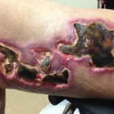
Update on Calciphylaxis Etiopathogenesis, Diagnosis, and Management
Calciphylaxis is associated with potentially severe complications and high mortality rates. This article highlights the challenges faced in...