Article
Improving Spanning-Knee External Fixator Stiffness: A Biomechanical Study
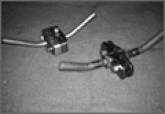
The purpose of this study was to test and compare external fixator construct stiffness using pin-to-bar clamps or multipin clamps across 2...
Article
Medicaid Insurance Is Associated With Larger Curves in Patients Who Require Scoliosis Surgery
Children with Medicaid may have difficulty accessing care for adolescent idiopathic scoliosis (AIS), a condition that may worsen with time. We...