Article

From smiling to smizing: Assessing the affect of a patient wearing a mask
Although the guidelines for masking in hospitals and other health care settings have been revised and face masks are no longer mandatory, it is...
Article
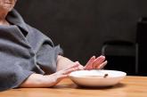
Food for thought: Dangerous weight loss in an older adult
CASE Fixated on health and nutrition
Ms. L, age 75, presents to the...
Article

Elaborate hallucinations, but is it a psychotic disorder?
Mr. B, age 93, has recurrent visual, auditory, and tactile hallucinations involving a man named ‘Harry,’ but no other psychotic symptoms. What...
News
Epilepsy or something else?
Ms. T, age 20, has a history of trauma and mood problems. She has seizure-like episodes but EEG and other tests do not find a cause. How would you...