Article
Purpura Fulminans in an Asplenic Intravenous Drug User
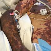
In patients with severe purpura fulminans and a gangrenous limb, it is important to allow adequate time for demarcation of gangrene and not rush...
Article
What’s Eating You? Human Body Lice (Pediculus humanus corporis)
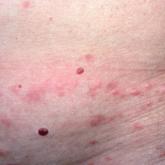
Compared to head and pubic lice, body lice carry increased morbidity in the form of greater body surface area involvement, possible infectious...