Article
Lumbar Degenerative Disc Disease and Tibiotalar Joint Arthritis: A 710-Specimen Postmortem Study
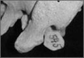
Research has associated lumbar spinal disease with lower extremity arthrosis. These studies focused solely on the lumbar spine’s connection with...