Article
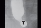
Role of barium esophagography in evaluating dysphagia
A 55-year-old woman presents with an intermittent sensation of food getting stuck in her mid to lower chest. How should her symptoms best be...
Article
Eosinophilic esophagitis: An increasingly recognized cause of dysphagia, food impaction, and refractory heartburn
This disease, which was not described as a distinct clinical entity until 1993, may be due to allergic and immune-mediated mechanisms similar to...
Article
Current challenges in Barrett's esophagus
The diagnosis and treatment of Barrett's esophagus are fraught with unresolved questions.
Article
Treatment of Helicobacter pylori in nonulcer dyspepsia: Should we or shouldn’t we?
Although it may be tempting to treat for Helicobacter pylori in every patient with dyspepsia, two recent trials indicate we should temper...
Article
H pylori 1997: Testing and treatment options
Which tests for H pylori are most useful? Which treatment regimens are most effective?