Stem-Based Repair of the Subscapularis in Total Shoulder Arthroplasty

Article Type
Changed
Thu, 09/19/2019 - 13:26
Display Headline
Stem-Based Repair of the Subscapularis in Total Shoulder Arthroplasty

Subscapularis integrity following total shoulder arthroplasty (TSA) is important to maintaining glenohumeral joint stability and functional outcome. In recent years increased emphasis has been placed on the management of the subscapularis during TSA. Options for management of the subscapularis during TSA include tenotomy, release of the tendon from the bone (peel technique), or a lesser tuberosity osteotomy (LTO). Several studies have demonstrated that subscapularis integrity is often impaired with a traditional tenotomy approach.1,2 Based on these studies, a subscapularis peel or LTO approach have gained popularity.3 This technical article describes a subscapularis peel repair technique that is integrated into a press-fit anatomical short-stem during TSA.

Technique

The repair technique demonstrated in this article features the Univers Apex (Arthrex) humeral stem, but it can be adapted to other stems with features that allow for the incorporation of sutures.

A standard deltopectoral approach is used to gain access to the shoulder. The biceps tendon is released or tenotomized to gain access to the bicipital groove. The rotator interval is then opened beginning at the superior subscapularis by following the course of the anterior side of the proximal biceps and then directing the release toward the base of the coracoid in order to protect the supraspinatus tendon. Next, the subscapularis is sharply released from the lesser tuberosity. The tendon and capsule are released as a unit and a 3-sided release of the subscapularis is performed.

The humeral canal is opened with a reamer and broached to accommodate an appropriately sized press-fit component. A polyethylene glenoid component is placed and then attention is returned to the humerus.

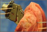
Prior to placement of the humeral stem, 6 No. 2 or No. 5 FiberWire (Arthrex) sutures are pre-placed through suture holes in the stem (Figure 1). Four sutures are passed by hand through the medial calcar component and 2 sutures are placed through holes in the lateral portion of the stem. A 2.0-mm or 2.5-mm drill is used to create 2 holes in the bicipital groove: 1 at the superior aspect of the lesser tuberosity, and 1 at the inferior aspect of the lesser tuberosity (Figure 2A). Prior to impacting the stem, the 4 lateral suture limbs (limbs A through D) are shuttled through the holes in the bicipital groove (Figure 2B). Then the stem is impacted and secured, the final humeral head is placed, the joint is reduced, and the subscapularis is repaired (Figure 2C).


The 4 sutures passing through the medial calcar of the stem result in 8 suture limbs (limbs 1 through 8). Each limb is separately passed through the subscapularis tendon with a free needle, moving obliquely from inferior-medial to superior-lateral (Figure 3). Note: A variation is to pass 2 suture limbs at a time, but this technique has not been biomechanically investigated at the time of this writing.

Prior to tying the sutures, it is helpful to place a stitch between the superolateral corner of the subscapularis and the anterior supraspinatus in order to facilitate reduction. The suture limbs are then tied with a specific sequence to create a suture-bridging construct with 2 additional medial mattress sutures as follows (Figures 4A, 4B):

1 to A

4 to C

5 to B

8 to D

2 to 3

6 to 7

In this technique, each suture limb is tied to a limb from another suture. When the last 2 pairs are tied (2 to 3 and 6 to 7), they are tensioned to remove any slack from the repair and equalize tension within all suture pairs. After the sutures are tied, the rotator interval may be closed with simple sutures if desired. The patient is immobilized in a sling for 4 to 6 weeks. Immediate passive forward flexion is allowed as well as external rotation to 30°. Strengthening is initiated at 8 weeks.

Discussion

The incidence of TSA has increased dramatically in the last decade and is projected to continue in the coming years.4 In the majority of cases, TSA leads to improvement in pain and function. However, failures continue to exist. In addition to glenoid loosening, prosthetic instability and rotator cuff insufficiency are the most common causes of failure.5 The latter 2 are intimately related since glenohumeral stability depends largely upon the rotator cuff. Therefore, optimization of outcome following TSA depends largely upon maintaining integrity of the rotator cuff. While the incidence of preoperative rotator cuff tears and fatty degeneration of the rotator are not modifiable, the management of the subscapularis is in the hands of the surgeon.

While subscapularis tenotomy has historically been used to access the glenohumeral joint during TSA, this approach is associated with an alarmingly high failure rate. Jackson and colleagues1 reported that 7 out of 15 (47%) of subscapularis tendons managed with tenotomy during TSA were completely torn on postoperative ultrasound. The patients with postoperative rupture had decreased internal rotation strength and DASH scores (4.6 intact vs. 25 ruptured; P = .04) compared to the patients with an intact tendon. Scalise and colleagues2 retrospectively compared a tenotomy approach to a LTO. They reported that 7 out of 15 subscapularis tenotomies were ruptured or attenuated postoperatively. By comparison, 18 out of 20 LTOs were healed. Regardless of approach, functional outcome was higher at 1 year postoperative when the subscapularis was intact.


The high failure rate with tendon-to-tendon healing following tenotomy has led to interest in a subscapularis peel to achieve tendon-to-bone healing or an LTO approach to achieve bone-to-bone healing. Lapner and colleagues3 compared a peel to an LTO in a randomized controlled trial of 87 patients. At 2 years postoperative, there was no difference in functional outcome between the 2 groups.

While both a peel and an LTO approach can be repaired with the technique described in this article, there are advantages to a peel approach. First, a peel approach may be considered more reproducible, particularly for surgeons who do a limited amount of shoulder arthroplasty. Whereas an LTO can vary in size, the subscapularis can nearly always be reproducibly peeled from the lesser tuberosity. Second, this technique uses a short stem, which relies upon proximal fixation. While this approach is bone-preserving, a large osteotomy has the potential to compromise fixation of the stem. Therefore, while one of us (PJD) uses a fleck LTO with a short stem, we advise a peel technique in most cases.

In summary, the subscapularis repair technique described here provides a reproducible and biomechanically sound approach to managing the subscapularis during TSA.

References


1.    Jackson JD, Cil A, Smith J, Steinmann SP. Integrity and function of the subscapularis after total shoulder arthroplasty. J Shoulder Elbow Surg. 2010;19(7):1085-1090.

2.    Scalise JJ, Ciccone J, Iannotti JP. Clinical, radiographic, and ultrasonographic comparison of subscapularis tenotomy and lesser tuberosity osteotomy for total shoulder arthroplasty. J Bone Joint Surg Am. 2010;92(7):1627-1634.

3.    Lapner PL, Sabri E, Rakhra K, Bell K, Athwal GS. Comparison of lesser tuberosity osteotomy to subscapularis peel in shoulder arthroplasty: a randomized controlled trial. J Bone Joint Surg Am. 2012;94(24):2239-2246.

4.    Kim SH, Wise BL, Zhang Y, Szabo RM. Increasing incidence of shoulder arthroplasty in the United States. J Bone Joint Surg Am. 2011;93(24):2249-2254.

5.    Australian Orthopaedic Association National Joint Replacement Registry. Shoulder Arthroplasty 2015 Annual Report. https://aoanjrr.sahmri.com/documents/10180/217645/Shoulder%20Arthroplasty. Accessed April 7, 2016.

Article PDF
Author and Disclosure Information


Patrick J. Denard, MD, Evan Lederman, MD, Reuben Gobezie, MD, and Bryan T. Hanypsiak, MD

Authors’ Disclosure Statement: Dr. Denard reports he is a paid consultant to and receives research support from Arthrex. Dr. Lederman reports he is a paid consultant to, on the speaker’s bureau for, and receives royalties from Arthrex. Dr. Gobezie reports he is a paid consultant to and receives research support from Arthrex. Dr. Hanypsiak reports he is a former employee of, and served on the speaker’s bureau for, Arthrex.

Issue
The American Journal of Orthopedics - 45(4)
Publications
Topics
Page Number
228-230
Legacy Keywords
total shoulder arthroplasty, TSA, shoulder, arthroplasty, stem, repair, subscapularis, denard, lederman, gobezie, hanypsiak
Sections
Author and Disclosure Information


Patrick J. Denard, MD, Evan Lederman, MD, Reuben Gobezie, MD, and Bryan T. Hanypsiak, MD

Authors’ Disclosure Statement: Dr. Denard reports he is a paid consultant to and receives research support from Arthrex. Dr. Lederman reports he is a paid consultant to, on the speaker’s bureau for, and receives royalties from Arthrex. Dr. Gobezie reports he is a paid consultant to and receives research support from Arthrex. Dr. Hanypsiak reports he is a former employee of, and served on the speaker’s bureau for, Arthrex.

Article PDF
Article PDF

Subscapularis integrity following total shoulder arthroplasty (TSA) is important to maintaining glenohumeral joint stability and functional outcome. In recent years increased emphasis has been placed on the management of the subscapularis during TSA. Options for management of the subscapularis during TSA include tenotomy, release of the tendon from the bone (peel technique), or a lesser tuberosity osteotomy (LTO). Several studies have demonstrated that subscapularis integrity is often impaired with a traditional tenotomy approach.1,2 Based on these studies, a subscapularis peel or LTO approach have gained popularity.3 This technical article describes a subscapularis peel repair technique that is integrated into a press-fit anatomical short-stem during TSA.

Technique

The repair technique demonstrated in this article features the Univers Apex (Arthrex) humeral stem, but it can be adapted to other stems with features that allow for the incorporation of sutures.

A standard deltopectoral approach is used to gain access to the shoulder. The biceps tendon is released or tenotomized to gain access to the bicipital groove. The rotator interval is then opened beginning at the superior subscapularis by following the course of the anterior side of the proximal biceps and then directing the release toward the base of the coracoid in order to protect the supraspinatus tendon. Next, the subscapularis is sharply released from the lesser tuberosity. The tendon and capsule are released as a unit and a 3-sided release of the subscapularis is performed.

The humeral canal is opened with a reamer and broached to accommodate an appropriately sized press-fit component. A polyethylene glenoid component is placed and then attention is returned to the humerus.

Prior to placement of the humeral stem, 6 No. 2 or No. 5 FiberWire (Arthrex) sutures are pre-placed through suture holes in the stem (Figure 1). Four sutures are passed by hand through the medial calcar component and 2 sutures are placed through holes in the lateral portion of the stem. A 2.0-mm or 2.5-mm drill is used to create 2 holes in the bicipital groove: 1 at the superior aspect of the lesser tuberosity, and 1 at the inferior aspect of the lesser tuberosity (Figure 2A). Prior to impacting the stem, the 4 lateral suture limbs (limbs A through D) are shuttled through the holes in the bicipital groove (Figure 2B). Then the stem is impacted and secured, the final humeral head is placed, the joint is reduced, and the subscapularis is repaired (Figure 2C).


The 4 sutures passing through the medial calcar of the stem result in 8 suture limbs (limbs 1 through 8). Each limb is separately passed through the subscapularis tendon with a free needle, moving obliquely from inferior-medial to superior-lateral (Figure 3). Note: A variation is to pass 2 suture limbs at a time, but this technique has not been biomechanically investigated at the time of this writing.

Prior to tying the sutures, it is helpful to place a stitch between the superolateral corner of the subscapularis and the anterior supraspinatus in order to facilitate reduction. The suture limbs are then tied with a specific sequence to create a suture-bridging construct with 2 additional medial mattress sutures as follows (Figures 4A, 4B):

1 to A

4 to C

5 to B

8 to D

2 to 3

6 to 7

In this technique, each suture limb is tied to a limb from another suture. When the last 2 pairs are tied (2 to 3 and 6 to 7), they are tensioned to remove any slack from the repair and equalize tension within all suture pairs. After the sutures are tied, the rotator interval may be closed with simple sutures if desired. The patient is immobilized in a sling for 4 to 6 weeks. Immediate passive forward flexion is allowed as well as external rotation to 30°. Strengthening is initiated at 8 weeks.

Discussion

The incidence of TSA has increased dramatically in the last decade and is projected to continue in the coming years.4 In the majority of cases, TSA leads to improvement in pain and function. However, failures continue to exist. In addition to glenoid loosening, prosthetic instability and rotator cuff insufficiency are the most common causes of failure.5 The latter 2 are intimately related since glenohumeral stability depends largely upon the rotator cuff. Therefore, optimization of outcome following TSA depends largely upon maintaining integrity of the rotator cuff. While the incidence of preoperative rotator cuff tears and fatty degeneration of the rotator are not modifiable, the management of the subscapularis is in the hands of the surgeon.

While subscapularis tenotomy has historically been used to access the glenohumeral joint during TSA, this approach is associated with an alarmingly high failure rate. Jackson and colleagues1 reported that 7 out of 15 (47%) of subscapularis tendons managed with tenotomy during TSA were completely torn on postoperative ultrasound. The patients with postoperative rupture had decreased internal rotation strength and DASH scores (4.6 intact vs. 25 ruptured; P = .04) compared to the patients with an intact tendon. Scalise and colleagues2 retrospectively compared a tenotomy approach to a LTO. They reported that 7 out of 15 subscapularis tenotomies were ruptured or attenuated postoperatively. By comparison, 18 out of 20 LTOs were healed. Regardless of approach, functional outcome was higher at 1 year postoperative when the subscapularis was intact.


The high failure rate with tendon-to-tendon healing following tenotomy has led to interest in a subscapularis peel to achieve tendon-to-bone healing or an LTO approach to achieve bone-to-bone healing. Lapner and colleagues3 compared a peel to an LTO in a randomized controlled trial of 87 patients. At 2 years postoperative, there was no difference in functional outcome between the 2 groups.

While both a peel and an LTO approach can be repaired with the technique described in this article, there are advantages to a peel approach. First, a peel approach may be considered more reproducible, particularly for surgeons who do a limited amount of shoulder arthroplasty. Whereas an LTO can vary in size, the subscapularis can nearly always be reproducibly peeled from the lesser tuberosity. Second, this technique uses a short stem, which relies upon proximal fixation. While this approach is bone-preserving, a large osteotomy has the potential to compromise fixation of the stem. Therefore, while one of us (PJD) uses a fleck LTO with a short stem, we advise a peel technique in most cases.

In summary, the subscapularis repair technique described here provides a reproducible and biomechanically sound approach to managing the subscapularis during TSA.

References


1.    Jackson JD, Cil A, Smith J, Steinmann SP. Integrity and function of the subscapularis after total shoulder arthroplasty. J Shoulder Elbow Surg. 2010;19(7):1085-1090.

2.    Scalise JJ, Ciccone J, Iannotti JP. Clinical, radiographic, and ultrasonographic comparison of subscapularis tenotomy and lesser tuberosity osteotomy for total shoulder arthroplasty. J Bone Joint Surg Am. 2010;92(7):1627-1634.

3.    Lapner PL, Sabri E, Rakhra K, Bell K, Athwal GS. Comparison of lesser tuberosity osteotomy to subscapularis peel in shoulder arthroplasty: a randomized controlled trial. J Bone Joint Surg Am. 2012;94(24):2239-2246.

4.    Kim SH, Wise BL, Zhang Y, Szabo RM. Increasing incidence of shoulder arthroplasty in the United States. J Bone Joint Surg Am. 2011;93(24):2249-2254.

5.    Australian Orthopaedic Association National Joint Replacement Registry. Shoulder Arthroplasty 2015 Annual Report. https://aoanjrr.sahmri.com/documents/10180/217645/Shoulder%20Arthroplasty. Accessed April 7, 2016.

Issue
The American Journal of Orthopedics - 45(4)
Issue
The American Journal of Orthopedics - 45(4)
Page Number
228-230
Page Number
228-230
Publications
Publications
Topics
Article Type
Display Headline
Stem-Based Repair of the Subscapularis in Total Shoulder Arthroplasty
Display Headline
Stem-Based Repair of the Subscapularis in Total Shoulder Arthroplasty
Legacy Keywords
total shoulder arthroplasty, TSA, shoulder, arthroplasty, stem, repair, subscapularis, denard, lederman, gobezie, hanypsiak
Legacy Keywords
total shoulder arthroplasty, TSA, shoulder, arthroplasty, stem, repair, subscapularis, denard, lederman, gobezie, hanypsiak
Sections
Disallow All Ads
Alternative CME
Article PDF Media

Dislocation and Instability After Arthroscopic Capsular Release for Refractory Frozen Shoulder

Article Type
Changed
Thu, 09/19/2019 - 14:13
Display Headline
Dislocation and Instability After Arthroscopic Capsular Release for Refractory Frozen Shoulder

Article PDF
Author and Disclosure Information

Reuben Gobezie, MD, Iván H. Pacheco, MD, Charles J. Petit, MD, and Peter J. Millett, MD, MSc

Dr. Gobezie is Assistant Professor, The Case Shoulder & Elbow Service, Case Western Reserve University School of Medicine, Cleveland, Ohio.

Dr. Pacheco is with Harvard Shoulder Service, Harvard Medical School, Brigham and Women's Hospital, and Massachusetts General Hospital, Boston, Massachusetts.

Dr. Petit is with Harvard Shoulder Fellowship, Harvard Combined Orthopaedic Residency Program, Brigham and Women's Hospital, Boston, Massachusetts.

Dr. Millett is Director of Shoulder Surgery, Shoulder, Knee, and Sports Medicine, Steadman Hawkins Clinic, Vail, Colorado.

Issue
The American Journal of Orthopedics - 36(12)
Publications
Topics
Page Number
672-674
Legacy Keywords
dislocation, instability, arthroscopy, capsular release, frozen shoulder, diabetes, shoulder, ajo, american journal of orthopedics
Sections
Author and Disclosure Information

Reuben Gobezie, MD, Iván H. Pacheco, MD, Charles J. Petit, MD, and Peter J. Millett, MD, MSc

Dr. Gobezie is Assistant Professor, The Case Shoulder & Elbow Service, Case Western Reserve University School of Medicine, Cleveland, Ohio.

Dr. Pacheco is with Harvard Shoulder Service, Harvard Medical School, Brigham and Women's Hospital, and Massachusetts General Hospital, Boston, Massachusetts.

Dr. Petit is with Harvard Shoulder Fellowship, Harvard Combined Orthopaedic Residency Program, Brigham and Women's Hospital, Boston, Massachusetts.

Dr. Millett is Director of Shoulder Surgery, Shoulder, Knee, and Sports Medicine, Steadman Hawkins Clinic, Vail, Colorado.

Article PDF
Article PDF

Issue
The American Journal of Orthopedics - 36(12)
Issue
The American Journal of Orthopedics - 36(12)
Page Number
672-674
Page Number
672-674
Publications
Publications
Topics
Article Type
Display Headline
Dislocation and Instability After Arthroscopic Capsular Release for Refractory Frozen Shoulder
Display Headline
Dislocation and Instability After Arthroscopic Capsular Release for Refractory Frozen Shoulder
Legacy Keywords
dislocation, instability, arthroscopy, capsular release, frozen shoulder, diabetes, shoulder, ajo, american journal of orthopedics
Legacy Keywords
dislocation, instability, arthroscopy, capsular release, frozen shoulder, diabetes, shoulder, ajo, american journal of orthopedics
Sections
Article Source

PURLs Copyright

Inside the Article

Article PDF Media