Firm Tumor Encasing the Left Second Toe of an Infant

Article Type
Changed
Mon, 06/08/2020 - 09:15
Display Headline
Firm Tumor Encasing the Left Second Toe of an Infant

The Diagnosis: Infantile Digital Fibromatosis

Infantile digital fibromatosis (IDF), or recurring digital fibrous tumor of childhood, is a benign juvenile myofibromatosis that presents as a firm, flesh-colored or slightly erythematous, dome-shaped papule or nodule on the dorsolateral aspects of the digits, usually sparing the thumbs and great toes.1 The tumor appears most commonly at birth and in infants younger than 1 year. It grows slowly over the first month, then rapidly over the next 10 to 14 months.1,2  

Although lesions usually regress spontaneously within a few years, excision may be necessary when functional impairment and joint deformity occur. Tumors, however, may recur locally.1,2  

Histologically, IDFs are composed of spindled myofibroblasts with characteristic round eosinophilic intracytoplasmic inclusion bodies, which represent actin and vimentin filaments.1 In our case, histopathologic evaluation showed a proliferation of fibrous spindle cells with pathognomonic eosinophilic intracytoplasmic inclusions consistent with IDF (Figure).  

Fibrous spindle cells with pathognomonic eosinophilic intracytoplasmic inclusions (H&E, original magnification ×600).

Fibrosarcomas are high-grade and low-grade soft-tissue neoplasms comprised of atypical spindle cells in a herringbone pattern with mitotic figures on pathology.3 They typically present as a slowly growing subcutaneous tumor on the lower extremities of young to middle-aged adults that may progress to become a palpable tender nodule. Infantile hemangiomas, the most common benign soft-tissue tumors of childhood, are vascular proliferations more commonly seen in low-birth-weight female white infants of multiple gestation pregnancies. Superficial hemangiomas present as bright red and lobular nodules or plaques. Deep hemangiomas present as ill-defined, blue-violaceous nodules with no overlying skin changes. Mixed hemangiomas present with features of both superficial and deep hemangiomas. Infantile hemangiomas experience a proliferative phase until 9 to 12 months of age, followed by a gradual involutional phase ending with possible residual telangiectases or fibrofatty change. Unlike IDFs, infantile hemangiomas favor the head and neck over other areas of the body. Keloids are firm, smooth, variably colored papules or plaques of haphazardly arranged thick dermal collagen bundles. They usually develop within a year of skin injury and extend beyond the original injury margin into normal tissue. Supernumerary digits present as small fleshy papules or larger nodules with a vestigial nail, most commonly on the ulnar side of the fifth finger or the radial side of the thumb. Histologically, they are composed of fascicles of nerve fibers.

Treatment in our patient included partial amputation of the left second toe and excision with local tissue rearrangement by a plastic surgeon. His postoperative course was complicated by a minor wound infection, which resolved with a 7-day course of cephalexin. Since then, the patient has healed well, with gradual toe tissue and nail growth. No recurrence was reported 11 months after surgery.  

References
  1. Heymann WR. Infantile digital fibromatosis. J Am Acad Dermatol. 2008;59:122-123. 
  2. Niamba P, Léauté-Labrèze C, Boralevi F, et al. Further documentation of spontaneous regression of infantile digital fibromatosis. Pediatr Dermatol. 2007;24:280-284. 
  3. Bolognia JL, Schaffer JV, Cerroni L, eds. Dermatology. Philadelphia, PA: Elsevier Limited; 2018. 
Article PDF
Author and Disclosure Information

Dr. Ren is from the Department of Dermatology, Baylor College of Medicine, Houston, Texas. Dr. Lee is from the Department of Dermatology, Baylor College of Medicine, Texas Children’s Hospital, Houston.

The authors report no conflict of interest.

Correspondence: Grace L. Lee, MD, Department of Dermatology, Baylor College of Medicine, Texas Children’s Hospital, Clinical Care Center, 6701 Fannin St, 8th Floor, Houston, TX 77030 (glee@texaschildrens.org).

Issue
Cutis - 105(1)
Publications
Topics
Page Number
16, 20
Sections
Author and Disclosure Information

Dr. Ren is from the Department of Dermatology, Baylor College of Medicine, Houston, Texas. Dr. Lee is from the Department of Dermatology, Baylor College of Medicine, Texas Children’s Hospital, Houston.

The authors report no conflict of interest.

Correspondence: Grace L. Lee, MD, Department of Dermatology, Baylor College of Medicine, Texas Children’s Hospital, Clinical Care Center, 6701 Fannin St, 8th Floor, Houston, TX 77030 (glee@texaschildrens.org).

Article PDF
Article PDF
Related Articles

The Diagnosis: Infantile Digital Fibromatosis

Infantile digital fibromatosis (IDF), or recurring digital fibrous tumor of childhood, is a benign juvenile myofibromatosis that presents as a firm, flesh-colored or slightly erythematous, dome-shaped papule or nodule on the dorsolateral aspects of the digits, usually sparing the thumbs and great toes.1 The tumor appears most commonly at birth and in infants younger than 1 year. It grows slowly over the first month, then rapidly over the next 10 to 14 months.1,2  

Although lesions usually regress spontaneously within a few years, excision may be necessary when functional impairment and joint deformity occur. Tumors, however, may recur locally.1,2  

Histologically, IDFs are composed of spindled myofibroblasts with characteristic round eosinophilic intracytoplasmic inclusion bodies, which represent actin and vimentin filaments.1 In our case, histopathologic evaluation showed a proliferation of fibrous spindle cells with pathognomonic eosinophilic intracytoplasmic inclusions consistent with IDF (Figure).  

Fibrous spindle cells with pathognomonic eosinophilic intracytoplasmic inclusions (H&E, original magnification ×600).

Fibrosarcomas are high-grade and low-grade soft-tissue neoplasms comprised of atypical spindle cells in a herringbone pattern with mitotic figures on pathology.3 They typically present as a slowly growing subcutaneous tumor on the lower extremities of young to middle-aged adults that may progress to become a palpable tender nodule. Infantile hemangiomas, the most common benign soft-tissue tumors of childhood, are vascular proliferations more commonly seen in low-birth-weight female white infants of multiple gestation pregnancies. Superficial hemangiomas present as bright red and lobular nodules or plaques. Deep hemangiomas present as ill-defined, blue-violaceous nodules with no overlying skin changes. Mixed hemangiomas present with features of both superficial and deep hemangiomas. Infantile hemangiomas experience a proliferative phase until 9 to 12 months of age, followed by a gradual involutional phase ending with possible residual telangiectases or fibrofatty change. Unlike IDFs, infantile hemangiomas favor the head and neck over other areas of the body. Keloids are firm, smooth, variably colored papules or plaques of haphazardly arranged thick dermal collagen bundles. They usually develop within a year of skin injury and extend beyond the original injury margin into normal tissue. Supernumerary digits present as small fleshy papules or larger nodules with a vestigial nail, most commonly on the ulnar side of the fifth finger or the radial side of the thumb. Histologically, they are composed of fascicles of nerve fibers.

Treatment in our patient included partial amputation of the left second toe and excision with local tissue rearrangement by a plastic surgeon. His postoperative course was complicated by a minor wound infection, which resolved with a 7-day course of cephalexin. Since then, the patient has healed well, with gradual toe tissue and nail growth. No recurrence was reported 11 months after surgery.  

References
  1. Heymann WR. Infantile digital fibromatosis. J Am Acad Dermatol. 2008;59:122-123. 
  2. Niamba P, Léauté-Labrèze C, Boralevi F, et al. Further documentation of spontaneous regression of infantile digital fibromatosis. Pediatr Dermatol. 2007;24:280-284. 
  3. Bolognia JL, Schaffer JV, Cerroni L, eds. Dermatology. Philadelphia, PA: Elsevier Limited; 2018. 
Issue
Cutis - 105(1)
Issue
Cutis - 105(1)
Page Number
16, 20
Page Number
16, 20
Publications
Publications
Topics
Article Type
Display Headline
Firm Tumor Encasing the Left Second Toe of an Infant
Display Headline
Firm Tumor Encasing the Left Second Toe of an Infant
Sections
Questionnaire Body

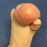
A 10-month-old infant boy presented to the dermatology clinic with a firm, nonpainful, 5.5.×5.6-cm tumor encasing the left second toe, with associated skin breakdown, gait impairment, and lateral displacement of the third toe. The lesion began as a small bump under the toenail at 2 months of age; it then grew rapidly without bleeding or ulceration. It was diagnosed as a hemangioma at 4 months of age, and oral propranolol was initiated for 3 months, without suppression of tumor growth. The patient was referred to the pediatric dermatology department where a clinical diagnosis was made, and the patient was subsequently referred to plastic surgery for excision.

Disallow All Ads
Content Gating
No Gating (article Unlocked/Free)
Alternative CME
Disqus Comments
Default
Gate On Date
Fri, 01/10/2020 - 11:30
Un-Gate On Date
Fri, 01/10/2020 - 11:30
Use ProPublica
CFC Schedule Remove Status
Fri, 01/10/2020 - 11:30
Hide sidebar & use full width
render the right sidebar.
Conference Recap Checkbox
Not Conference Recap
Article PDF Media