User login
Antibody can treat HSCT-TMA and GVHD, case suggests
GRANADA, SPAIN—A monoclonal antibody can resolve co-existing hematopoietic stem cell transplant-associated thrombotic microangiopathy (HSCT-TMA) and graft-versus-host disease (GVHD), according to a case study.
The antibody is OMS721, and it targets MASP-2, a pro-inflammatory protein target involved in activation of the complement system.
The case of OMS721 ameliorating HSCT-TMA and GVHD was presented at the European Society for Blood and Marrow Transplantation Crash Course on Diagnosis and Treatment of Noninfectious Complications after HCT.
The patient was a participant in an ongoing phase 2 trial of thrombotic microangiopathies, including HSCT-TMA. The trial is sponsored by Omeros Corporation, the company developing OMS721.
The patient was an adult male with post-transplant TMA persisting at least 2 weeks following calcineurin inhibitor modification.
The patient had undergone HSCT for T-cell acute lymphoblastic leukemia. His post-transplant course was complicated by multiple episodes of steroid-refractory grade 4 GVHD, cytomegalovirus infection, and HSCT-TMA.
After 2 prior episodes of GVHD, the patient presented with bloody diarrhea. Intestinal biopsy demonstrated both HSCT-TMA and GVHD. No infections were identified.
The patient also had new-onset neurological symptoms of paresthesias, tetraparesis, and a neurogenic bladder, which have been reported as neurological manifestations of GVHD and TMA.
The patient was unable to walk due to the tetraparesis and required blood transfusions at least once daily. Hematological markers demonstrated HSCT-TMA with thrombocytopenia, elevated lactate dehydrogenase, and schistocytes.
Two weeks prior to starting OMS721, the patient’s immunosuppression (cyclosporine) had been decreased, and, given his history of steroid-refractory GVHD, he was receiving only low-dose corticosteroids. He received no other GVHD treatment.
After 2 OMS721 doses, the patient’s bloody diarrhea resolved, and his hematological markers improved. After 4 OMS721 doses, he was able to walk with help.
The patient completed 8 weeks of OMS721 treatment and has been doing well at home. All signs and symptoms of HSCT-TMA and all clinical symptoms of GVHD have resolved. His neurological symptoms have continued to improve.
“This patient’s marked response to OMS721 treatment was very gratifying,” said Anna Grassi, MD, of Azienda Ospedaliera Papa Giovanni XXIII in Bergamo, Italy.
“The cause of his neurological symptoms is not clear but may be a manifestation of GVHD or other endothelial injury. Prior to OMS721 treatment, this patient was deteriorating and at high risk for early death. The improvement of GVHD, H[S]CT-TMA, and the neurological symptoms following OMS721 treatment is promising.” ![]()
EMA recommends orphan designation for G100 to treat FL
The European Medicines Agency’s (EMA’s) Committee for Orphan Medicinal Products has recommended orphan designation for G100 for the treatment of follicular lymphoma (FL).
G100 contains the synthetic small molecule toll-like receptor-4 agonist glucopyranosyl lipid A.
G100 works by activating innate and adaptive immunity in the tumor microenvironment to generate an immune response against the tumor’s pre-existing antigens.
Clinical and preclinical data have demonstrated G100’s ability to activate tumor-infiltrating lymphocytes, macrophages, and dendritic cells, and promote antigen-presentation and the recruitment of T cells to the tumor.
The induction of local and systemic immune responses has been shown in preclinical studies to result in local and abscopal tumor control.
Immune Design, the company developing G100, is currently evaluating G100 plus local radiation, with or without pembrolizumab, in a phase 1/2 trial of FL patients.
Results from this trial were presented at the 2017 ASCO Annual Meeting (abstract 7537). Nine patients who received G100 (3 patients each at the 5, 10, or 20 μg dose) with radiation (but not pembrolizumab) were evaluable for safety and efficacy.
The overall response rate was 44%, and all of these were partial responses (n=4). Thirty-three percent of patients had stable disease (n=3).
Among the responders, tumor regression ranged from 58% to 89%, which included up to 56% shrinkage of abscopal sites. Tumor biopsies showed increased inflammatory responses and T-cell infiltrates in abscopal tumors.
An additional 13 patients treated at the 10 μg dose were evaluable for safety. There were no dose-limiting toxicities, serious adverse events (AEs), or grade 3/4 AEs observed.
Common AEs included injection site disorders, abdominal pain/discomfort, nausea, pruritus, and decrease in lymphocytes.
Immune Design said that, if this trial produces a sufficiently robust clinical benefit for patients, the company may pursue FL as the first indication for regulatory approval of G100.
About orphan designation
Orphan designation provides regulatory and financial incentives for companies to develop and market therapies that treat life-threatening or chronically debilitating conditions affecting no more than 5 in 10,000 people in the European Union, and where no satisfactory treatment is available.
Orphan designation provides a 10-year period of marketing exclusivity if the drug receives regulatory approval.
The designation also provides incentives for companies seeking protocol assistance from the EMA during the product development phase and direct access to the centralized authorization procedure.
The EMA’s Committee for Orphan Medicinal Products adopts an opinion on the granting of orphan drug designation, and that opinion is submitted to the European Commission for a final decision. The commission typically makes a decision within 30 days of the submission. ![]()
New assay may aid diagnosis, treatment of DLBCL
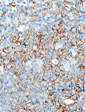
A new assay may help improve the diagnosis and treatment of diffuse large B-cell lymphoma (DLBCL), according to researchers.
The gene expression signature assay can be used to classify subtypes of DLBCL and may enhance disease management by helping to match tumors with the appropriate targeted therapy.
Researchers described the assay in the Journal of Molecular Diagnostics.
The assay is a novel gene expression profiling DLBCL classifier based on reverse transcriptase multiplex ligation-dependent probe amplification (RT-MLPA).
It can simultaneously evaluate the expression of 21 markers, allowing differentiation of the 3 subtypes of DLBCL—germinal center B-cell-like (GCB), activated B-cell-like (ABC), and primary mediastinal B-cell lymphoma (PMBL)—as well as other individualized disease characteristics, such as Epstein-Barr infection status.
Researchers used the RT-MLPA assay to test 150 samples from DLBCL patients. Forty-two percent of the samples were the ABC subtype, 37% the GCB subtype, and 10% molecular PMBL. Eleven percent of the samples could not be classified.
Overall, the RT-MLPA assay correctly assigned 85.0% of the cases into the expected subtypes, compared to 78.8% of samples assigned via immunohistochemistry.
The RT-MLPA assay was also able to detect the MYD88 L265P mutation, one of the most common genetic abnormalities found in ABC DLBCLs. This information can influence treatment, since the presence of the mutation is thought to be predictive of ibrutinib sensitivity.
The researchers said RT-MLPA is a robust, efficient, rapid, and cost-effective alternative to current methods used in the clinic to establish the cell-of-origin classification of DLBCLs.
RT-MLPA requires only common laboratory equipment and can be applied to formalin-fixed, paraffin-embedded samples. Other types of diagnostic methods may not provide the level of detail needed and may also be limited by poor reproducibility and lack of adaptability to routine use in standard laboratories.
“Because we have provided the classification algorithms, other laboratories will be able to verify our results and adjust the procedures to suit their environment,” said study author Philippe Ruminy, PhD, of the Henri Becquerel Cancer Treatment Center, INSERM U1245 in Rouen, France.
“It is our hope that the assay we have developed, which addresses an important recommendation of the recent WHO classifications, will contribute to better management of these tumors and improved patient outcomes.” ![]()
Preserving normal hematopoietic function in AML
Preclinical research has revealed a treatment that might preserve normal hematopoietic function in patients with acute myeloid leukemia (AML).
Researchers found that AML suppresses adipocytes in the bone marrow, which leads to imbalanced regulation of endogenous hematopoietic stem and progenitor cells and impaired myelo-erythroid maturation.
However, a PPARγ agonist can induce adipogenesis, which suppresses AML cells and stimulates the regeneration of healthy blood cells.
Researchers reported these findings in Nature Cell Biology.
The team found that adipocytes in the bone marrow support myelo-erythroid maturation of hematopoietic stem and progenitor cells. But AML has a negative effect on the maturation of adipocytes, which explains why deficient myelo-erythropoiesis is “a consistent feature” of AML.
The researchers also found that pro-adipogenesis therapy—treatment with the PPARγ agonist GW1929—protects healthy myelo-erythropoiesis and limits leukemic self-renewal.
“Our approach represents a different way of looking at leukemia and considers the entire bone marrow as an ecosystem, rather than the traditional approach of studying and trying to directly kill the diseased cells themselves,” said study author Allison Boyd, PhD, of McMaster University in Hamilton, Ontario, Canada.
“These traditional approaches have not delivered enough new therapeutic options for patients. The standard of care for this disease hasn’t changed in several decades.”
“The focus of chemotherapy and existing standard of care is on killing cancer cells, but, instead, we took a completely different approach, which changes the environment the cancer cells live in,” said study author Mick Bhatia, PhD, of McMaster University.
“This not only suppressed the ‘bad’ cancer cells but also bolstered the ‘good’ healthy cells, allowing them to regenerate in the new drug-induced environment. The fact that we can target one cell type in one tissue using an existing drug makes us excited about the possibilities of testing this in patients.”
“We can envision this becoming a potential new therapeutic approach that can either be added to existing treatments or even replace others in the near future. The fact that this drug activates blood regeneration may provide benefits for those waiting for bone marrow transplants by activating their own healthy cells.” ![]()
System automates classification of RBCs
Scientists say they have developed an automated system that can identify shapes of red blood cells (RBCs).
The team found their system could classify sickled RBCs “with high accuracy,” which suggests it could be used to help monitor patients with sickle cell disease.
“We have developed the first deep learning tool that can automatically identify and classify red blood cell alteration, hence providing direct quantitative evidence of the severity of the disease,” said George Karniadakis, PhD, of Brown University in Providence, Rhode Island.
Dr Karniadakis and his colleagues described their tool in PLOS Computational Biology.
The researchers wanted to automate the process of identifying RBC shape. So they developed a computational framework that employs a machine-learning tool known as a deep convolutional neural network (CNN).
The framework uses 3 steps to classify the shapes of RBCs in microscopic images of blood.
First, it distinguishes RBCs from the background of each image and from each other. Then, for each cell detected, it zooms in or out until all cell images are a uniform size. Finally, it uses deep CNNs to categorize RBCs by shape.
The researchers validated their new tool using 7000 microscopy images from 8 patients with sickle cell disease. The method successfully classified RBC shape for both oxygenated and deoxygenated cells.
The researchers plan to further improve their deep CNN tool and test it in other diseases that alter the shape and size of RBCs, such as diabetes and HIV. They also plan to explore its usefulness in characterizing cancer cells. ![]()
Eltrombopag can control ITP long-term, study suggests
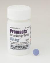
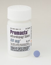
Eltrombopag can provide long-term disease control for chronic/persistent immune thrombocytopenia (ITP), according to research published in Blood.
In the EXTEND study, investigators evaluated patients exposed to eltrombopag for a median of 2.4 years.
Most patients achieved a response to the drug, and more than half of them maintained that response for at least 25 weeks.
More than a third of patients were able to discontinue at least 1 concomitant ITP medication.
Most adverse events (AEs) were grade 1 or 2. However, 32% of patients had serious AEs, and 14% of patients withdrew from the study due to AEs.
This research was sponsored by GlaxoSmithKline, the company that previously owned eltrombopag. Now, the drug is a product of Novartis.
Patients
EXTEND is an open-label extension study of 4 trials (TRA100773A, TRA100773B, TRA102537/RAISE, and TRA108057/REPEAT), which enrolled 302 adults with chronic/persistent ITP.
Patients had completed the treatment and follow-up periods as defined in their previous study protocol and did not experience eltrombopag-related toxicity or other drug intolerance on a prior eltrombopag study. Patients who discontinued a previous study due to toxicity were only eligible if they had received a placebo.
The patients’ median time from diagnosis to enrollment in EXTEND was 58.8 months (range, 9-552). Their median age was 50 (range, 18-86), and 67% were female.
Most patients (70%) had a baseline platelet count below 30×109/L. Thirty-three percent of patients were using concomitant ITP medications, 53% had received at least 3 prior ITP treatments, and 38% had undergone splenectomy.
Treatment
Eltrombopag was started at a dose of 50 mg/day and titrated to 25-75 mg/day or less often based on platelet counts. Maintenance dosing continued after minimization of concomitant ITP medication and optimization of eltrombopag dosing.
The overall median duration of eltrombopag exposure was 2.37 years (range, 2 days to 8.76 years), and the mean average daily dose was 50.2 mg/day (range, 1-75).
One hundred and thirty-five patients (45%) completed the study, and 75 patients (25%) were treated for 4 or more years. The most common reasons for study withdrawal included AEs (n=41), patient decision (n=39), lack of efficacy (n=32), and “other” reasons (n=39).
Safety
AEs leading to study withdrawal (occurring at least twice) included hepatobiliary AEs (n=7), cataracts (n=4), deep vein thrombosis (n=3), cerebral infarction (n=2), headache (n=2), and myelofibrosis (n=2).
The overall incidence of AEs was 92%. The most frequent AEs were headache (28%), nasopharyngitis (25%), and upper respiratory tract infection (23%).
Twenty-six percent of patients had grade 3 AEs, 6% had grade 4 AEs, and 32% had serious AEs. Serious AEs included cataracts (5%), pneumonia (3%), anemia (2%), ALT increase (2%), epistaxis (1%), AST increase, (1%), bilirubin increase (1%), and deep vein thrombosis (1%).
Three percent of patients reported a malignancy while on study, including basal cell carcinoma, intramucosal adenocarcinoma, breast cancer, metastases to the lung, ovarian cancer, squamous cell carcinoma, transitional cell carcinoma, lymphoma, unclassifiable B-cell lymphoma (low grade), and Hodgkin lymphoma.
Efficacy
In all, 85.8% (259/302) of patients had a response to eltrombopag, which was defined as achieving a platelet count of at least 50×109/L at least once without rescue therapy.
Fifty-two percent (133/257) of patients achieved a continuous response lasting at least 25 weeks.
Thirty-four percent (34/101) of patients who were on concomitant ITP medication discontinued at least 1 medication. Thirty-nine percent (39/101) reduced or permanently stopped at least 1 ITP medication without receiving rescue therapy.
Fifty-seven percent of patients (171/302) had bleeding symptoms at baseline. This decreased to 16% (13/80) at 1 year.
“The EXTEND data published in Blood validate [eltrombopag] as an important oral treatment option that, by often increasing platelet counts, significantly decreased bleeding rates and reduced the need for concurrent therapies in certain patients with chronic/persistent immune thrombocytopenia,” said study author James Bussel, MD, of Weill Cornell Medicine in New York, New York.
“With this information, physicians can better optimize long-term disease management for appropriate patients living with this chronic disease.” ![]()
What we don’t know about BIA-ALCL
Results of a systematic review suggest a need for more research and long-term follow-up of patients with breast implant-associated anaplastic large-cell lymphoma (BIA-ALCL).
Although data suggest BIA-ALCL is likely associated with textured implants and may result from chronic inflammation, neither of these theories has been confirmed.
Furthermore, researchers have yet to establish optimal prognostic and treatment guidelines for BIA-ALCL.
Dino Ravnic, DO, of Penn State Health Milton S. Hershey Medical Center in Hershey, Pennsylvania, and his colleagues highlighted these areas of need in an article published in JAMA Surgery.
The team conducted a literature review to learn more about the development, risk factors, diagnosis, and treatment of BIA-ALCL. They reviewed data from 115 articles and 95 patients.
The researchers noted that the incidence of BIA-ALCL is unknown. The Association of Breast Surgery estimates an incidence of 1 in 300,000 breast implants, while the Australian Therapeutic Goods Administration estimates BIA-ALCL could affect between 1 in 1000 and 1 in 10,000 women with breast implants.
“We’re seeing that this cancer is likely very underreported, and, as more information on this type of cancer comes to light, the number of cases is likely to increase in the coming years,” Dr Ravnic said.
He and his colleagues noted that almost all documented cases of BIA-ALCL have been associated with textured implants. These implants rose in popularity in the 1990s, and the first case of BIA-ALCL was documented in 1997.
The researchers said that because they could find no incidents of BIA-ALCL prior to the introduction of textured implants, this suggests a causal relationship, but more research is needed to confirm this theory.
“We’re still exploring the exact causes, but according to current knowledge, this cancer only really started to appear after textured implants came on the market in the 1990s,” Dr Ravnic said.
“All manufacturers of textured implants have had cases linked to this type of lymphoma, and we haven’t seen cases linked to smooth implants. But, in many of these cases, the implant was removed without testing the surrounding fluid and tissue for lymphoma cells, so it’s difficult to definitively correlate the two.”
The researchers also said the evidence suggests BIA-ALCL may occur as a result of inflammation surrounding the breast implant, and tissue that grows into pores in the textured implant may prolong inflammation.
Chronic inflammation may lead to malignant transformation of T cells that are anaplastic lymphoma kinase-negative and CD30-positive.
The data also suggest BIA-ALCL tends to develop slowly. The mean time to BIA-ALCL presentation in the 95 patients analyzed was about 10 years after the patients received their implants.
The researchers said treatment of BIA-ALCL must include removal of the implant and surrounding capsule. However, patients with advanced disease—including a tumor mass (stage II), lymph node involvement (stage II/III), or distant disease (stage IV)—may require chemotherapy, radiotherapy, or both. Brentuximab vedotin has also been used.
Overall, the patients included in this review appeared to have a good prognosis, with only 5 patients experiencing disease recurrence and dying of BIA-ALCL.
However, the researchers noted that it was difficult to calculate the mean overall survival and disease-free survival of these patients due to a lack of data and inadequate follow-up. ![]()
Results of a systematic review suggest a need for more research and long-term follow-up of patients with breast implant-associated anaplastic large-cell lymphoma (BIA-ALCL).
Although data suggest BIA-ALCL is likely associated with textured implants and may result from chronic inflammation, neither of these theories has been confirmed.
Furthermore, researchers have yet to establish optimal prognostic and treatment guidelines for BIA-ALCL.
Dino Ravnic, DO, of Penn State Health Milton S. Hershey Medical Center in Hershey, Pennsylvania, and his colleagues highlighted these areas of need in an article published in JAMA Surgery.
The team conducted a literature review to learn more about the development, risk factors, diagnosis, and treatment of BIA-ALCL. They reviewed data from 115 articles and 95 patients.
The researchers noted that the incidence of BIA-ALCL is unknown. The Association of Breast Surgery estimates an incidence of 1 in 300,000 breast implants, while the Australian Therapeutic Goods Administration estimates BIA-ALCL could affect between 1 in 1000 and 1 in 10,000 women with breast implants.
“We’re seeing that this cancer is likely very underreported, and, as more information on this type of cancer comes to light, the number of cases is likely to increase in the coming years,” Dr Ravnic said.
He and his colleagues noted that almost all documented cases of BIA-ALCL have been associated with textured implants. These implants rose in popularity in the 1990s, and the first case of BIA-ALCL was documented in 1997.
The researchers said that because they could find no incidents of BIA-ALCL prior to the introduction of textured implants, this suggests a causal relationship, but more research is needed to confirm this theory.
“We’re still exploring the exact causes, but according to current knowledge, this cancer only really started to appear after textured implants came on the market in the 1990s,” Dr Ravnic said.
“All manufacturers of textured implants have had cases linked to this type of lymphoma, and we haven’t seen cases linked to smooth implants. But, in many of these cases, the implant was removed without testing the surrounding fluid and tissue for lymphoma cells, so it’s difficult to definitively correlate the two.”
The researchers also said the evidence suggests BIA-ALCL may occur as a result of inflammation surrounding the breast implant, and tissue that grows into pores in the textured implant may prolong inflammation.
Chronic inflammation may lead to malignant transformation of T cells that are anaplastic lymphoma kinase-negative and CD30-positive.
The data also suggest BIA-ALCL tends to develop slowly. The mean time to BIA-ALCL presentation in the 95 patients analyzed was about 10 years after the patients received their implants.
The researchers said treatment of BIA-ALCL must include removal of the implant and surrounding capsule. However, patients with advanced disease—including a tumor mass (stage II), lymph node involvement (stage II/III), or distant disease (stage IV)—may require chemotherapy, radiotherapy, or both. Brentuximab vedotin has also been used.
Overall, the patients included in this review appeared to have a good prognosis, with only 5 patients experiencing disease recurrence and dying of BIA-ALCL.
However, the researchers noted that it was difficult to calculate the mean overall survival and disease-free survival of these patients due to a lack of data and inadequate follow-up.
NCCN releases new guidelines for patients with MPNs
The National Comprehensive Cancer Network® (NCCN) has released new guidelines for patients with myeloproliferative neoplasms (MPNs).
The guidelines include information on the diagnosis and treatment of polycythemia vera, essential thrombocythemia, and myelofibrosis.
NCCN Guidelines for Patients® are written in plain language and include tools such as suggested questions for doctors, a glossary of terms, and medical illustrations of anatomy, tests, and treatment.
NCCN also provides accompanying Quick Guide™ sheets, which are short summaries of key points in the guidelines.
The patient guidelines and Quick Guide sheet for MPNs are available to read and download for free from the NCCN website and via the NCCN Patient Guides for Cancer mobile app. Printed editions can be ordered from Amazon.com for a fee.
“As a physician, I find it makes a difference when patients and caregivers have access to the information they need when making treatment decisions, to complement what they’re hearing from me,” said Brady L. Stein, MD, an associate professor at Northwestern University in Chicago, Illinois, and a member of the NCCN Clinical Practice Guidelines in Oncology Panel for MPN.
“Sitting in the hematologists’ office can be an overwhelming experience. These patient guidelines provide the most comprehensive at-home resource available for people with these rare diseases. They cover everything from basic explanations to complicated decision-making around diagnostic confirmation, supportive care techniques, treatment sequencing, adverse effects, and more.”
NCCN also has patient guidelines on acute lymphoblastic leukemia, adolescents and young adults with cancer, chronic lymphocytic leukemia, chronic myelogenous leukemia*, distress/supportive care, Hodgkin lymphoma, multiple myeloma*, myelodysplastic syndromes*, nausea and vomiting/supportive care, non-Hodgkin lymphomas, Waldenström’s macroglobulinemia, and a range of solid tumor malignancies.
*Guidelines with new updates coming soon.
CAR T-cell therapy approved to treat lymphomas
The US Food and Drug Administration (FDA) has approved axicabtagene ciloleucel (Yescarta™, formerly KTE-C19) for use in adults with relapsed or refractory large B-cell lymphoma who have received 2 or more lines of systemic therapy.
Axicabtagene ciloleucel is the first chimeric antigen receptor (CAR) T-cell therapy approved to treat lymphomas.
The approval encompasses diffuse large B-cell lymphoma not otherwise specified, primary mediastinal large B-cell lymphoma, high-grade B-cell lymphoma, and transformed follicular lymphoma.
Axicabtagene ciloleucel is not approved to treat primary central nervous system lymphoma.
The FDA’s approval of axicabtagene ciloleucel was based on results from the phase 2 ZUMA-1 trial. Updated results from this trial were presented at the AACR Annual Meeting 2017.
Risks
Axicabtagene ciloleucel has a Boxed Warning in its product label noting that the therapy can cause cytokine release syndrome (CRS) and neurologic toxicities. Full prescribing information for axicabtagene ciloleucel is available at https://www.yescarta.com/.
Because of the risk of CRS and neurologic toxicities, axicabtagene ciloleucel was approved with a risk evaluation and mitigation strategy (REMS), which includes elements to assure safe use. The FDA is requiring that hospitals and clinics that dispense axicabtagene ciloleucel be specially certified.
As part of that certification, staff who prescribe, dispense, or administer axicabtagene ciloleucel are required to be trained to recognize and manage CRS and nervous system toxicities. In addition, patients must be informed of the potential serious side effects associated with axicabtagene ciloleucel and of the importance of promptly returning to the treatment site if side effects develop.
Additional information about the REMS program can be found at https://www.yescartarems.com/.
To further evaluate the long-term safety of axicabtagene ciloleucel, the FDA is requiring the manufacturer—Kite, a Gilead company—to conduct a post-marketing observational study of patients treated with axicabtagene ciloleucel.
Access and cost
The list price of axicabtagene ciloleucel is $373,000.
The product will be manufactured in Kite’s commercial manufacturing facility in El Segundo, California.
In 2017, Kite established a multi-disciplinary field team focused on providing education and logistics training for medical centers. Now, this team has provided final site certification to 16 centers, enabling them to make axicabtagene ciloleucel available to appropriate patients.
Kite is working to train staff at more than 30 additional centers, with an eventual target of 70 to 90 centers across the US. The latest information on authorized centers is available at https://www.yescarta.com/authorized-treatment-centers/.
Blood donors’ pregnancy history may impact recipients’ risk of death
In a large study, men who received blood from women with a history of pregnancy had an increased risk of death after transfusion.
However, receiving blood from a woman who was never pregnant did not carry the same risk.
And female recipients of blood transfusions had a similar risk of death whether they received blood from women with or without a history of pregnancy.
Rutger A. Middelburg, PhD, of Sanquin Research in Leiden, Netherlands, and his colleagues reported these results in JAMA.
The researchers noted that the most common cause of transfusion-related mortality is transfusion-related acute lung injury, which has been associated with transfusions from female donors, specifically those with a history of pregnancy.
Therefore, Dr Middleburg and his colleagues set out to determine whether an increased risk of mortality after red blood cell transfusions could depend on the donor’s history of pregnancy.
The team studied first-time transfusion recipients at 6 major Dutch hospitals.
When the researchers looked only at patients who received blood from a single type of donor (male/female with pregnancy history/female without), there was a significantly higher risk of death among men who received blood from females with a history of pregnancy.
Male recipients
There were 1722 deaths among the 12,212 males who only received blood from male donors (hazard ratio [HR]=1.00).
There were 1873 deaths among the 13,669 males who only received blood from females with a history of pregnancy (HR=1.13, P=0.03).
And there were 1831 deaths among the 13,538 men who only received blood from females without a history of pregnancy (HR=0.93, P=0.29).
Female recipients
There were 1752 deaths among the 13,332 females who only received blood from male donors (HR=1.00).
There were 1871 deaths among the 14,770 females who only received blood from females with a history of pregnancy (HR=0.99, P=0.92).
And there were 1868 deaths among the 14,685 females who only received blood from females with no history of pregnancy (HR=1.01, P=0.92).
Role of age
The researchers also found the association between donor pregnancy history and recipient death was only observed for men younger than 51.
There were 107 deaths among the 2251 males ages 0 to 17 who received blood from male donors (HR=1.00). And there were 124 deaths among the 2556 males ages 0 to 17 who received blood from females with a history of pregnancy (HR=1.63, P=0.04).
There were 84 deaths among the 1170 males ages 18 to 50 who received blood from male donors (HR=1.00). And there were 94 deaths among the 1296 males ages 18 to 50 who received blood from females with a history of pregnancy (HR=1.50, P=0.06).
There were 598 deaths among the 4292 males ages 51 to 70 who received blood from male donors (HR=1.00). And there were 645 deaths among the 4775 males ages 51 to 70 who received blood from females with a history of pregnancy (HR=1.10, P=0.31).
There were 933 deaths among the 4499 males ages 71 and older who received blood from male donors (HR=1.00). And there were 1010 deaths among the 5042 males ages 71 and older who received blood from females with a history of pregnancy (HR=1.06, P=0.47).
The researchers said more work is required to replicate these findings, determine their clinical significance, and identify the underlying mechanism.
