User login
Study shows similar safety with DOACs and warfarin
New research suggests patients with venous thromboembolism (VTE) have similar safety outcomes whether they receive direct oral anticoagulants (DOACs) or warfarin.
The population-based study showed there was no significant difference in the risk of major bleeding and all-cause mortality at 90 days whether patients received warfarin or DOACs (apixaban, dabigatran, and rivaroxaban).
Brenda R. Hemmelgarn, MD, PhD, of the University of Calgary in Alberta, Canada, and her colleagues reported results from this study in The BMJ.
The researchers noted that recent clinical trials have shown similar effectiveness and a reduced or similar risk of major bleeding complications for DOACs compared with warfarin. However, clinical trials involve a highly selected group of patients, so the rate of safety events reported in trials may not reflect those seen in everyday clinical practice.
With this in mind, Dr Hemmelgarn and her colleagues conducted a population-based study to determine the safety of DOAC and warfarin use in adults diagnosed with VTE between January 1, 2009, and March 31, 2016.
Using healthcare data from 6 jurisdictions in Canada and the US, the researchers identified 59,525 adults with a new diagnosis of VTE and a prescription for a DOAC (n=12,489) or warfarin (n=47,036) within 30 days of diagnosis.
Participants were followed for an average of 85.2 days. Of the 59,525 participants, 1967 (3.3%) had a major bleed, and 1029 (1.7%) died during the follow-up period.
Bleeding rates at 30 days ranged between 0.2% and 2.9% for DOACs and 0.2% and 2.9% for warfarin.
Bleeding rates at 60 days ranged between 0.4% and 4.3% for DOACs and 0.4% and 4.3% for warfarin.
The hazard ratio for major bleeding at 90 days was 0.92 (favoring DOACs). The hazard ratio for all-cause mortality at 90 days was 0.99.
The researchers said there was no evidence of heterogeneity across treatment centers, between patients with and without chronic kidney disease, across age groups, or between male and female patients.
The team noted that this is an observational study, so no firm conclusions can be drawn about cause and effect. And they could not rule out the possibility that their results may be due to confounding factors. ![]()
Women may ask fewer questions at scientific conferences
Women may ask fewer questions than men at scientific conferences, according to research published in PLOS ONE.
Researchers studied question-asking behavior at a large international conference and found that men asked 80% more questions than women.
“Previous research has shown that men are more likely to be invited to speak at conferences, which is likely to lead to them having a higher social reputation than their female peers,” said study author Amy Hinsley, PhD, of the University of Oxford in the UK.
“If women feel that they are low-status and have suffered discrimination and bias throughout their career, then they may be less likely to participate in public discussions, which will, in turn, affect their scientific reputation. This negative feedback loop can affect women and men, but the evidence in this study suggests that women are affected more.”
For this study, Dr Hinsley and her colleagues looked at question-asking behavior at the 2015 International Congress for Conservation Biology. The conference had a clear code of conduct for its 2000 attendees, which promoted equality and prohibited discrimination.
The authors observed 31 sessions across the 4-day conference, counting how many questions were asked and whether men or women were asking them.
Accounting for the number of men and women in the audience, men asked 1.8 questions for each question asked by a woman.
The same pattern was observed in younger researchers (1.8 to 1), suggesting it is not simply due to senior researchers, a large proportion of whom are men, asking all the questions.
Dr Hinsley and her colleagues feel this study should be used as an opportunity to raise awareness of the disparity in question-asking behavior between men and women and inspire discussion about why it is happening.
“We want our research to inspire conference organizers to encourage participation among all attendees,” said Alison Johnston, PhD, of Cambridge University in the UK.
“For example, questions over Twitter or other creative solutions could be tested. Session chairs could also be encouraged to pick participants that represent the gender in the audience. However, these patterns of behavior we observed are only a symptom of the bigger issue. Addressing this alone will not solve the problem [of gender inequality].”
“We should continue to research and investigate the underlying causes so we can implement actions that change the bigger picture for women in science. If we are to level the playing field for women in STEM, the complex issue of gender inequality has to stay on the agenda.” ![]()
Flu vaccine appears ineffective in young leukemia patients
Vaccination may fail to protect young leukemia patients from developing influenza during cancer treatment, according to research published in the Journal of Pediatrics.
Researchers found that young patients with acute leukemia who received flu shots were just as likely as their unvaccinated peers to develop the flu.
The team said these results are preliminary, but they suggest a need for more research and additional efforts to prevent flu in young patients with leukemia.
“The annual flu shot, whose side effects are generally mild and short-lived, is still recommended for patients with acute leukemia who are being treated for their disease,” said study author Elisabeth Adderson, MD, of St. Jude Children’s Research Hospital in Memphis, Tennessee.
“However, the results do highlight the need for additional research in this area and for us to redouble our efforts to protect our patients through other means.”
In this retrospective study, Dr Adderson and her colleagues looked at rates of flu infection during 3 successive flu seasons (2010-2013) in 498 patients treated for acute leukemia at St. Jude.
The patients’ median age was 6 years (range, 1-21). Most patients had acute lymphoblastic leukemia (ALL, 94%), though some had acute myeloid leukemia (4.8%) or mixed-lineage leukemia (1.2%).
Most patients (n=354) received flu shots, including 98 patients who received booster doses. The remaining 144 patients were not vaccinated.
The vaccinated patients received the trivalent vaccine, which is designed to protect against 3 flu strains predicted to be in wide circulation during a particular flu season. The vaccine was a fairly good match for circulating flu viruses during the flu seasons included in this analysis.
Demographic characteristic were largely similar between vaccinated and unvaccinated patients. The exceptions were that more vaccinated patients had ALL (95.5% vs 90.3%; P=0.034) and vaccinated patients were more likely to be in a low-intensity phase of cancer therapy (90.7% vs 73.6%, P<0.0001).
Results
There were no significant differences between vaccinated and unvaccinated patients when it came to flu rates or rates of flu-like illnesses.
There were 37 episodes of flu in vaccinated patients and 16 episodes in unvaccinated patients. The rates (per 1000 patient days) were 0.73 and 0.70, respectively (P=0.874).
There were 123 cases of flu-like illnesses in vaccinated patients and 55 cases in unvaccinated patients. The rates were 2.44 and 2.41, respectively (P=0.932).
Likewise, there was no significant difference in the rates of flu or flu-like illnesses between patients who received 1 dose of flu vaccine and those who received 2 doses.
The flu rates were 0.60 and 1.02, respectively (P=0.107). And the rates of flu-like illnesses were 2.42 and 2.73, respectively (P=0.529).
Dr Adderson said additional research is needed to determine if a subset of young leukemia patients may benefit from vaccination.
She added that patients at risk of flu should practice good hand hygiene and avoid crowds during the flu season. Patients may also benefit from “cocooning,” a process that focuses on getting family members, healthcare providers, and others in close contact with at-risk patients vaccinated. ![]()
Gel shows promise for treating early stage MF
LONDON—Results of a phase 2 trial suggest the topical histone deacetylase (HDAC) inhibitor remetinostat can elicit responses in patients with early stage mycosis fungoides (MF).
At the highest dose level tested, remetinostat gel reduced the severity of skin lesions in 40% of patients and reduced the severity of pruritus in 80% of patients.
In addition, the HDAC inhibitor was considered well tolerated and did not produce systemic adverse effects.
These results were presented at the European Organization for Research and Treatment of Cancer Cutaneous Lymphoma Task Force meeting, which took place October 13-15.
The study was sponsored by Medivir AB, which purchased remetinostat from TetraLogic Pharmaceuticals last year.
The trial of remetinostat enrolled 60 patients with stage IA-IIA MF across 5 clinical sites in the US. Patients were randomized to receive remetinostat gel 0.5% twice daily, remetinostat gel 1% once daily, or remetinostat gel 1% twice daily for up to 12 months.
The study’s primary endpoint was the proportion of patients with a confirmed response to therapy, assessed using the Composite Assessment of Index Lesion Severity.
The researchers observed a dose response, with patients in the 1% twice-daily arm having the highest proportion of confirmed responses.
Based on an intent-to-treat analysis, confirmed response rates were as follows:
| Dose arm | Number of patients per arm | Number of responders (complete responders) | % of patients with a response |
| 1% twice daily | 20 | 8 (1) | 40% |
| 0.5% twice daily | 20 | 5 (0) | 25% |
| 1% once daily | 20 | 4 (0) | 20% |
The researchers also assessed the effect of remetinostat gel on the severity of pruritus. This was assessed monthly for the duration of the study using the visual analogue scale.
Among patients with clinically significant pruritus at baseline, those who received remetinostat gel 1% twice daily were most likely to have a clinically significant reduction in pruritus. This was defined as at least a 30 mm reduction in the visual analogue scale score sustained for more than 4 weeks.
The proportion of patients who had confirmed, clinically significant reductions in pruritus from baseline was 80% in the 1% twice-daily arm, 50% in the 0.5% twice-daily arm, and 37.5% in the 1% once-daily arm.
The researchers said remetinostat was generally well tolerated, with adverse events evenly distributed across the treatment arms. The most common adverse events were skin-related and mostly grade 1-2.
There were no signs of systemic adverse effects related to remetinostat, including those associated with systemic HDAC inhibitors.
Most patients remained on study for the maximum possible duration, and the median treatment time was 350 days.
Based on the outcomes of this study, Medivir expects to meet with regulatory authorities to discuss the design of a pivotal clinical program for remetinostat in MF. ![]()
Team devises new method to analyze cells
Biophysicists have developed a new method to determine a cell’s mechanical properties, and they believe this method could provide insights regarding cancers, sickle cell anemia, and other diseases.
The method allows researchers to make standardized measurements of single cells, determine each cell’s stiffness, and assign it a number, generally between 10 and 20,000, in pascals.
“Measuring cells with our calibrated instrument is like measuring time with a standardized clock,” said Amy Rowat, PhD, of the University of California Los Angeles.
“Our method can be used to obtain stiffness measurements of hundreds of cells per second.”
Dr Rowat and her colleagues described their method in Biophysical Journal.
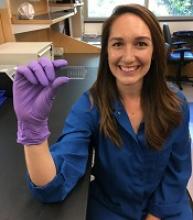
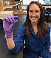
The method is called quantitative deformability cytometry (q-DC). It involves a small device (about 1 inch by 2 inches) made of a soft, flexible rubber that has integrated circuit chips like those in computers.
The researchers use gel particles containing molecules derived from seaweed to force cells through tiny pores in the device. As the cells flow through the device, the researchers take videos at thousands of frames per second—more than 100 times faster than standard video.
Dr Rowat and her colleagues used the device to analyze promyelocytic leukemia cells (HL-60) and breast cancer cells.
The researchers believe this work will provide scientists with a more precise, standardized method to distinguish cancer cells from normal cells.
The team thinks that, in the future, their method could be used to track a cancer patient over time to see how a drug is affecting the patient’s cancer cells.
“By using q-DC, we can very rapidly assess how specific drug treatments affect physical properties of single cells—such as shape, size, and stiffness—and achieve calibrated, quantitative measurements,” Dr Rowat said.
She and her colleagues believe q-DC might also help predict how invasive a cancer cell could be and which drugs might be most effective in fighting the cancer, as well as revealing which proteins are important in regulating the invasion of a cancer cell.
The researchers are now applying q-DC to other types of cancer cells. The team would like to better understand the relationship between a cancer cell’s physical properties and how easily cancer cells can spread through the body.
Dr Rowat’s hypothesis is that properties such as stiffness, size, and a cell’s ability to change shape are important in enabling cancer cells to maneuver.
The researchers said they can also use q-DC to measure other types of cells, such as normal and sickled red blood cells. ![]()
Compound induces selective apoptosis in AML
Researchers say they have discovered a compound that can overcome resistance to apoptosis in acute myeloid leukemia (AML).
The compound, BTSA1, works by activating the BCL-2 family protein BAX.
BTSA1 prompted apoptosis in leukemia cells while sparing healthy cells. It also suppressed AML in mice without producing side effects.
Evripidis Gavathiotis, PhD, of Albert Einstein College of Medicine in Bronx, New York, and his colleagues described these results in Cancer Cell.
The team knew that apoptosis occurs when BAX is activated by pro-apoptotic proteins. However, cancer cells can avoid apoptosis by producing anti-apoptotic proteins that suppress BAX and the proteins that activate it.
“Our novel compound revives suppressed BAX molecules in cancer cells by binding with high affinity to BAX’s activation site,” Dr Gavathiotis said. “BAX can then swing into action, killing cancer cells while leaving healthy cells unscathed.”
Dr Gavathiotis was the lead author of a paper published in Nature in 2008 that first described the structure and shape of BAX’s activation site. He has since looked for small molecules that can activate BAX strongly enough to overcome cancer cells’ resistance to apoptosis.
His team initially screened more than 1 million compounds to reveal those with BAX-binding potential. The most promising 500 compounds were then evaluated in the lab.
“A compound dubbed BTSA1 (short for BAX Trigger Site Activator 1) proved to be the most potent BAX activator, causing rapid and extensive apoptosis when added to several different human AML cell lines,” said Denis Reyna, a doctoral student in Dr Gavathiotis’s lab.
The researchers also tested BTSA1 in blood samples from patients with high-risk AML. BTSA1 induced apoptosis in the patients’ AML cells but did not affect healthy hematopoietic stem cells.
Finally, the researchers generated mouse models of AML. BTSA1 was given to half the mice, while the other half served as controls.
On average, the BTSA1-treated mice survived significantly longer than the control mice—55 days and 40 days, respectively (P=0.0009). In fact, 43% of BTSA1-treated mice were still alive after 60 days and showing no signs of AML.
In addition, the mice treated with BTSA1 showed no evidence of toxicity.
“BTSA1 activates BAX and causes apoptosis in AML cells while sparing healthy cells and tissues, probably because the cancer cells are primed for apoptosis,” Dr Gavathiotis said.
He and his colleagues found that AML cells contained significantly higher BAX levels than normal blood cells from healthy subjects.
“With more BAX available in AML cells, even low BTSA1 doses will trigger enough BAX activation to cause apoptotic death, while sparing healthy cells that contain low levels of BAX or none at all,” Dr Gavathiotis said.
He and his team plan to determine if BTSA1 will elicit similar results in other cancer types. ![]()
CHMP recommends new formulation of pegaspargase
The European Medicines Agency’s Committee for Medicinal Products for Human Use (CHMP) is recommending marketing authorization for lyophilized pegaspargase (ONCASPAR).
If approved, the product would be used as a component of antineoplastic therapy in patients of all ages who have acute lymphoblastic leukemia (ALL).
The product is a freeze-dried formulation of liquid pegaspargase, which is already approved for the aforementioned indication.
The CHMP’s recommendation regarding lyophilized pegaspargase will be submitted to the European Commission (EC).
The EC typically adheres to the CHMP’s recommendations and delivers its final decision within 67 days of the CHMP’s recommendation.
The EC’s decision will be applicable to the entire European Economic Area—all member states of the European Union plus Iceland, Liechtenstein, and Norway.
The CHMP’s recommendation regarding lyophilized pegaspargase is based on analytical and nonclinical studies, which indicate that lyophilized pegaspargase is comparable to the liquid formulation.
Once reconstituted, lyophilized pegaspargase demonstrates similar pharmacokinetics and pharmacodynamics as liquid pegaspargase.
“Lyophilized ONCASPAR builds on more than a decade of data and research with liquid ONCASPAR, and, with no change in dosing regimen, it offers a 3-times longer shelf life,” said Howard B. Mayer, MD, of Shire, the company that developed lyophilized pegaspargase.
“Prolonging shelf life to 24 months for this critically important therapy facilitates management of product inventory by enabling greater flexibility and longer-term planning. Once approved, with the extended shelf life of lyophilized ONCASPAR, we also hope to improve access to the medicine for ALL patients in countries currently not offering liquid ONCASPAR.”
Lyophilized pegaspargase works in the same way as the liquid formulation. It rapidly depletes serum L-asparagine levels and interferes with protein synthesis, thereby depriving lymphoblasts of asparaginase and resulting in cell death. ![]()
The European Medicines Agency’s Committee for Medicinal Products for Human Use (CHMP) is recommending marketing authorization for lyophilized pegaspargase (ONCASPAR).
If approved, the product would be used as a component of antineoplastic therapy in patients of all ages who have acute lymphoblastic leukemia (ALL).
The product is a freeze-dried formulation of liquid pegaspargase, which is already approved for the aforementioned indication.
The CHMP’s recommendation regarding lyophilized pegaspargase will be submitted to the European Commission (EC).
The EC typically adheres to the CHMP’s recommendations and delivers its final decision within 67 days of the CHMP’s recommendation.
The EC’s decision will be applicable to the entire European Economic Area—all member states of the European Union plus Iceland, Liechtenstein, and Norway.
The CHMP’s recommendation regarding lyophilized pegaspargase is based on analytical and nonclinical studies, which indicate that lyophilized pegaspargase is comparable to the liquid formulation.
Once reconstituted, lyophilized pegaspargase demonstrates similar pharmacokinetics and pharmacodynamics as liquid pegaspargase.
“Lyophilized ONCASPAR builds on more than a decade of data and research with liquid ONCASPAR, and, with no change in dosing regimen, it offers a 3-times longer shelf life,” said Howard B. Mayer, MD, of Shire, the company that developed lyophilized pegaspargase.
“Prolonging shelf life to 24 months for this critically important therapy facilitates management of product inventory by enabling greater flexibility and longer-term planning. Once approved, with the extended shelf life of lyophilized ONCASPAR, we also hope to improve access to the medicine for ALL patients in countries currently not offering liquid ONCASPAR.”
Lyophilized pegaspargase works in the same way as the liquid formulation. It rapidly depletes serum L-asparagine levels and interferes with protein synthesis, thereby depriving lymphoblasts of asparaginase and resulting in cell death.
Cryotherapy can reduce signs of CIPN
A new study suggests cryotherapy can reduce symptoms of chemotherapy-induced peripheral neuropathy (CIPN).
Researchers found that having chemotherapy patients wear frozen gloves and socks for 90-minute periods significantly reduced the incidence of CIPN symptoms.
Hiroshi Ishiguro, MD, PhD, of International University of Health and Welfare Hospital in Tochigi, Japan, and colleagues reported these findings in the Journal of the National Cancer Institute.
The researchers prospectively evaluated the efficacy of cryotherapy for preventing CIPN. Breast cancer patients treated weekly with paclitaxel (80 mg/m2 for 1 hour) wore frozen gloves and socks on one side of their bodies for 90 minutes, including the entire duration of drug infusion.
The researchers then compared symptoms on the treated sides with those on the untreated sides.
The primary endpoint was CIPN incidence assessed by changes in tactile sensitivity from a pretreatment baseline. The researchers also assessed subjective symptoms, as reported in the Patient Neuropathy Questionnaire, and patients' manual dexterity.
Among the 40 patients studied, 4 did not reach the cumulative dose due to the occurrence of pneumonia, severe fatigue, liver dysfunction, and macular edema. Of the 36 remaining patients, none dropped out due to cold intolerance.
The incidence of objective and subjective signs of CIPN was clinically and statistically significantly lower on the intervention side than on the control side for most measurements, which includes (among other measures):
- Hand tactile sensitivity—27.8% and 80.6%, respectively (odds ratio[OR]= 20.00, P<0.001)
- Foot tactile sensitivity—25.0% and 63.9%, respectively (OR=infinite, P<0.001)
- Hand warm sense—8.8% and 32.4%, respectively (OR=9.00, P=0.02)
- Foot warm sense—33.4% and 57.6%, respectively (OR=5.00, P=0.04)
- Hand cold sense—2.8% and 13.9%, respectively (OR=infinite, P=0.13)
- Foot cold sense—12.6% and 18.8%, respectively (OR=2.00, P=0.69)
- Severe CIPN in the hand according to the Patient Neuropathy Questionnaire—2.8% and 41.7%, respectively (OR=infinite, P<0.001)
- Severe CIPN in the foot according to the Patient Neuropathy Questionnaire—2.8% and 36.1%, respectively (OR=infinite, P<0.001).
Natural selection opportunities tied to cancer rates
Countries with the lowest opportunities for natural selection have higher cancer rates than countries with the highest opportunities for natural selection, according to a study published in Evolutionary Applications.
Researchers said this is because modern medicine is enabling people to survive cancers, and their genetic backgrounds are passing from one generation to the next.
The team said the rate of some cancers has doubled and even quadrupled over the past 100 to 150 years, and human evolution has moved away from “survival of the fittest.”
“Modern medicine has enabled the human species to live much longer than would otherwise be expected in the natural world,” said study author Maciej Henneberg, PhD, DSc, of the University of Adelaide in South Australia.
“Besides the obvious benefits that modern medicine gives, it also brings with it an unexpected side-effect—allowing genetic material to be passed from one generation to the next that predisposes people to have poor health, such as type 1 diabetes or cancer.”
“Because of the quality of our healthcare in western society, we have almost removed natural selection as the ‘janitor of the gene pool.’ Unfortunately, the accumulation of genetic mutations over time and across multiple generations is like a delayed death sentence.”
Country comparison
The researchers studied global cancer data from the World Health Organization as well as other health and socioeconomic data from the United Nations and the World Bank of 173 countries. The team compared the top 10 countries with the highest opportunities for natural selection to the 10 countries with the lowest opportunities for natural selection.
“We looked at countries that offered the greatest opportunity to survive cancer compared with those that didn’t,” said study author Wenpeng You, a PhD student at the University of Adelaide. “This does not only take into account factors such as socioeconomic status, urbanization, and quality of medical services but also low mortality and fertility rates, which are the 2 distinguishing features in the ‘better’ world.”
“Countries with low mortality rates may allow more people with cancer genetic background to reproduce and pass cancer genes/mutations to the next generation. Meanwhile, low fertility rates in these countries may not be able to have diverse biological variations to provide the opportunity for selecting a naturally fit population—for example, people without or with less cancer genetic background. Low mortality rate and low fertility rate in the ‘better’ world may have formed a self-reinforcing cycle which has accumulated cancer genetic background at a greater rate than previously thought.”
Based on the researchers’ analysis, the 20 countries are:
| Lowest opportunities for natural selection | Highest opportunities for natural selection |
| Iceland | Burkina Faso |
| Singapore | Chad |
| Japan | Central African Republic |
| Switzerland | Afghanistan |
| Sweden | Somalia |
| Luxembourg | Sierra Leone |
| Germany | Democratic Republic of the Congo |
| Italy | Guinea-Bissau |
| Cyprus | Burundi |
| Andorra | Cameroon |
Cancer incidence
The researchers found the rates of most cancers were higher in the 10 countries with the lowest opportunities for natural selection. The incidence of all cancers was 2.326 times higher in the low-opportunity countries than the high-opportunity ones.
The increased incidences of hematologic malignancies were as follows:
- Non-Hodgkin lymphoma—2.019 times higher in the low-opportunity countries
- Hodgkin lymphoma—3.314 times higher in the low-opportunity countries
- Leukemia—3.574 times higher in the low-opportunity countries
- Multiple myeloma—4.257 times higher in the low-opportunity countries .
Dr Henneberg said that, having removed natural selection as the “janitor of the gene pool,” our modern society is faced with a controversial issue.
“It may be that the only way humankind can be rid of cancer once and for all is through genetic engineering—to repair our genes and take cancer out of the equation,” he said.
Study supports prophylaxis in kids with ALL
Results of an observational study support targeted antibacterial prophylaxis in children undergoing induction therapy for acute lymphoblastic leukemia (ALL).
Prophylaxis effectively prevented febrile neutropenia and systemic infection in the children studied.
Prophylaxis with the drug levofloxacin reduced the use of treatment antibiotics and the incidence of Clostridium difficile infection.
“This research provides the first major evidence supporting targeted use of antibacterial prophylaxis for at-risk pediatric ALL patients, particularly use of the broad-spectrum antibiotic levofloxacin,” said study author Joshua Wolf, MD, of St. Jude Children’s Research Hospital in Memphis, Tennessee.
“Prophylactic antibiotic therapy with levofloxacin is routine for at-risk adult ALL patients, but it has remained controversial in children. Until this study, evidence supporting the safety and efficacy of prophylactic antibiotic therapy in children with ALL has been sparse.”
Dr Wolf and his colleagues described their study in Clinical Infectious Diseases.
The study included 344 patients newly diagnosed with ALL who were enrolled in the St. Jude Total XVI clinical trial (NCT00549848). Patients were enrolled from 2007 to 2016.
Until July 2014, the patients received prophylactic antibiotic therapy at the discretion of their physicians. Patients typically received cefepime, ciprofloxacin, or vancomycin plus cefepime or ciprofloxacin. And prophylaxis was typically started at the onset of neutropenia after chemotherapy.
Beginning in August 2014, hospital treatment guidelines changed to recommend prophylactic levofloxacin during induction for ALL patients who develop neutropenia expected to last at least 7 days.
Dr Wolf and his colleagues used the change to compare infection rates and other questions in the following patient groups.
| Patient characteristics | No prophylaxis (n=173) | Levofloxacin prophylaxis (n=69) | Other prophylaxis (n=102) |
| Median age in years (range) | 5.8 (3-11.9) | 6.8 (3.9-11.1) | 7 (3.6-11.9) |
| B-ALL | 83% | 78% | 79% |
| Low-risk ALL | 51% | 54% | 50% |
| Standard-risk ALL | 47% | 41% | 42% |
| High-risk ALL | 2% | 6% | 8% |
| Median duration of neutropenia in days (range) | 17 (11-24) | 18 (12-23) | 20 (17-25) |
| Median duration of profound neutropenia in days (range) | 6 (2-13) | 7 (4-12) | 11 (5-16) |
Results
Researchers reported that patients with neutropenia who received any prophylactic therapy were far less likely than those who did not to develop fever, documented or likely infections, or bloodstream infections.
In a multivariate analysis, the adjusted odds ratios in patients who received prophylaxis, compared to those who did not, were as follows.
- Febrile neutropenia—0.23, P<0.001
- Febrile neutropenia with clinically documented infection—0.30, P=0.002
- Febrile neutropenia with microbiologically documented infection—0.25, P<0.001
- Clinically documented infection—0.54, P=0.02
- Microbiologically documented infection—0.40, P<0.001
- Bloodstream infection—0.30, P=0.008
- C difficile infection—0.38, P=0.04
- Likely bacterial infection—0.26, P<0.001
- Any enterocolitis—0.44, P=0.03.
Analysis also revealed that patients who received levofloxacin had a greater reduction in C difficile infection than patients who received other prophylaxis. The adjusted odds ratio was 0.04 (P<0.001).
However, there was no significant difference between the prophylaxis groups when it came to other infections.
Patients who received levofloxacin prophylaxis had significantly less exposure to other antibiotics than patients who received other prophylaxis or no prophylaxis.
This included exposure to cefepime/ceftazidime (P<0.001 for both comparisons), vancomycin (P<0.001 for both), meropenem (P<0.001 for both), and aminoglycosides (P=0.002 for no prophylaxis, P=0.04 for other prophylaxis).
The reduction in exposure to other antibiotics may partly explain why C difficile infections declined in levofloxacin-treated patients, Dr Wolf said.
He also noted that antibiotic resistance did not significantly increase in this study, despite the greater use of levofloxacin to prevent infections.
“We are cautiously optimistic that any impact of levofloxacin on antibacterial resistance will be balanced by the reduction in use of other antibiotics,” Dr Wolf said, “but long-term monitoring of antibiotic resistance patterns in young ALL patients will be needed to prove this.”
