Article
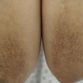
Symmetric Lichen Amyloidosis: An Atypical Location on the Bilateral Extensor Surfaces of the Arms
Lichen amyloidosis (LA) classically presents as a pruritic and papular eruption localized to the pretibial surface of the legs. This report...
Article
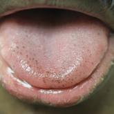
Pigmented Fungiform Papillae of the Tongue in an Indian Male
Pigmented fungiform papillae of the tongue are common lingual hyperpigmented macules in patients with skin of color. It is important to be aware...