Article

Hyperpigmented Flexural Plaques, Hypohidrosis, and Hypotrichosis
A 61-year-old woman with a history of hypohidrosis and deafness presented with a pruritic rash on the neck and antecubital fossae of several years...
Article
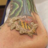
Verruciform Plaques Within a Tattoo of an HIV-Positive Patient
A 40-year-old man with a medical history of human immunodeficiency virus infection managed with highly active antiretroviral therapy, psoriasis,...
Article
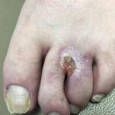
Nonhealing Eroded Plaque on an Interdigital Web Space of the Foot
A 53-year-old man with a history of numerous basal cell carcinomas and odontogenic keratocysts presented with a nonhealing erosion between the...