Article
Bariatric Surgery + Medical Therapy: Effective Tx for T2DM?
Short-term studies have indicated “Yes.” But does a long-term randomized controlled trial give this combination a thumbs up?
Article

Bariatric surgery + medical therapy: Effective Tx for T2DM?
Short-term studies have indicated “Yes,” but does a long-term randomized controlled trial give it a thumbs up?
Article
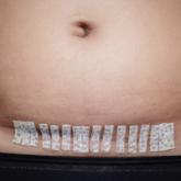
Does Azithromycin Have a Role in Cesarean Sections?
Adding azithromycin to the usual antibiotic protocol in nonelective c-sections reduces infections, research shows. But not everyone can take...
Article

Does azithromycin have a role in cesarean sections?
Yes, adding azithromycin to the usual antibiotic protocol in nonelective c-sections reduces infections. But not everyone can take advantage of it...
Article
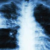
When to “CAP” Off Pneumonia Treatment
Let's be frank: Practitioners often ignore guidelines recommending a five-day course of antibiotics for hospitalized patients with community-...
Article

When to “CAP” off treatment for pneumonia
Is 5 days of antibiotic therapy really sufficient for adults hospitalized with community-acquired pneumonia?
Article
Oral Agent Offers Relief From Generalized Hyperhidrosis
This common problem involving localized sweating can significantly lower quality of life for affected patients. A new treatment option goes down...
Article

Oral agent offers relief from generalized hyperhidrosis
An inexpensive and well-tolerated anticholinergic reduces sweating in those with localized—and generalized—hyperhidrosis.
Article
When Can Exercise Supplant Surgery for Degenerative Meniscal Tears?
Should patients with medial degenerative meniscal tears always undergo surgery? Not according to a recent study, which suggests physical therapy...
Article

When can exercise supplant surgery for degenerative meniscal tears?
Patients with a medial, degenerative meniscal tear and a minimal history of osteoarthritis make good candidates for physical therapy—and there is...
Article
Oral Rehydration Therapy for Kids: A More Palatable Alternative
Getting kids to drink electrolyte solutions following episodes of mild gastroenteritis can be a challenge. Thankfully, parents can rest easy—a new...
Article

A more palatable alternative to oral rehydration therapy for kids
Parents no longer need to struggle to get their kids to drink electrolyte solutions during episodes of mild gastroenteritis; apple juice works...
Article
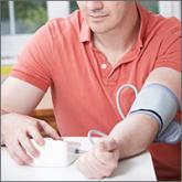
Monitoring home BP readings just got easier
This novel method of identifying patients with uncontrolled hypertension correlates well with ambulatory BP monitoring.