User login
Surgery, dose-escalated chemoradiotherapy yield similar outcomes in NSCLC
After induction chemotherapy and concurrent chemoradiotherapy, patients with non–small cell lung cancer (NSCLC) who underwent surgery versus dose-escalated chemoradiotherapy had similar 5-year overall survival rates.
With a median follow-up of 78 months, the 5-year overall survival for the surgery arm was 44% compared with 40% for the chemoradiotherapy arm, and progression-free survival (PFS) rates were 32% vs. 35%, respectively. In addition, the study showed no significant improvement for the surgery group in the median overall survival or median PFS.

Although the results did not demonstrate a benefit for surgery versus chemoradiotherapy, the 5-year overall survival data for all randomly assigned patients, “are among the best reported so far from prospective trials with definitive chemoradiotherapy,” wrote Dr. Wilfried Eberhardt, of the department of medical oncology, University Hospital Essen, Germany, and colleagues. “Both trimodality treatment that includes surgery and bimodality treatment without surgery but with a definitive chemoradiotherapy boost lead to excellent long-term [overall survival] and PFS,” the researchers wrote (J Clin Oncol 2015 Oct 26. doi:10.1200/JCO.2015.62.6812).
The multicenter phase III trial enrolled 246 patients with NSCLC stage IIIA-UICC6 (n = 75) or IIIB-UICC6 (n = 171) from 2004 to 2013. Overall, 161 patients completed induction therapy with three cycles of cisplatin and paclitaxel and concurrent chemoradiotherapy with 45 Gy, hyperfractionated-accelerated radiotherapy. Patients who had complete, partial, or minor responses after the third cycle were randomly assigned to surgery (n = 81) or chemoradiotherapy (n = 80).
Toxicities of grades 3 and 4 were similar for both arms, as well as for the patients not randomly assigned.
Dr. Eberhardt reported having financial relationships with Eli Lilly, Boehringer Ingelheim, Pfizer, Novartis, Roche, and many other pharmaceutical companies. Several of his coauthors reported ties to industry sources.
After induction chemotherapy and concurrent chemoradiotherapy, patients with non–small cell lung cancer (NSCLC) who underwent surgery versus dose-escalated chemoradiotherapy had similar 5-year overall survival rates.
With a median follow-up of 78 months, the 5-year overall survival for the surgery arm was 44% compared with 40% for the chemoradiotherapy arm, and progression-free survival (PFS) rates were 32% vs. 35%, respectively. In addition, the study showed no significant improvement for the surgery group in the median overall survival or median PFS.
Although the results did not demonstrate a benefit for surgery versus chemoradiotherapy, the 5-year overall survival data for all randomly assigned patients, “are among the best reported so far from prospective trials with definitive chemoradiotherapy,” wrote Dr. Wilfried Eberhardt, of the department of medical oncology, University Hospital Essen, Germany, and colleagues. “Both trimodality treatment that includes surgery and bimodality treatment without surgery but with a definitive chemoradiotherapy boost lead to excellent long-term [overall survival] and PFS,” the researchers wrote (J Clin Oncol 2015 Oct 26. doi:10.1200/JCO.2015.62.6812).
The multicenter phase III trial enrolled 246 patients with NSCLC stage IIIA-UICC6 (n = 75) or IIIB-UICC6 (n = 171) from 2004 to 2013. Overall, 161 patients completed induction therapy with three cycles of cisplatin and paclitaxel and concurrent chemoradiotherapy with 45 Gy, hyperfractionated-accelerated radiotherapy. Patients who had complete, partial, or minor responses after the third cycle were randomly assigned to surgery (n = 81) or chemoradiotherapy (n = 80).
Toxicities of grades 3 and 4 were similar for both arms, as well as for the patients not randomly assigned.
Dr. Eberhardt reported having financial relationships with Eli Lilly, Boehringer Ingelheim, Pfizer, Novartis, Roche, and many other pharmaceutical companies. Several of his coauthors reported ties to industry sources.
FROM JOURNAL OF CLINICAL ONCOLOGY
Key clinical point: After a complex induction protocol, patients with non–small cell lung cancer (NSCLC) who underwent surgery vs. dose-escalated chemoradiotherapy had similar 5-year overall survival rates.
Major finding: Five-year overall survival for the surgery arm was 44% compared with 40% for the chemoradiotherapy arm; progression-free survival rates were 32% vs. 35%, respectively.
Data source: The multicenter phase III ESPATUE trial enrolled 246 patients with NSCLC stage IIIA-UICC6 (n = 75) or IIIB-UICC6 (n = 171).
Disclosures: Dr. Eberhardt reported having financial relationships with Eli Lilly, Boehringer Ingelheim, Pfizer, and Roche, and many other pharmaceutical companies. Several of his coauthors reported ties to industry sources.

Higher risk of basal cell carcinoma linked to menopausal hormone therapy
Late age of natural menopause and use of menopausal hormone therapy were associated with significantly increased risk of basal cell carcinoma (BCC), according to results of a large, nationwide cohort study.
Women who experienced menopause at age 55 years or later, compared with age 50-54 years, had higher BCC risk (hazard ratio, 1.50; 95% confidence interval, 1.04-2.17; P for trend = .017). Among all women, those who had used menopausal hormone therapy had a higher risk (HR, 1.16; 95% confidence interval, 1.03-1.30) (J Clin Oncol. 2015 Nov 2. doi: 10.1200/JCO.2015.62.0625).
The findings are consistent with the hypothesis that exposure to endogenous and exogenous estrogens is photocarcinogenic, according to Dr. Elizabeth Cahoon, research fellow in the Radiation Epidemiology Branch of the National Cancer Institute, Bethesda, Md., and colleagues. Women with the highest observed BCC risk were those who experienced natural menopause and used menopausal hormone therapy for 10 or more years (HR, 1.97; 95% CI, 1.35-2.87; P for trend less than .001).
“In contrast to surgical menopause, which is associated with an immediate and dramatic drop in estrogen levels, women undergoing natural menopause may be exposed to a nontrivial amount of total and unopposed estrogen during the menopausal transition,” they wrote. “In this group, the combination of endogenous and exogenous estrogens may have increased the cumulative estrogen exposure (and, thus, the cumulative phototoxic effects).
The prospective study included 46,100 female, white participants of the US Radiologic Technologists Study, an occupational cohort composed of radiologic technologists. All participants were cancer free at baseline, and 4% had an incident BCC during the approximately 10-year follow-up.
Results showed no association between oral contraceptives and BCC, which may be due a to lower susceptibility in young adults to photosensitizing medications or to the lower doses typical of oral contraceptives. The ethinyl estradiol level in oral contraceptives typically ranges from 20 to 50 mcg, compared with a typical equine estrogen dose in menopausal therapy of 625 mcg.
Most reproductive factors evaluated, including age at menarche, number of live births, and age at first birth, showed no association with BCC.
Dr. Cahoon and coauthors reported having no disclosures.
Key clinical point: Risk of basal cell carcinoma was higher in women with late age of natural menopause and in women who used menopausal hormone therapy.
Major finding: Women who experienced menopause at a late age (55 years or older vs. 50-54 years) had higher BCC risk (HR, 1.50; 95% CI, 1.04-2.17; P for trend = .017), as did women who had ever used menopausal hormone therapy (HR, 1.16; 95% CI, 1.03-1.30).
Data source: The prospective cohort included 46,100 female, white participants of the US Radiologic Technologists Study who were cancer free at baseline; 4% had an incident BCC during the approximately 10-year follow-up.
Disclosures: Dr. Cahoon and coauthors reported having no disclosures.

Dose-intensive, multiagent regimen improves outcomes from low-risk rhabdomyosarcoma
A dose-intensive multiagent regimen including dose-compressed cycles of ifosfamide/etoposide and vincristine/doxorubicin/cyclophosphamide, irinotecan, and radiation resulted in improved outcomes for patients with low-risk stage IV rhabdomyosarcoma (RMS), but not for patients with high-risk disease, according to results from the Children’s Oncology Group study.
For all patients with stage IV rhabdomyosarcoma, the 3-year event-free and overall survival rates were 38% and 56%, respectively. Patients with stage IV RMS with one or fewer Oberlin risk factors had 3-year event-free survival and overall survival rates of 69% and 79%, respectively; patients with two or more Oberlin risk factors had rates of 20% and 14%, respectively (Jour Clin Oncol. 2015 Oct 26. doi: 10.1200/JCO.2015.63.4048).
The study identified an expanded group of patients with low-risk metastatic RMS that included patients with embryonal RMS aged 10 years and older but with an Oberlin score of less than 2. The results in this group represent an improvement over previous study results. However, for the remainder of high-risk patients with alveolar RMS, different approaches are needed, according to Dr. Brenda Weigel of the University of Minnesota, Minneapolis, and her colleagues.
“Unfortunately, alveolar RMS has fewer genetic aberrations than embryonal RMS and no known recurrently mutated cancer consensus genes, which limits genetic targets available for therapeutic approaches,” they wrote.
The Children’s Oncology Group study ARST0431 enrolled 109 patients with metastatic RMS who had no prior chemotherapy or radiation treatment from 2006 to 2008.
The study combined three treatment strategies: dose intensification by interval compression, use of active agents identified in previous phase II window studies, and use of irinotecan as a radiation sensitizer. The 54-week treatment schedule began with two cycles of vincristine/irinotecan followed by interval-compressed vincristine/doxorubicin/cyclophosphamide and ifosfamide/etoposide (cycles began every 14 days), and finished with four cycles of standard vincristine/actinomycin/cyclophosphamide (VAC) and two more cycles of vincristine/irinotecan. The treatment plan also included radiation of primary and metastatic sites at week 19.
The most common nonhematologic adverse event of grade 3 or higher was diarrhea, reported in 20% of patients during the time period when they received irinotecan. Febrile neutropenia occurred in 63% of patients.
Dr. Weigel reported financial relationships with Genentech and Eli Lilly/ImClone System. Several of her coauthors reported having ties to industry sources.
Key clinical point: A dose-intensive, multiagent regimen including dose-compressed cycles of ifosfamide/etoposide and vincristine/doxorubicin/cyclophosphamide, irinotecan, and radiation resulted in improved outcomes for patients with low-risk stage IV rhabdomyosarcoma (RMS), but not for patients with high-risk disease.
Major finding: Patients with stage IV rhabdomyosarcoma with one or fewer Oberlin risk factors had 3-year event-free survival and overall survival rates of 69% and 79%, respectively; patients with two or more Oberlin risk factors had rates of 20% and 14%, respectively.
Data source: The Children’s Oncology Group study ARST0431 involving 109 patients with metastatic RMS who had no prior chemotherapy or radiation treatment from 2006 to 2008.
Disclosures: Dr. Weigel reported financial relationships with Genentech and Eli Lilly/ImClone System. Several of her coauthors reported having ties to industry sources.

Gene mutations had little impact on everolimus benefit in advanced breast cancer
Genetic alterations affecting phosphoinositide 3-kinase (PI3K) and fibroblast growth factor receptor (FGFR), and associated pathways, only minimally affected the efficacy of everolimus in patients with hormone receptor–positive (HR+), human epidermal growth factor receptor 2–negative (EGFR-) advanced breast cancer.
To determine whether everolimus yielded greater treatment benefits in tumors dependent on a hyperactive PI3K/mTOR pathway, progression-free survival (PFS) in the presence and absence of mutated PIK3CA (catalytic subunit of PI3K) was examined. Median PFS was longer for all patients with wild-type PIK3CA (catalytic subunit of PI3K); everolimus was associated with a slightly greater reduction in risk than placebo: the hazard ratio was 0.37 (95% CI, 0.25 to 0.55) for wild-type PIK3CA and 0.51 (95% CI, 0.34 to 0.77) for mutated. The everolimus benefit was greater in patients with exon-9 mutations (HR, 0.26; 95% CI, 0.12 to 0.54) than exon-20 mutations (HR, 0.56; 95% CI, 0.31 to 1.00).

The lack of an appreciable effect of activating mutations on everolimus benefit, “highlighted the oversimplification of both the hypothesis and the analytic method of pooling all genetic alterations in this frequently mutated gene or hyperactive pathway,” wrote Dr. Gabriel N. Hortobagyi, professor of medicine, University of Texas MD Anderson Cancer Center, Houston, and his colleagues (J Clin Oncol. 2015 Oct. 26. doi:10.1200/JCO.2014.60.1971).
The data showed that PIK3CA exon-9 mutations were associated with more benefit than were exon-20 mutations, pointing to the importance of clinically evaluating mutations in terms of their specific functional effects, the investigators said.
Similar to observations with the PI3K pathway, genetic alterations in FGFR and cell-cycle control genes had minimal effect on PFS gains with everolimus.

The study also used NextGen sequencing to derive a contrast-induced nephropathy scoring system to assess the influence of chromosomal instability on everolimus benefit. Patients with low chromosomal instability (below the 75th percentile) derived a median PFS gain of 5.5 months with everolimus, compared with 1.5 months for patients with high chromosomal instability (above the 75th percentile). The effect of chromosomal instability on PFS was evident only in the everolimus arm, suggesting that tumors with lower chromosomal instability may derive greater benefit from the addition of everolimus.
Using data acquired from a medium-size cancer panel to measure chromosomal instability expands the utility of the NextGen sequencing data increasingly collected in oncology practice, Dr. Hortobagyi and his associates wrote.
Genetic alterations affecting phosphoinositide 3-kinase (PI3K) and fibroblast growth factor receptor (FGFR), and associated pathways, only minimally affected the efficacy of everolimus in patients with hormone receptor–positive (HR+), human epidermal growth factor receptor 2–negative (EGFR-) advanced breast cancer.
To determine whether everolimus yielded greater treatment benefits in tumors dependent on a hyperactive PI3K/mTOR pathway, progression-free survival (PFS) in the presence and absence of mutated PIK3CA (catalytic subunit of PI3K) was examined. Median PFS was longer for all patients with wild-type PIK3CA (catalytic subunit of PI3K); everolimus was associated with a slightly greater reduction in risk than placebo: the hazard ratio was 0.37 (95% CI, 0.25 to 0.55) for wild-type PIK3CA and 0.51 (95% CI, 0.34 to 0.77) for mutated. The everolimus benefit was greater in patients with exon-9 mutations (HR, 0.26; 95% CI, 0.12 to 0.54) than exon-20 mutations (HR, 0.56; 95% CI, 0.31 to 1.00).
The lack of an appreciable effect of activating mutations on everolimus benefit, “highlighted the oversimplification of both the hypothesis and the analytic method of pooling all genetic alterations in this frequently mutated gene or hyperactive pathway,” wrote Dr. Gabriel N. Hortobagyi, professor of medicine, University of Texas MD Anderson Cancer Center, Houston, and his colleagues (J Clin Oncol. 2015 Oct. 26. doi:10.1200/JCO.2014.60.1971).
The data showed that PIK3CA exon-9 mutations were associated with more benefit than were exon-20 mutations, pointing to the importance of clinically evaluating mutations in terms of their specific functional effects, the investigators said.
Similar to observations with the PI3K pathway, genetic alterations in FGFR and cell-cycle control genes had minimal effect on PFS gains with everolimus.
The study also used NextGen sequencing to derive a contrast-induced nephropathy scoring system to assess the influence of chromosomal instability on everolimus benefit. Patients with low chromosomal instability (below the 75th percentile) derived a median PFS gain of 5.5 months with everolimus, compared with 1.5 months for patients with high chromosomal instability (above the 75th percentile). The effect of chromosomal instability on PFS was evident only in the everolimus arm, suggesting that tumors with lower chromosomal instability may derive greater benefit from the addition of everolimus.
Using data acquired from a medium-size cancer panel to measure chromosomal instability expands the utility of the NextGen sequencing data increasingly collected in oncology practice, Dr. Hortobagyi and his associates wrote.
Key clinical point: Mutations in genes involved in the PI3K/mTOR pathway, FGFR signaling, and cell cycle regulation had little impact on everolimus benefit in hormone receptor–positive (HR+), human epidermal growth factor receptor 2–negative (EGFR-) advanced breast cancer.
Major finding: For patients treated with either everolimus or placebo, median PFS was longer if they carried wild-type PIK3CA. For patients treated with everolimus, hazard ratios for wild-type vs. mutated PIK3CA were 0.37 (95% CI, 0.25 to 0.55) and 0.51 (95% CI, 0.34 to 0.77), respectively.
Data source: The BOLERO-2 trial included 724 patients with HR+, HER2- advanced breast cancer; samples from 302 (42%) were evaluated with NextGen sequencing.
Disclosures: Dr. Hortobagyi reported consulting or advisory roles with Pfizer, Antigen Express, Novartis, Galena, Genentech, and Amgen. Several of his coauthors reported having ties to industry sources.

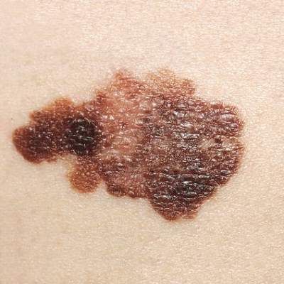
Intermittent interferon falls short for stage III melanoma patients
For patients with resected stage III melanoma, intermittent intravenous high-dose IFN–alpha-2b (iHDI) is not superior to conventional 1-year high-dose IFN–alpha-2b therapy, according to results from a phase III study. Relapse-free survival was significantly inferior with the intermittent regimen.
“These data preclude speculation about a possible equivalence of the regimens. Taken together, the approved schedules for adjuvant IFN [interferon] treatment of patients with melanoma are likely to remain the standard of care until new approaches, such as with ipilimumab or other drugs, show durable improvements in RFS or OS and acceptable safety profiles,” wrote Dr. Peter Mohr of Elbe-Klinikum Buxtehude, Germany, and his colleagues (J Clin. Onc. 2015 Oct. 26. doi: 10.1200/JCO.2015.59.6932).
The prospective, multicenter, phase III randomized trial included 649 patients with stage III melanoma who received either three courses of iHDI or the conventional HDI 1-year regimen.After a median follow-up time of 55 months, distant metastasis-free survival and overall survival for the two treatment schedules were similar. The 5-year distant metastasis-free survival for iHDI, compared with HDI, was 49.2% vs. 53.1%; 5-year overall survival was 62.9% vs. 64.1%, respectively. However, relapse-free survival was significantly worse for iHDI than for HDI (hazard ratio, 1.27; 95% confidence interval, 1.02-1.59; P = .03).
Adverse events (AEs) were similar for the two arms, except the HDI group experienced more anemia than the iHDI group (48.1% vs. 30.9%; P less than .001) and neutropenia of grade 3 or higher was more frequent in the iHDI group (38.9% vs. 30.1%; P = .004). The intermittent schedule resulted in fewer cumulative AEs, particularly grade 3 or 4 fatigue. Fatigue is an expected but often debilitating AE of HDI therapy, the investigators noted.
Reduced fatigue was associated with 40% less early discontinuation with iHDI than with standard HDI. In the iHDI group, quality of life measures returned to or exceeded baseline values within 4 weeks of administration. However, improvements in quality of life should be weighed against potential loss of efficacy: at least 50% of patients would tolerate moderate or severe IFN–alpha-2b toxicity for improvement in 5-year, disease-free survival, the investigators said.
Key clinical point: Distant metastasis-free survival and overall survival were similar in patients with stage III melanoma treated with intermittent high-dose intravenous IFN–alpha-2b (iHDI) vs. standard 1-year high-dose IFN–alpha-2b (HDI), but the difference for relapse-free survival was significant (HR, 1.27; P = .03), favoring standard HDI.
Major finding: The 5-year distant metastasis-free survival for iHDI, compared with HDI was 49.2% vs. 53.1%; 5-year overall survival was 62.9% vs. 64.1%, respectively. Relapse-free survival was significantly worse for iHDI than HDI (HR, 1.27; 95% CI, 1.02-1.59; P = .03).
Data source: The prospective, multicenter, phase III trial randomized 649 patients with stage III melanoma to receive either three courses of iHDI or the conventional HDI 1-year regimen.
Disclosures: Dr. Mohr reported financial relationships with Merck, Bristol-Myers Squibb, Roche, GlaxoSmithKline, LEO Pharma, Novartis, Merck Sharp & Dohme, and Roche. Several of his coauthors reported having ties to industry sources.
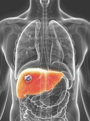
High-radiation doses improve survival with inoperable intrahepatic cholangiocarcinoma
Using recent advances in radiotherapy (RT) planning and delivery, high-dose radiation delivered to hepatic tumors produced major survival benefits in patients with inoperable intrahepatic cholangiocarcinoma (IHCC), investigators reported online in Journal of Clinical Oncology.
“Treatment with ablative doses of RT using high-quality daily CT image guidance with inspiration breath-hold gating can achieve survival times comparable to those achieved with resection,” wrote Dr. Randa Tao, radiation oncologist at the University of Texas MD Anderson Cancer Center, Houston, and her colleagues (Jour Clin Onc. 2015 Oct 26 [doi: 10.1200/JCO.2015.61.3778]).

From 2002 to 2014, 79 patients with inoperable IHCC were treated with definitive RT. The median survival time was 30 months; 1-, 2-, and 3-year overall survival rates were 87%, 61%, and 44%, respectively. Median progression-free survival was 30 months, and 1-, 2-, and 3-year progression-free survival rates were 88%, 61%, and 39%, respectively.
After completion of RT, 38 patients (48%) had primary tumor progression. Actuarial 1-, 2-, and 3-year local control rates were 81%, 45%, and 27%, respectively, with median duration of 23 months. The majority of patients (34) had recurrence within the high-dose radiation region, three had both in-field and marginal progression, and one had recurrence at the margin.
RT dose was the most important prognostic factor for overall survival and local control. Patients treated with doses higher than the conventional 50.4 Gy had a median survival of 43 months, compared with 23 months for patients treated with doses 50.4 Gy or less (P = .01).
Total biologically effective dose (BED) affected outcomes also. The 2- and 3-year overall survival rates for patients treated with BED greater than 80.5 Gy were both 73%, compared with 58% and 38% for those treated with BED of 80.5 Gy or less.
The treatment was well tolerated, with no cases of radiation induced liver disease observed.
The investigators recommend that higher total RT doses and higher doses delivered per fraction to achieve BED greater than 80.5 Gy should be considered for all patients as long as image guidance is used to ensure that the dose is delivered safely, and dose constraints to the liver, bile duct, stomach, and bowel can be met. The findings support the use of 67.5 Gy in 15 fractions (BED, 97.88 Gy).
Dr. Tao reported having no disclosures. Several of her coauthors reported having ties to industry sources.
Using recent advances in radiotherapy (RT) planning and delivery, high-dose radiation delivered to hepatic tumors produced major survival benefits in patients with inoperable intrahepatic cholangiocarcinoma (IHCC), investigators reported online in Journal of Clinical Oncology.
“Treatment with ablative doses of RT using high-quality daily CT image guidance with inspiration breath-hold gating can achieve survival times comparable to those achieved with resection,” wrote Dr. Randa Tao, radiation oncologist at the University of Texas MD Anderson Cancer Center, Houston, and her colleagues (Jour Clin Onc. 2015 Oct 26 [doi: 10.1200/JCO.2015.61.3778]).
From 2002 to 2014, 79 patients with inoperable IHCC were treated with definitive RT. The median survival time was 30 months; 1-, 2-, and 3-year overall survival rates were 87%, 61%, and 44%, respectively. Median progression-free survival was 30 months, and 1-, 2-, and 3-year progression-free survival rates were 88%, 61%, and 39%, respectively.
After completion of RT, 38 patients (48%) had primary tumor progression. Actuarial 1-, 2-, and 3-year local control rates were 81%, 45%, and 27%, respectively, with median duration of 23 months. The majority of patients (34) had recurrence within the high-dose radiation region, three had both in-field and marginal progression, and one had recurrence at the margin.
RT dose was the most important prognostic factor for overall survival and local control. Patients treated with doses higher than the conventional 50.4 Gy had a median survival of 43 months, compared with 23 months for patients treated with doses 50.4 Gy or less (P = .01).
Total biologically effective dose (BED) affected outcomes also. The 2- and 3-year overall survival rates for patients treated with BED greater than 80.5 Gy were both 73%, compared with 58% and 38% for those treated with BED of 80.5 Gy or less.
The treatment was well tolerated, with no cases of radiation induced liver disease observed.
The investigators recommend that higher total RT doses and higher doses delivered per fraction to achieve BED greater than 80.5 Gy should be considered for all patients as long as image guidance is used to ensure that the dose is delivered safely, and dose constraints to the liver, bile duct, stomach, and bowel can be met. The findings support the use of 67.5 Gy in 15 fractions (BED, 97.88 Gy).
Dr. Tao reported having no disclosures. Several of her coauthors reported having ties to industry sources.
Key clinical point: High-radiation doses delivered to hepatic tumors were well tolerated and survival outcomes were comparable to surgical resection.
Major finding: The median survival time was 30 months; 1-, 2-, and 3-year overall survival rates were 87%, 61%, and 44%, respectively.
Data source: From 2002 to 2014, 79 patients with IHCC were treated with definitive radiotherapy at the University of Texas MD Anderson Cancer Center.
Disclosures: Dr. Tao reported having no disclosures. Several of her coauthors reported having ties to industry sources.
Low BMI predicted worse survival in mCRC
In patients with metastatic colorectal cancer (mCRC), body mass index (BMI) was prognostic for overall survival (OS) and progression-free survival (PFS), investigators reported online in Journal of Clinical Oncology.
Risks were highest at the lowest BMI values, decreased as BMI increased to 28 kg/m2, and plateaued at higher BMI values.
By pooling data from more than 21,000 patients enrolled worldwide in 25 randomized trials for frontline treatment, “we have shown that BMI is prognostic for OS and PFS in this population, but with a shape of the risk curve across the BMI spectrum, different than that observed in the adjuvant setting,” wrote Lindsay Renfro, Ph.D., of the Mayo Clinic, Rochester, Minn., and her colleagues (Jour Clin Onc. 2015 Oct 26 [doi: 10.1200/JCO.2015.61.6441]).
Patients with a BMI of 18.5 kg/m2 had a 50% increased risk of death (95% confidence interval, 43%-56%). After researchers adjusted for age, sex, performance status, and clinical characteristics, the prognostic significance of BMI remained (P less than .001).
Previous studies showed that obese patients with stage II or III colon cancer were at increased risk for disease recurrence or death, but results of the current study showed obese patients with mCRC were not at increased risk.
Men with low BMIs had a greater risk of death than did women. Both men and women with moderate and higher BMIs had similar risks. Previous studies have shown that the prognosis for women with colorectal cancer is improved over men, possibly because of the protective effect of estrogen.
The results suggest that patients with mCRC and low BMI are likely cachectic, a condition that affects approximately 50% of patients with colon cancer and is associated with a 20% mortality rate, the authors noted.
FROM JOURNAL OF CLINICAL ONCOLOGY
Key clinical point: Low BMI predicted worse overall and progression-free survival in patients with mCRC.
Major finding: Risks of death and disease progression were highest for patients with the lowest BMI, decreased as BMI increased to 28 kg/m2, and plateaued at higher BMI values.
Data source: Retrospective analysis of data from 25 first-line clinical trials that included 21,149 patients with metastatic colorectal cancer.
Disclosures: Dr. Renfro reported having no disclosures. Several of her coauthors reported having ties to industry sources.

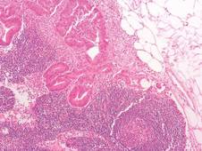
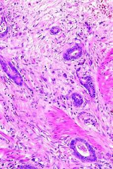
Blacks receive inferior care for localized prostate cancer
Black patients with localized prostate cancer who underwent radical prostatectomy received inferior surgical care, compared with whites, as evidenced by fewer lymph node dissections and longer delays from diagnosis to treatment, among other indicators, researchers reported.
The time from diagnosis to treatment was longer in blacks than whites (79 vs. 71 days, P = .001). Overall, 57.7% of blacks had surgery without adjuvant therapy, compared with 61.3% of whites (P = .001). Blacks were less likely to undergo lymph node dissection (52.8% vs. 61.5%, P less than .001). The difference persisted but was not significant when the lymph node dissection analysis was restricted to patients with intermediate- and high-risk disease. Blacks were more likely to visit the emergency department within 30 days (P = .04) and beyond 30 days (P = .006) (JAMA Oncol. 2015 Oct 22. doi: 10.1001/jamaoncol/2015.3384).
When the lymph node dissection analysis was adjusted for geographic location (health service area), there were no differences between groups, which suggests that geographic variation in quality of care is tightly linked to racial disparities and may account for a large proportion of the differences.
The retrospective analysis of Medicare billing records evaluated 26,482 patients (2,020 blacks [7.6%] and 24,462 non-Hispanic whites [92.4%]) who underwent radical prostatectomy from 1992 through 2009.
Prostate cancer–specific mortality was not significantly different between blacks and whites. Unadjusted overall mortality was increased in blacks, but after adjustment for geographic location, overall mortality was similar between groups.
“Despite important constellations of poor quality of care for blacks undergoing [radical prostatectomy], we did not detect significant differences in overall and cancer-specific survival,” wrote Dr. Marianne Schmid of Brigham and Women’s Hospital, Harvard Medical School, Boston, and her colleagues. Furthermore, the research revealed no regional variation in cancer-specific mortality among patients who underwent surgery.
“A possible interpretation of our findings is that the biological differences in tumor aggressiveness among blacks may have been exaggerated, and that the perceived gap in survival is a result of lack of access or cultural perceptions with regard to surgical care for [prostate cancer] or other factors that differentiate who makes it to the operating table,” the researchers noted.
Median total calculated costs were lower for blacks ($13,015 vs. $15,758), but surgical treatment was associated with higher incremental annual costs, with the top 50% spending $1,185 more. Increased costs are likely due to the higher prevalence of radiotherapy and androgen deprivation therapy, and increased rates of emergency department visits, rather than the use of newer, more expensive technologies.
The research indicates that unfavorable quality of care for black patients did not translate to worse overall survival and cancer-specific survival. Rather, lower survival among black patients with prostate cancer may result from barriers to definitive treatment, according to the investigators.
Dr. Schmid reported having no relevant financial disclosures. Several of her coauthors reported financial ties to several industry sources.
The findings of the study by Dr. Schmid and her colleagues point to racial health inequalities in the United States and are applicable to medical care of the American population in general, not just to men with prostate cancer.
 |
Dr. Otis Brawley |
Despite the fact that black patients in this study had insurance and access to care and were considered healthy enough for surgery, there was still a disparity in quality of care. Interestingly, there were no differences between the groups in all-cause or cancer-specific mortality. This is hard evidence that the biology of prostate cancer is similar for black and white men with localized disease.
Is the disparity in quality of care due to racism on the part of physicians? Probably not. More likely, a higher proportion of black men have physicians who do not routinely perform radical prostatectomies, and a higher proportion of blacks are treated at hospitals that have a low volume of prostate surgery.
Even though a significant proportion of blacks received inferior treatment, similar outcomes for the two groups suggests that some patients with localized prostate cancer are overtreated, and the medical community should be more discerning in who receives treatment.
Dr. Otis Brawley is the chief medical officer for the American Cancer Society and professor of hematology, oncology, medicine, and epidemiology at Emory University, Atlanta. These remarks were part of an editorial accompanying the report (JAMA Oncol. 2015 Oct 22. doi: 10.1001/jamaoncol/2015.3384). Dr. Brawley reported having no relevant financial disclosures.
The findings of the study by Dr. Schmid and her colleagues point to racial health inequalities in the United States and are applicable to medical care of the American population in general, not just to men with prostate cancer.
|
Dr. Otis Brawley |
Despite the fact that black patients in this study had insurance and access to care and were considered healthy enough for surgery, there was still a disparity in quality of care. Interestingly, there were no differences between the groups in all-cause or cancer-specific mortality. This is hard evidence that the biology of prostate cancer is similar for black and white men with localized disease.
Is the disparity in quality of care due to racism on the part of physicians? Probably not. More likely, a higher proportion of black men have physicians who do not routinely perform radical prostatectomies, and a higher proportion of blacks are treated at hospitals that have a low volume of prostate surgery.
Even though a significant proportion of blacks received inferior treatment, similar outcomes for the two groups suggests that some patients with localized prostate cancer are overtreated, and the medical community should be more discerning in who receives treatment.
Dr. Otis Brawley is the chief medical officer for the American Cancer Society and professor of hematology, oncology, medicine, and epidemiology at Emory University, Atlanta. These remarks were part of an editorial accompanying the report (JAMA Oncol. 2015 Oct 22. doi: 10.1001/jamaoncol/2015.3384). Dr. Brawley reported having no relevant financial disclosures.
Black patients with localized prostate cancer who underwent radical prostatectomy received inferior surgical care, compared with whites, as evidenced by fewer lymph node dissections and longer delays from diagnosis to treatment, among other indicators, researchers reported.
The time from diagnosis to treatment was longer in blacks than whites (79 vs. 71 days, P = .001). Overall, 57.7% of blacks had surgery without adjuvant therapy, compared with 61.3% of whites (P = .001). Blacks were less likely to undergo lymph node dissection (52.8% vs. 61.5%, P less than .001). The difference persisted but was not significant when the lymph node dissection analysis was restricted to patients with intermediate- and high-risk disease. Blacks were more likely to visit the emergency department within 30 days (P = .04) and beyond 30 days (P = .006) (JAMA Oncol. 2015 Oct 22. doi: 10.1001/jamaoncol/2015.3384).
When the lymph node dissection analysis was adjusted for geographic location (health service area), there were no differences between groups, which suggests that geographic variation in quality of care is tightly linked to racial disparities and may account for a large proportion of the differences.
The retrospective analysis of Medicare billing records evaluated 26,482 patients (2,020 blacks [7.6%] and 24,462 non-Hispanic whites [92.4%]) who underwent radical prostatectomy from 1992 through 2009.
Prostate cancer–specific mortality was not significantly different between blacks and whites. Unadjusted overall mortality was increased in blacks, but after adjustment for geographic location, overall mortality was similar between groups.
“Despite important constellations of poor quality of care for blacks undergoing [radical prostatectomy], we did not detect significant differences in overall and cancer-specific survival,” wrote Dr. Marianne Schmid of Brigham and Women’s Hospital, Harvard Medical School, Boston, and her colleagues. Furthermore, the research revealed no regional variation in cancer-specific mortality among patients who underwent surgery.
“A possible interpretation of our findings is that the biological differences in tumor aggressiveness among blacks may have been exaggerated, and that the perceived gap in survival is a result of lack of access or cultural perceptions with regard to surgical care for [prostate cancer] or other factors that differentiate who makes it to the operating table,” the researchers noted.
Median total calculated costs were lower for blacks ($13,015 vs. $15,758), but surgical treatment was associated with higher incremental annual costs, with the top 50% spending $1,185 more. Increased costs are likely due to the higher prevalence of radiotherapy and androgen deprivation therapy, and increased rates of emergency department visits, rather than the use of newer, more expensive technologies.
The research indicates that unfavorable quality of care for black patients did not translate to worse overall survival and cancer-specific survival. Rather, lower survival among black patients with prostate cancer may result from barriers to definitive treatment, according to the investigators.
Dr. Schmid reported having no relevant financial disclosures. Several of her coauthors reported financial ties to several industry sources.
FROM JAMA ONCOLOGY
Key clinical point: Compared with whites, black patients with localized prostate cancer received lower quality of care.
Major finding: Blacks were less likely to undergo lymph node dissection than whites (52.8% vs. 61.5%; OR, 0.76; P less than .001), and had more postoperative complications, emergency department visits, and readmissions (P less than .05 for all comparisons).
Data source: A retrospective analysis of Medicare billing records of 26,482 patients (7.6% blacks and 92.4% non-Hispanic whites) who underwent radical prostatectomy from 1992 through 2009.
Disclosures: Dr. Schmid reported having no relevant financial disclosures. Several of her coauthors reported financial ties to several industry sources.

Small nucleolar RNA signals colorectal cancer outcomes
Expression of a set of small nucleolar RNAs (snoRNAs) was found to be significantly elevated in cancer tissue, compared with normal tissue, and expression of a specific snoRNA, SNORA42, was associated with poor overall and disease-free survival in patients with colorectal cancer (CRC).
Elevated expression of the specific snoRNA SNORA42 was an independent prognostic indicator for overall survival (hazard ratio, 2.11; P = .021), disease-free survival (HR, 3.17; P = .011), and distant metastasis (HR, 2.66; P .023). In a subset of patients with stage II CRC, the significant association between high expression of SNORA42 and poor prognosis remained.
The majority of patients with stage II CRC are cured by surgery alone, but a significant proportion experience disease progression. According to Dr. Yoshinaga Okugawa of the Center for Gastrointestinal Cancer Research at the Baylor University Medical Center, Dallas, and colleagues, adjuvant chemotherapy treatment for all patients with stage II cancer is controversial.
“Therefore, identification of such high-risk patients with CRC using molecular biomarkers such as SNORA42 expression will allow use of adjuvant chemotherapy after surgery only in a select subgroup of high-risk patients to improve their prognosis,” they wrote (Gut 2015 Oct 15. doi: 10.1136/gutjnl-2015.309359).
The research examined expression levels of RNA extracted from formalin-fixed paraffin-embedded (FFPE) tissue samples from 250 patients with colorectal cancer and 24 matched controls. The initial focus was on four snoRNAs that were reported to be dysregulated in other cancers. The study found that all four of the snoRNAs were significantly upregulated in CRC tissue, compared with normal tissue. Receiver operating characteristic (ROC) curves showed that expression levels of all four snoRNAs distinguished between cancer and noncancer tissue; area under the ROC curves values ranged from 0.75 to 0.88.
SnoRNAs are single-stranded, noncoding RNAs that have recently emerged as important components in the control of cell fate and oncogenesis in various cancers. In addition, studies have suggested the potential of snoRNAs as prognostic indicators.
While the expression levels of all four snoRNAs distinguished cancer tissue from normal tissue, only one of the snoRNAs, SNORA42, had expression levels that correlated with disease outcomes (overall survival, disease-free survival, and distant metastasis).
To characterize the biologic role of SNORN42 in CRC progression, the research team investigated its expression and impact on cancer cell lines and animal models. Overexpressed SNORN42 enhanced cell proliferation and tumorigenicity in cultured cells and in an animal model of CRC.
Taken together, the results suggest potential clinical usefulness of SNORA42 as a diagnostic and predictive biomarker in patients with CRC.
Dr. Okugawa and coauthors reported having no disclosures.
FROM GUT
Key clinical point: Elevated expression of a small nucleolar RNA was associated with decreased overall and disease-free survival in patients with colorectal cancer.
Major finding: Elevated expression of the specific snoRNA SNORA42 was an independent prognostic indicator for overall survival (HR, 2.11; P = .021), disease-free survival (HR, 3.17; P = .011), and distant metastasis (HR, 2.66; P = .023).
Data source: RNA was extracted from FFPE tissue samples from 250 patients with colorectal cancer and 24 samples from matched controls.
Disclosures: Dr. Okugawa and coauthors reported having no disclosures.

Cladribine plus cytarabine yields 85% 5-year survival in Langerhans cell histiocytosis
After standard therapy failed, cladribine plus cytosine-arabinoside (cytarabine [Ara-C]) was effective in treating patients with high-risk, organ-positive Langerhans cell histiocytosis; however, all patients experienced hematologic toxicity, based on the results of a phase II study.
The overall response rate was 92% and the 5-year survival rate was 85% (95% confidence interval, 65%-94%). The study included 27 patients, median age 0.73 years (range, 0.1-3.3 years), who did not respond to standard therapy and had involvement of at least one risk organ (spleen, liver, or bone marrow), reported Dr. Jean Donadieu of Service d’Hemato-Oncologie Pediatrique, Hopital Trousseau, Paris, and colleagues (Blood. 2015;126[12]:1415-23).
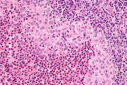
The study had strict inclusion criteria, and treatment was only given to children with severe Langerhans cell histiocytosis (LCH), which historically has had long-term survival rates of 20% to 30%.
After one course of combination therapy, all of the patients experienced pancytopenia (grade 4, median duration 23 days) complicated by fever, and 30% had severe infections. In the two patients who died as a result of protocol-related toxicity, the cumulative doses of cladribine were 250 mg/m2 and 225 mg/m2.
Given that cladribine is known to cause severe lymphopenia and prolonged pancytopenia, the researchers suggested limiting the cumulative dose to 200 mg/m2 and limiting cladribine/Ara-C treatment to a maximum of three courses. The researchers further cautioned that the treatment should be performed at institutions trained in providing acute myeloid leukemia chemotherapy regimens or hematopoietic transplantation.
The response to cladribine/Ara-C treatment was slow in LCH patients. Those who responded to the therapy, but had not achieved a complete response after two to three courses, achieved a complete response after continuation with a less toxic therapy.
“Accordingly, we recommend that care be taken at this time to avoid potential toxicity on the one hand but also, on the other hand, to avoid prematurely deciding that the therapy is not working and possibly discontinuing treatment that might help longer-term,” wrote Dr. Donadieu and associates.
A comprehensive LCH activity scoring system determined the initial severity of disease and assessed treatment efficacy. With successful therapy, the median disease activity score decreased from 12 (range, 6-20) to 3 (range, 0-18; P less than .0001).
Dr. Donadieu and coauthors reported having no disclosures.
In previous collaborative trials, patients with the worst prognosis (overall 2-year survival of less than 30%) had disease involving the bone marrow, liver, or spleen; did not respond to routine induction treatment; or had reactivation in one of the high-risk organs. The bottom line in this report is the overall response rate of 92% and long-term survival rate of 85% among 27 patients with severe LCH.
This report demonstrates that children who have disease of the bone marrow, liver, or spleen and who do not respond to standard induction therapy can be treated with intensive chemotherapy.
The next step is the LCH-IV study, launched by the Histiocyte Society to test a slightly modified treatment plan that attempts to decrease the toxicity yet maintain the improved overall survival seen in this phase II trial.
Dr. Kimo C. Stine is a pediatric hematologist-oncologist at Arkansas Children’s Hospital, Little Rock, and professor of pediatrics at the University of Arkansas for Medical Sciences. These remarks were part of an editorial (Blood. 2015;126[12]:1399-1400) accompanying the report. Dr. Stine had no disclosures to report.
After standard therapy failed, cladribine plus cytosine-arabinoside (cytarabine [Ara-C]) was effective in treating patients with high-risk, organ-positive Langerhans cell histiocytosis; however, all patients experienced hematologic toxicity, based on the results of a phase II study.
The overall response rate was 92% and the 5-year survival rate was 85% (95% confidence interval, 65%-94%). The study included 27 patients, median age 0.73 years (range, 0.1-3.3 years), who did not respond to standard therapy and had involvement of at least one risk organ (spleen, liver, or bone marrow), reported Dr. Jean Donadieu of Service d’Hemato-Oncologie Pediatrique, Hopital Trousseau, Paris, and colleagues (Blood. 2015;126[12]:1415-23).
The study had strict inclusion criteria, and treatment was only given to children with severe Langerhans cell histiocytosis (LCH), which historically has had long-term survival rates of 20% to 30%.
After one course of combination therapy, all of the patients experienced pancytopenia (grade 4, median duration 23 days) complicated by fever, and 30% had severe infections. In the two patients who died as a result of protocol-related toxicity, the cumulative doses of cladribine were 250 mg/m2 and 225 mg/m2.
Given that cladribine is known to cause severe lymphopenia and prolonged pancytopenia, the researchers suggested limiting the cumulative dose to 200 mg/m2 and limiting cladribine/Ara-C treatment to a maximum of three courses. The researchers further cautioned that the treatment should be performed at institutions trained in providing acute myeloid leukemia chemotherapy regimens or hematopoietic transplantation.
The response to cladribine/Ara-C treatment was slow in LCH patients. Those who responded to the therapy, but had not achieved a complete response after two to three courses, achieved a complete response after continuation with a less toxic therapy.
“Accordingly, we recommend that care be taken at this time to avoid potential toxicity on the one hand but also, on the other hand, to avoid prematurely deciding that the therapy is not working and possibly discontinuing treatment that might help longer-term,” wrote Dr. Donadieu and associates.
A comprehensive LCH activity scoring system determined the initial severity of disease and assessed treatment efficacy. With successful therapy, the median disease activity score decreased from 12 (range, 6-20) to 3 (range, 0-18; P less than .0001).
Dr. Donadieu and coauthors reported having no disclosures.
FROM BLOOD
Key clinical point: In children with high-risk, refractory Langerhans cell histiocytosis (LCH), treatment with cladribine plus cytarabine was effective but highly toxic.
Major finding: The overall response rate was 92%, and the 5-year survival rate was 85% (95% CI, 65%-94%).
Data source: The phase II study included 27 patients (mean age, 0.73 years) who had involvement of at least one risk organ (spleen, liver, bone marrow) and had failed standard therapy.
Disclosures: Dr. Donadieu and coauthors reported having no disclosures.