Article
Nodular Extramammary Paget Disease With Fibroepitheliomatous Hyperplasia
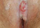
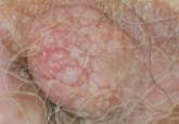
Extramammary Paget disease (EMPD) is a rare skin condition usually found in the anogenital region. Histologically, EMPD may be associated with...
Article
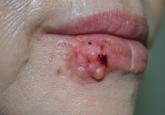
An Amelanotic Malignant Melanoma of the Lip: Unusual Shape and Atypical Location
Amelanotic malignant melanoma (AMM) is characterized by little or no visible pigment. The diagnosis of AMM is a challenge for clinicians because...