Article
Reinforcing a Spica Cast With a Fiberglass Bar
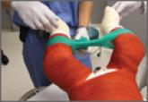
Hip spica casting is used in the treatment of femur fractures and hip dysplasia in children 1 to 6 years old. A bar connecting the legs of the...
Hip spica casting is used in the treatment of femur fractures and hip dysplasia in children 1 to 6 years old. A bar connecting the legs of the...