Skip that repeat DXA scan in these postmenopausal women

Article Type
Changed
Wed, 12/08/2021 - 17:51
Display Headline
Skip that repeat DXA scan in these postmenopausal women

ILLUSTRATIVE CASE

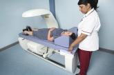
A 70-year-old White woman with a history of type 2 diabetes and a normal body mass index (BMI) presents to your office for a preventive care exam. She is otherwise doing well, without concerns. Her first dual-energy x-ray absorptiometry (DXA) scan, completed at age 67, demonstrated normal bone density. Should you recommend a repeat DXA scan today?

As many as 1 in 2 postmenopausal women are at risk for an ­osteoporosis-related fracture.2 Each year, about 2 million fragility fractures occur in the United States.2,3 The US Preventive Services Task Force (USPSTF) recommends bone mineral density (BMD) measurement in all women ages 65 years and older, as well as in younger postmenopausal women with certain clinical risk factors.4 The USPSTF does not make a recommendation regarding the interval for follow-up BMD testing.

Two prospective cohort studies determined that repeat BMD testing 4 to 8 years after baseline screening did not improve fracture risk prediction.5,6 Limitations of these studies included no analysis of high-risk subgroups, as well as failure to include many younger postmenopausal women in the studies.5,6 An additional longitudinal study that followed postmenopausal women for up to 15 years estimated that the interval for at least 10% of women to develop osteoporosis after initial screening was more than 15 years for women with normal BMD and about 5 years for those with moderate osteopenia.7

STUDY SUMMARY

No added predictive benefit found in 3-year repeat scan

The current study examined data from the Women’s Health Initiative Extension 1 Study, a large prospective cohort that included a broader range of postmenopausal women (N = 7419) than the previous studies. The purpose of this study was to determine if a second BMD measurement, about 3 years after the baseline BMD screening, would be useful in predicting risk for major osteoporotic fracture (MOF), compared with baseline BMD measurement alone. It analyzed data from prespecified subgroups defined by age, BMI, race/ethnicity, presence or absence of diabetes, and baseline BMD T score.1

Study participants averaged 66 years of age, with a mean BMI of 29, and 23% were non-White. In addition, 97% had either normal BMD or osteopenia (T score ≥ −2.4). Participants were excluded from the study if they had been treated with bone-active medications other than vitamin D and calcium, reported a history of MOF (fracture of the hip, spine, radius, ulna, wrist, upper arm, or shoulder) at baseline or between BMD tests, missed follow-up visits after the Year 3 BMD scan, or had missing covariate data. Participants self-reported fractures on annual patient questionnaires, and hip fractures were confirmed through medical records.

During the mean follow-up period of 9 years after the second BMD test, 139 women (1.9%) had 1 or more hip fractures, and 732 women (9.9%) had 1 or more MOFs.

Area under the receiver operating characteristic curve (AU-ROC) values for baseline BMD screening and baseline plus 3-year BMD measurement were similar in their ability to discriminate between women who had a hip fracture or MOF and women who did not. AU-ROC values communicate the usefulness of a diagnostic or screening test. An AU-ROC value of 1 would be considered perfect (100% sensitive and 100% specific), whereas an AU-ROC of 0.5 suggests a test with no ability to discriminate at all. Values between 0.7 and 0.8 would be considered acceptable, and those between 0.8 and 0.9, excellent.

Continue to: The AU-ROCs in this study...


The AU-ROCs in this study were 0.71 (95% CI, 0.67-0.75) for baseline total hip BMD, 0.61 (95% CI, 0.56-0.65) for change in total hip BMD between baseline and 3-year BMD scan, and 0.73 (95% CI, 0.69-0.77) for the combined baseline total hip BMD and change in total hip BMD. For femoral neck and lumbar spine BMD, AU-ROC values demonstrated comparable discrimination of hip fracture and MOF as those for total hip BMD. The AU-ROC values among age subgroups (< 65 years, 65-74 years, and ≥ 75 years) were also similar. Associations between change in bone density and fracture risk did not change when adjusted for factors such as BMI, race/ethnicity, diabetes, or baseline BMD.

WHAT’S NEW

Results can be applied to a wider range of patients

This study found that for postmenopausal women, a repeat BMD measurement obtained 3 years after the initial assessment did not improve risk discrimination for hip fracture or MOF beyond the baseline BMD value and should not be routinely performed. Additionally, evidence from this study allows this recommendation to apply to younger postmenopausal women and a variety of high-risk subgroups.

CAVEATS

Possible bias due to self-reporting of fractures

This study suggests that for women without a diagnosis of osteoporosis at initial screening, repeat testing is unlikely to affect future risk stratification. Repeat BMD testing should still be considered when the results are likely to influence clinical management.

However, an important consideration is that fractures were self-reported in this study, introducing a possible source of bias. Additionally, although this study supports foregoing repeat screening at a 3-year interval, there is still no agreed-upon determination of when (or if) to repeat BMD screening in women without osteoporosis.

A large subset of the study population was younger than 65 (44%), the age when family physicians typically recommend screening for osteoporosis. However, the age-adjusted AU-ROC values for fracture risk prediction were the same, and this should not invalidate the conclusions for the study population at large.

CHALLENGES TO IMPLEMENTATION

No challenges seen

We see no challenges in implementing this recommendation.

ACKNOWLEDGEMENT

The PURLs Surveillance System was supported in part by Grant Number UL1RR024999 from the National Center for Research Resources, a Clinical Translational Science Award to the University of Chicago. The content is solely the responsibility of the authors and does not necessarily represent the official views of the National Center for Research Resources or the National Institutes of Health.

Files
References

1. Crandall CJ, Larson J, Wright NC, et al. Serial bone density measurement and incident fracture risk discrimination in postmenopausal women. JAMA Intern Med. 2020;180:1232-1240. doi: 10.1001/jamainternmed.2020.2986

2. US Preventive Services Task Force. Screening for osteoporosis: US Preventive Services Task Force recommendation statement. Ann Intern Med. 2011;154:356-364. doi: 10.7326/0003-4819-154-5-201103010-00307

3. Burge R, Dawson-Hughes B, Solomon DH, et al. Incidence and economic burden of osteoporosis-related fractures in the United States, 2005-2025. J Bone Miner Res. 2007;22:465-475. doi: 10.1359/jbmr.061113

4. US Preventive Services Task Force; Curry SJ, Krist AH, Owens DK, et al. Screening for osteoporosis to prevent fractures: US Preventive Services Task Force recommendation statement. JAMA. 2018;319:2521-2531. doi: 10.1001/jama.2018.7498

5. Hillier TA, Stone KL, Bauer DC, et al. Evaluating the value of repeat bone mineral density measurement and prediction of fractures in older women: the study of osteoporotic fractures. Arch Intern Med. 2007;167:155-160. doi: 10.1001/archinte.167.2.155

6. Berry SD, Samelson EJ, Pencina MJ, et al. Repeat bone mineral density screening and prediction of hip and major osteoporotic fracture. JAMA. 2013;310:1256-1262. doi: 10.1001/jama.2013.277817

7. Gourlay ML, Fine JP, Preisser JS, et al; Study of Osteoporotic Fractures Research Group. Bone-density testing interval and transition to osteoporosis in older women. N Engl J Med. 2012;366:225-233. doi: 10.1056/NEJMoa1107142

Article PDF
Author and Disclosure Information

Department of Family and Community Medicine, University of Missouri, Columbia

DEPUTY EDITOR
Shailendra Prasad, MBBS, MPH

University of Minnesota North Memorial Family Medicine Residency Program, Minneapolis

Issue
The Journal of Family Practice - 70(10)
Publications
Topics
Page Number
510-512
Sections
Files
Files
Author and Disclosure Information

Department of Family and Community Medicine, University of Missouri, Columbia

DEPUTY EDITOR
Shailendra Prasad, MBBS, MPH

University of Minnesota North Memorial Family Medicine Residency Program, Minneapolis

Author and Disclosure Information

Department of Family and Community Medicine, University of Missouri, Columbia

DEPUTY EDITOR
Shailendra Prasad, MBBS, MPH

University of Minnesota North Memorial Family Medicine Residency Program, Minneapolis

Article PDF
Article PDF

ILLUSTRATIVE CASE

A 70-year-old White woman with a history of type 2 diabetes and a normal body mass index (BMI) presents to your office for a preventive care exam. She is otherwise doing well, without concerns. Her first dual-energy x-ray absorptiometry (DXA) scan, completed at age 67, demonstrated normal bone density. Should you recommend a repeat DXA scan today?

As many as 1 in 2 postmenopausal women are at risk for an ­osteoporosis-related fracture.2 Each year, about 2 million fragility fractures occur in the United States.2,3 The US Preventive Services Task Force (USPSTF) recommends bone mineral density (BMD) measurement in all women ages 65 years and older, as well as in younger postmenopausal women with certain clinical risk factors.4 The USPSTF does not make a recommendation regarding the interval for follow-up BMD testing.

Two prospective cohort studies determined that repeat BMD testing 4 to 8 years after baseline screening did not improve fracture risk prediction.5,6 Limitations of these studies included no analysis of high-risk subgroups, as well as failure to include many younger postmenopausal women in the studies.5,6 An additional longitudinal study that followed postmenopausal women for up to 15 years estimated that the interval for at least 10% of women to develop osteoporosis after initial screening was more than 15 years for women with normal BMD and about 5 years for those with moderate osteopenia.7

STUDY SUMMARY

No added predictive benefit found in 3-year repeat scan

The current study examined data from the Women’s Health Initiative Extension 1 Study, a large prospective cohort that included a broader range of postmenopausal women (N = 7419) than the previous studies. The purpose of this study was to determine if a second BMD measurement, about 3 years after the baseline BMD screening, would be useful in predicting risk for major osteoporotic fracture (MOF), compared with baseline BMD measurement alone. It analyzed data from prespecified subgroups defined by age, BMI, race/ethnicity, presence or absence of diabetes, and baseline BMD T score.1

Study participants averaged 66 years of age, with a mean BMI of 29, and 23% were non-White. In addition, 97% had either normal BMD or osteopenia (T score ≥ −2.4). Participants were excluded from the study if they had been treated with bone-active medications other than vitamin D and calcium, reported a history of MOF (fracture of the hip, spine, radius, ulna, wrist, upper arm, or shoulder) at baseline or between BMD tests, missed follow-up visits after the Year 3 BMD scan, or had missing covariate data. Participants self-reported fractures on annual patient questionnaires, and hip fractures were confirmed through medical records.

During the mean follow-up period of 9 years after the second BMD test, 139 women (1.9%) had 1 or more hip fractures, and 732 women (9.9%) had 1 or more MOFs.

Area under the receiver operating characteristic curve (AU-ROC) values for baseline BMD screening and baseline plus 3-year BMD measurement were similar in their ability to discriminate between women who had a hip fracture or MOF and women who did not. AU-ROC values communicate the usefulness of a diagnostic or screening test. An AU-ROC value of 1 would be considered perfect (100% sensitive and 100% specific), whereas an AU-ROC of 0.5 suggests a test with no ability to discriminate at all. Values between 0.7 and 0.8 would be considered acceptable, and those between 0.8 and 0.9, excellent.

Continue to: The AU-ROCs in this study...


The AU-ROCs in this study were 0.71 (95% CI, 0.67-0.75) for baseline total hip BMD, 0.61 (95% CI, 0.56-0.65) for change in total hip BMD between baseline and 3-year BMD scan, and 0.73 (95% CI, 0.69-0.77) for the combined baseline total hip BMD and change in total hip BMD. For femoral neck and lumbar spine BMD, AU-ROC values demonstrated comparable discrimination of hip fracture and MOF as those for total hip BMD. The AU-ROC values among age subgroups (< 65 years, 65-74 years, and ≥ 75 years) were also similar. Associations between change in bone density and fracture risk did not change when adjusted for factors such as BMI, race/ethnicity, diabetes, or baseline BMD.

WHAT’S NEW

Results can be applied to a wider range of patients

This study found that for postmenopausal women, a repeat BMD measurement obtained 3 years after the initial assessment did not improve risk discrimination for hip fracture or MOF beyond the baseline BMD value and should not be routinely performed. Additionally, evidence from this study allows this recommendation to apply to younger postmenopausal women and a variety of high-risk subgroups.

CAVEATS

Possible bias due to self-reporting of fractures

This study suggests that for women without a diagnosis of osteoporosis at initial screening, repeat testing is unlikely to affect future risk stratification. Repeat BMD testing should still be considered when the results are likely to influence clinical management.

However, an important consideration is that fractures were self-reported in this study, introducing a possible source of bias. Additionally, although this study supports foregoing repeat screening at a 3-year interval, there is still no agreed-upon determination of when (or if) to repeat BMD screening in women without osteoporosis.

A large subset of the study population was younger than 65 (44%), the age when family physicians typically recommend screening for osteoporosis. However, the age-adjusted AU-ROC values for fracture risk prediction were the same, and this should not invalidate the conclusions for the study population at large.

CHALLENGES TO IMPLEMENTATION

No challenges seen

We see no challenges in implementing this recommendation.

ACKNOWLEDGEMENT

The PURLs Surveillance System was supported in part by Grant Number UL1RR024999 from the National Center for Research Resources, a Clinical Translational Science Award to the University of Chicago. The content is solely the responsibility of the authors and does not necessarily represent the official views of the National Center for Research Resources or the National Institutes of Health.

References

1. Crandall CJ, Larson J, Wright NC, et al. Serial bone density measurement and incident fracture risk discrimination in postmenopausal women. JAMA Intern Med. 2020;180:1232-1240. doi: 10.1001/jamainternmed.2020.2986

2. US Preventive Services Task Force. Screening for osteoporosis: US Preventive Services Task Force recommendation statement. Ann Intern Med. 2011;154:356-364. doi: 10.7326/0003-4819-154-5-201103010-00307

3. Burge R, Dawson-Hughes B, Solomon DH, et al. Incidence and economic burden of osteoporosis-related fractures in the United States, 2005-2025. J Bone Miner Res. 2007;22:465-475. doi: 10.1359/jbmr.061113

4. US Preventive Services Task Force; Curry SJ, Krist AH, Owens DK, et al. Screening for osteoporosis to prevent fractures: US Preventive Services Task Force recommendation statement. JAMA. 2018;319:2521-2531. doi: 10.1001/jama.2018.7498

5. Hillier TA, Stone KL, Bauer DC, et al. Evaluating the value of repeat bone mineral density measurement and prediction of fractures in older women: the study of osteoporotic fractures. Arch Intern Med. 2007;167:155-160. doi: 10.1001/archinte.167.2.155

6. Berry SD, Samelson EJ, Pencina MJ, et al. Repeat bone mineral density screening and prediction of hip and major osteoporotic fracture. JAMA. 2013;310:1256-1262. doi: 10.1001/jama.2013.277817

7. Gourlay ML, Fine JP, Preisser JS, et al; Study of Osteoporotic Fractures Research Group. Bone-density testing interval and transition to osteoporosis in older women. N Engl J Med. 2012;366:225-233. doi: 10.1056/NEJMoa1107142

Issue
The Journal of Family Practice - 70(10)
Issue
The Journal of Family Practice - 70(10)
Page Number
510-512
Page Number
510-512
Publications
Publications
Topics
Article Type
Display Headline
Skip that repeat DXA scan in these postmenopausal women
Display Headline
Skip that repeat DXA scan in these postmenopausal women
Sections
PURLs Copyright
Copyright © 2021. The Family Physicians Inquiries Network. All rights reserved.
Inside the Article

PRACTICE CHANGER

Do not routinely repeat bone density testing 3 years after initial screening in postmenopausal patients who do not have osteoporosis.

STRENGTH OF RECOMMENDATION

A: Based on several large, good-quality prospective cohort studies1

Crandall CJ, Larson J, Wright NC, et al. Serial bone density measurement and incident fracture risk discrimination in postmenopausal women. JAMA Intern Med. 2020;180:1232-1240. doi: 10.1001/jamainternmed.2020.2986

Disallow All Ads
Content Gating
No Gating (article Unlocked/Free)
Alternative CME
Disqus Comments
Default
Use ProPublica
Hide sidebar & use full width
render the right sidebar.
Conference Recap Checkbox
Not Conference Recap
Clinical Edge
Display the Slideshow in this Article
Medscape Article
Display survey writer
Reuters content
Disable Inline Native ads
WebMD Article
Article PDF Media
Media Files

An Easy Approach to Obtaining Clean-catch Urine From Infants

Article Type
Changed
Wed, 08/01/2018 - 12:17
Display Headline
An Easy Approach to Obtaining Clean-catch Urine From Infants

A fussy 6-month-old infant is brought to the emergency department (ED) with a rectal temperature of 101.5°F. She is consolable, breathing normally, and appears well hydrated. You find no clear etiology for her fever and suspect that a urinary tract infection (UTI) may be the source of her illness. How do you proceed with obtaining a urine sample?

A  febrile infant in a family practice office or ED is a familiar clinical situation that may require an invasive diagnostic workup. Up to 7% of infants ages 2 to 24 months with fever of unknown origin may have a UTI.2 Collecting a urine sample from pre–toilet-trained children can be time consuming. In fact, in one RCT, obtaining a clean-catch urine sample in this age group took more than an hour, on average.3 But more convenient methods of urine collection, such as placing a cotton ball in the diaper or using a perineal collection bag, have contamination rates of up to 63%.4

In its guidelines for evaluating possible UTI in a febrile child younger than age 2, the American Academy of Pediatrics (AAP) recommends obtaining a sample for urinalysis “through the most convenient means.”5 If urinalysis is positive, only urine obtained by catheterization or suprapubic aspiration should be cultured. Guidelines from the National Institute for Health and Care Excellence in the United Kingdom are similar, but allow for culture of clean-catch urine samples.6

A recent prospective cohort study examined a noninvasive alternating lumbar-bladder tapping method to stimulate voiding in infants ages 6 months or younger.7 Within five minutes, 49% of the infants provided a clean-catch sample, with contamination rates similar to those of samples obtained using invasive methods.7 Younger infants were more likely to void within the time allotted. Another trial of bladder tapping conducted in hospitalized infants younger than 30 days old showed similar results.8 There are, however, no previously reported randomized trials demonstrating the efficacy of a noninvasive urine collection technique in the outpatient setting.

Use of invasive collection methods requires skilled personnel and may cause significant discomfort for patients (and parents). Noninvasive methods, such as bag urine collection, have unacceptable contamination rates. In addition, waiting to catch a potentially cleaner urine sample is time consuming, so better strategies to collect urine from infants are needed. This RCT is the first to examine the efficacy of a unique stimulation technique to obtain a clean-catch urine sample from infants ages 1 to 12 months.

STUDY SUMMARY

Noninvasive stimulation triggers faster samples

A nonblinded, single-center RCT conducted in Australia compared two methods for obtaining a clean-catch urine sample within five minutes: the Quick-Wee method (suprapubic stimulation with gauze soaked in cold fluid) or usual care (waiting for spontaneous voiding with no stimulation).1 A total of 354 infants (ages 1-12 mo) who required urine sample collection were randomized in a 1:1 ratio; allocation was concealed. Infants with anatomic or neurologic abnormalities and those needing immediate antibiotic therapy were excluded.

The most common reasons for obtaining the urine sample were fever of unknown origin and “unsettled baby,” followed by poor feeding and suspected UTI. The primary outcome was voiding within five minutes; secondary outcomes included time to void, whether urine was successfully caught, contamination rate, and parent/clinician satisfaction.

Study personnel removed the diaper, then cleaned the genitals of all patients with room temperature sterile water. A caregiver or clinician was ready and waiting to catch urine when the patient voided. In the Quick-Wee group, a clinician rubbed the patient’s suprapubic area in a circular fashion with gauze soaked in refrigerated saline (2.8°C). At five minutes, clinicians recorded the voiding status and decided how to proceed.

Using intention-to-treat analysis, 31% of the patients in the Quick-Wee group voided within five minutes, compared with 12% of the usual-care patients. Similarly, 30% of patients in the Quick-Wee group provided a successful clean-catch sample within five minutes, compared with 9% in the usual-care group (number needed to treat, 4.7).

Contamination rates were no different between the Quick-Wee and usual-care samples. Both parents and clinicians were more satisfied with the Quick-Wee method than with usual care (median score of 2 vs 3 on a 5-point Likert scale, in which 1 is “most satisfied”). There was no difference when results were adjusted for age or sex. No adverse events occurred.

Continue to: WHAT'S NEW


WHAT’S NEW

Method could reduce need for invasive sampling

A simple suprapubic stimulation technique increased the number of infants who provided a clean-catch voided urine sample within five minutes—a clinically relevant and satisfying outcome. In appropriate patients, use of the Quick-Wee method to obtain a clean-catch voided sample for initial urinalysis, rather than attempting methods with known high contamination rates, may potentially reduce the need for invasive sampling using catheterization or suprapubic aspiration.

Credit: Shutterstock/Atstock Productions

CAVEATS

Complete age range & ideal storage temperature are unknown

Neonates and precontinent children older than 12 months were not included in this trial, so these conclusions do not apply to those groups. The intervention period lasted only five minutes, but other published studies suggest that this amount of time is adequate for voiding to occur.6,7 Although this study used soaking fluid stored at 2.8°C, the ideal storage temperature is unknown.

CHALLENGES TO IMPLEMENTATION

AAP doesn’t endorse clean-catch urine samples

The Quick-Wee method is simple and easy to implement, and requires no specialized training or equipment. AAP guidelines do not endorse the use of clean-catch voided urine for culture, which may be a barrier to changing urine collection practices in some settings.

ACKNOWLEDGEMENT

The PURLs Surveillance System was supported in part by Grant Number UL1RR024999 from the National Center For Research Resources, a Clinical Translational Science Award to the University of Chicago. The content is solely the responsibility of the authors and does not necessarily represent the official views of the National Center For Research Resources or the National Institutes of Health.

Copyright © 2018. The Family Physicians Inquiries Network. All rights reserved.

Reprinted with permission from the Family Physicians Inquiries Network and The Journal of Family Practice (2018;67[3]: 166, 168-169).

References

1. Kaufman J, Fitzpatrick P, Tosif S, et al. Faster clean catch urine collection (Quick-Wee method) from infants: randomised controlled trial. BMJ. 2017;357:j1341.
2. Shaikh N, Morone NE, Bost JE, Farrell MH. Prevalence of urinary tract infection in childhood: a meta-analysis. Pediatr Infect Dis J. 2008;27(4):302-308.
3. Davies P, Greenwood R, Benger J. Randomised trial of a vibrating bladder stimulator—the time to pee study. Arch Dis Child. 2008;93(5):423-424.
4. Al-Orifi F, McGillivray D, Tange S, Kramer MS. Urine culture from bag specimens in young children: are the risks too high? J Pediatr. 2000;137(2):221-226.
5. Roberts KB, Downs SM, Finnell SM, et al; Subcommittee on Urinary Tract Infection. Reaffirmation of AAP clinical practice guideline: the diagnosis and management of the initial urinary tract infection in febrile infants and young children 2-24 months of age. Pediatrics. 2016;138(6): e20163026.
6. National Institute for Health and Care Excellence. Urinary tract infection in under 16s: diagnosis and management [clinical guideline CG54]. www.nice.org.uk/guidance/cg54/chapter/1-guidance. Accessed March 1, 2018.
7. Labrosse M, Levy A, Autmizguine J, Gravel J. Evaluation of a new strategy for clean-catch urine in infants. Pediatrics. 2016;138(3):e20160573.
8. Herreros Fernández ML, González Merino N, Tagarro García A, et al. A new technique for fast and safe collection of urine in newborns. Arch Dis Child. 2013;98(1):27-29.

Article PDF
Author and Disclosure Information

Laura Morris is an Associate Professor of Clinical Family and Community Medicine at the University of Missouri, Columbia.

Issue
Clinician Reviews - 28(4)
Publications
Topics
Page Number
22-23,26
Sections
Author and Disclosure Information

Laura Morris is an Associate Professor of Clinical Family and Community Medicine at the University of Missouri, Columbia.

Author and Disclosure Information

Laura Morris is an Associate Professor of Clinical Family and Community Medicine at the University of Missouri, Columbia.

Article PDF
Article PDF

A fussy 6-month-old infant is brought to the emergency department (ED) with a rectal temperature of 101.5°F. She is consolable, breathing normally, and appears well hydrated. You find no clear etiology for her fever and suspect that a urinary tract infection (UTI) may be the source of her illness. How do you proceed with obtaining a urine sample?

A  febrile infant in a family practice office or ED is a familiar clinical situation that may require an invasive diagnostic workup. Up to 7% of infants ages 2 to 24 months with fever of unknown origin may have a UTI.2 Collecting a urine sample from pre–toilet-trained children can be time consuming. In fact, in one RCT, obtaining a clean-catch urine sample in this age group took more than an hour, on average.3 But more convenient methods of urine collection, such as placing a cotton ball in the diaper or using a perineal collection bag, have contamination rates of up to 63%.4

In its guidelines for evaluating possible UTI in a febrile child younger than age 2, the American Academy of Pediatrics (AAP) recommends obtaining a sample for urinalysis “through the most convenient means.”5 If urinalysis is positive, only urine obtained by catheterization or suprapubic aspiration should be cultured. Guidelines from the National Institute for Health and Care Excellence in the United Kingdom are similar, but allow for culture of clean-catch urine samples.6

A recent prospective cohort study examined a noninvasive alternating lumbar-bladder tapping method to stimulate voiding in infants ages 6 months or younger.7 Within five minutes, 49% of the infants provided a clean-catch sample, with contamination rates similar to those of samples obtained using invasive methods.7 Younger infants were more likely to void within the time allotted. Another trial of bladder tapping conducted in hospitalized infants younger than 30 days old showed similar results.8 There are, however, no previously reported randomized trials demonstrating the efficacy of a noninvasive urine collection technique in the outpatient setting.

Use of invasive collection methods requires skilled personnel and may cause significant discomfort for patients (and parents). Noninvasive methods, such as bag urine collection, have unacceptable contamination rates. In addition, waiting to catch a potentially cleaner urine sample is time consuming, so better strategies to collect urine from infants are needed. This RCT is the first to examine the efficacy of a unique stimulation technique to obtain a clean-catch urine sample from infants ages 1 to 12 months.

STUDY SUMMARY

Noninvasive stimulation triggers faster samples

A nonblinded, single-center RCT conducted in Australia compared two methods for obtaining a clean-catch urine sample within five minutes: the Quick-Wee method (suprapubic stimulation with gauze soaked in cold fluid) or usual care (waiting for spontaneous voiding with no stimulation).1 A total of 354 infants (ages 1-12 mo) who required urine sample collection were randomized in a 1:1 ratio; allocation was concealed. Infants with anatomic or neurologic abnormalities and those needing immediate antibiotic therapy were excluded.

The most common reasons for obtaining the urine sample were fever of unknown origin and “unsettled baby,” followed by poor feeding and suspected UTI. The primary outcome was voiding within five minutes; secondary outcomes included time to void, whether urine was successfully caught, contamination rate, and parent/clinician satisfaction.

Study personnel removed the diaper, then cleaned the genitals of all patients with room temperature sterile water. A caregiver or clinician was ready and waiting to catch urine when the patient voided. In the Quick-Wee group, a clinician rubbed the patient’s suprapubic area in a circular fashion with gauze soaked in refrigerated saline (2.8°C). At five minutes, clinicians recorded the voiding status and decided how to proceed.

Using intention-to-treat analysis, 31% of the patients in the Quick-Wee group voided within five minutes, compared with 12% of the usual-care patients. Similarly, 30% of patients in the Quick-Wee group provided a successful clean-catch sample within five minutes, compared with 9% in the usual-care group (number needed to treat, 4.7).

Contamination rates were no different between the Quick-Wee and usual-care samples. Both parents and clinicians were more satisfied with the Quick-Wee method than with usual care (median score of 2 vs 3 on a 5-point Likert scale, in which 1 is “most satisfied”). There was no difference when results were adjusted for age or sex. No adverse events occurred.

Continue to: WHAT'S NEW


WHAT’S NEW

Method could reduce need for invasive sampling

A simple suprapubic stimulation technique increased the number of infants who provided a clean-catch voided urine sample within five minutes—a clinically relevant and satisfying outcome. In appropriate patients, use of the Quick-Wee method to obtain a clean-catch voided sample for initial urinalysis, rather than attempting methods with known high contamination rates, may potentially reduce the need for invasive sampling using catheterization or suprapubic aspiration.

Credit: Shutterstock/Atstock Productions

CAVEATS

Complete age range & ideal storage temperature are unknown

Neonates and precontinent children older than 12 months were not included in this trial, so these conclusions do not apply to those groups. The intervention period lasted only five minutes, but other published studies suggest that this amount of time is adequate for voiding to occur.6,7 Although this study used soaking fluid stored at 2.8°C, the ideal storage temperature is unknown.

CHALLENGES TO IMPLEMENTATION

AAP doesn’t endorse clean-catch urine samples

The Quick-Wee method is simple and easy to implement, and requires no specialized training or equipment. AAP guidelines do not endorse the use of clean-catch voided urine for culture, which may be a barrier to changing urine collection practices in some settings.

ACKNOWLEDGEMENT

The PURLs Surveillance System was supported in part by Grant Number UL1RR024999 from the National Center For Research Resources, a Clinical Translational Science Award to the University of Chicago. The content is solely the responsibility of the authors and does not necessarily represent the official views of the National Center For Research Resources or the National Institutes of Health.

Copyright © 2018. The Family Physicians Inquiries Network. All rights reserved.

Reprinted with permission from the Family Physicians Inquiries Network and The Journal of Family Practice (2018;67[3]: 166, 168-169).

References

1. Kaufman J, Fitzpatrick P, Tosif S, et al. Faster clean catch urine collection (Quick-Wee method) from infants: randomised controlled trial. BMJ. 2017;357:j1341.
2. Shaikh N, Morone NE, Bost JE, Farrell MH. Prevalence of urinary tract infection in childhood: a meta-analysis. Pediatr Infect Dis J. 2008;27(4):302-308.
3. Davies P, Greenwood R, Benger J. Randomised trial of a vibrating bladder stimulator—the time to pee study. Arch Dis Child. 2008;93(5):423-424.
4. Al-Orifi F, McGillivray D, Tange S, Kramer MS. Urine culture from bag specimens in young children: are the risks too high? J Pediatr. 2000;137(2):221-226.
5. Roberts KB, Downs SM, Finnell SM, et al; Subcommittee on Urinary Tract Infection. Reaffirmation of AAP clinical practice guideline: the diagnosis and management of the initial urinary tract infection in febrile infants and young children 2-24 months of age. Pediatrics. 2016;138(6): e20163026.
6. National Institute for Health and Care Excellence. Urinary tract infection in under 16s: diagnosis and management [clinical guideline CG54]. www.nice.org.uk/guidance/cg54/chapter/1-guidance. Accessed March 1, 2018.
7. Labrosse M, Levy A, Autmizguine J, Gravel J. Evaluation of a new strategy for clean-catch urine in infants. Pediatrics. 2016;138(3):e20160573.
8. Herreros Fernández ML, González Merino N, Tagarro García A, et al. A new technique for fast and safe collection of urine in newborns. Arch Dis Child. 2013;98(1):27-29.

Issue
Clinician Reviews - 28(4)
Issue
Clinician Reviews - 28(4)
Page Number
22-23,26
Page Number
22-23,26
Publications
Publications
Topics
Article Type
Display Headline
An Easy Approach to Obtaining Clean-catch Urine From Infants
Display Headline
An Easy Approach to Obtaining Clean-catch Urine From Infants
Sections
Disallow All Ads
Content Gating
No Gating (article Unlocked/Free)
Alternative CME
Disqus Comments
Default
Use ProPublica
Article PDF Media

An easy approach to obtaining clean-catch urine from infants

Article Type
Changed
Fri, 01/18/2019 - 08:53
Display Headline
An easy approach to obtaining clean-catch urine from infants

ILLUSTRATIVE CASE

A fussy 6-month-old infant is brought into the emergency department (ED) with a rectal temperature of 101.5° F. She is consolable, breathing normally, and appears well hydrated. You find no clear etiology for her fever and suspect that a urinary tract infection (UTI) may be the source of her illness. How do you proceed with obtaining a urine sample?

Afebrile infant in the family physician’s office or ED is a familiar clinical situation that may require an invasive diagnostic work-up. Up to 7% of infants ages 2 to 24 months with fever of unknown origin may have a UTI.2 Collecting a urine sample from pre-toilet-trained children can be time consuming. In fact, obtaining a clean-catch urine sample in this age group took an average of more than one hour in one randomized controlled trial (RCT).3 More convenient methods of urine collection, such as placing a cotton ball in the diaper or using a perineal collection bag, have contamination rates of up to 63%.4

The American Academy of Pediatrics (AAP) guidelines for evaluating possible UTI in a febrile child <2 years of age recommend obtaining a sample for urinalysis “through the most convenient means.”5 If urinalysis is positive, only urine obtained by catheterization or suprapubic aspiration should be cultured. Guidelines from the National Institute for Health and Care Excellence in the United Kingdom are similar, but allow for culture of clean-catch urine samples.6

A recent prospective cohort study examined a noninvasive alternating lumbar-bladder tapping method to stimulate voiding in infants ages 0 to 6 months.7 Within 5 minutes, 49% of the infants provided a clean-catch sample, with contamination rates similar to those of samples obtained using invasive methods.7 Younger infants were more likely to void within the time allotted. Another trial of bladder tapping conducted in hospitalized infants <30 days old showed similar results.8

There are, however, no previously reported randomized trials demonstrating the efficacy of a noninvasive urine collection technique in the outpatient setting.

Use of invasive collection methods requires skilled personnel and may cause significant discomfort for patients (and parents). Noninvasive methods, such as bag urine collection, have unacceptable contamination rates. In addition, waiting to catch a potentially cleaner urine sample is time-consuming, so better strategies to collect urine from infants are needed. This RCT is the first to examine the efficacy of a unique stimulation technique to obtain a clean-catch urine sample from infants ages 1 to 12 months.

STUDY SUMMARY

Noninvasive stimulation method triggers faster clean urine samples

A nonblinded, single-center RCT conducted in Australia compared 2 methods for obtaining a clean-catch urine sample within 5 minutes: the Quick-Wee method (suprapubic stimulation with gauze soaked in cold fluid) or usual care (waiting for spontaneous voiding with no stimulation).1 Three hundred fifty-four infants (ages 1-12 months) who required urine sample collection were randomized in a 1:1 ratio; allocation was concealed. Infants with anatomic or neurologic abnormalities and those needing immediate antibiotic therapy were excluded.

Almost one-third of patients provided successful clean-catch samples within 5 minutes.

The most common reasons for obtaining the urine sample were fever of unknown origin and “unsettled baby,” followed by poor feeding and suspected UTI. The primary outcome was voiding within 5 minutes; secondary outcomes included time to void, whether urine was successfully caught, contamination rate, and parent/clinician satisfaction.

Study personnel removed the diaper, then cleaned the genitals of all patients with room temperature sterile water. A caregiver or clinician was ready and waiting to catch urine when the patient voided. In the Quick-Wee group, a clinician rubbed the patient’s suprapubic area in a circular fashion with gauze soaked in refrigerated saline (2.8° C). At 5 minutes, clinicians recorded the voiding status and decided how to proceed.

Using intention-to-treat analysis, 31% of the patients in the Quick-Wee group voided within 5 minutes, compared with 12% of the usual-care patients. Similarly, 30% of patients in the Quick-Wee group provided a successful clean-catch sample within 5 minutes compared with 9% in the usual-care group (P<.001; number needed to treat=4.7; 95% CI, 3.4-7.7). Contamination rates were no different between the Quick-Wee and usual-care samples. Both parents and clinicians were more satisfied with the Quick-Wee method than with usual care (median score of 2 vs 3 on a 5-point Likert scale, in which 1 is most satisfied; P<.001). There was no difference when results were adjusted for age or sex. No adverse events occurred.


WHAT’S NEW

New method could reduce the need for invasive sampling

A simple suprapubic stimulation technique increased the number of infants who provided a clean-catch voided urine sample within 5 minutes—a clinically relevant and satisfying outcome. In appropriate patients, use of the Quick-Wee method to obtain a clean-catch voided sample for initial urinalysis, rather than attempting methods with known high contamination rates, may potentially reduce the need for invasive sampling using catheterization or suprapubic aspiration.

CAVEATS

Complete age range and ideal storage temperature are unknown

Neonates and pre-continent children older than 12 months were not included in this trial, so these conclusions do not apply to those groups of patients. The intervention period lasted only 5 minutes, but other published studies suggest that this amount of time is adequate for voiding to occur.6,7 Although this study used soaking fluid stored at 2.8° C, the ideal storage temperature is unknown.

CHALLENGES TO IMPLEMENTATION

AAP doesn’t endorse clean-catch urine samples for culture

The Quick-Wee method is simple and easy to implement, and requires no specialized training or equipment. AAP guidelines do not endorse the use of clean-catch voided urine for culture, which may be a barrier to changing urine collection practices in some settings.

ACKNOWLEDGEMENT

The PURLs Surveillance System was supported in part by Grant Number UL1RR024999 from the National Center For Research Resources, a Clinical Translational Science Award to the University of Chicago. The content is solely the responsibility of the authors and does not necessarily represent the official views of the National Center For Research Resources or the National Institutes of Health.

Files
References

1. Kaufman J, Fitzpatrick P, Tosif S, et al. Faster clean catch urine collection (Quick-Wee method) from infants: randomised controlled trial. BMJ. 2017;357:j1341.

2. Shaikh N, Morone NE, Bost JE, et al. Prevalence of urinary tract infection in childhood: a meta-analysis. Pediatr Infect Dis J. 2008;27:302-308.

3. Davies P, Greenwood R, Benger J. Randomised trial of a vibrating bladder stimulator—the time to pee study. Arch Dis Child. 2008;93:423-424.

4. Al-Orifi F, McGillivray D, Tange S, et al. Urine culture from bag specimens in young children: are the risks too high? J Pediatr. 2000;137:221-226.

5. Reaffirmation of AAP clinical practice guideline: the diagnosis and management of the initial urinary tract infection in febrile infants and young children 2-24 months of age. Pediatrics. 2016;138:e20163026.

6. National Institute for Health and Care Excellence. Urinary tract infection in under 16s: diagnosis and management. Clinical guideline CG54. Published August 2007. Available at: https://www.nice.org.uk/guidance/cg54/chapter/1-guidance. Accessed May 30, 2017.

7. Labrosse M, Levy A, Autmizguine J, et al. Evaluation of a new strategy for clean-catch urine in infants. Pediatrics. 2016;138:e20160573.

8. Herreros Fernández ML, González Merino N, Tagarro García A, et al. A new technique for fast and safe collection of urine in newborns. Arch Dis Child. 2013;98:27-29.

Article PDF
Author and Disclosure Information

University of Missouri, Columbia 

DEPUTY EDITOR
Anne Mounsey, MD

University of North Carolina at Chapel Hill

Issue
The Journal of Family Practice - 67(3)
Publications
Topics
Page Number
166,168-169
Sections
Files
Files
Author and Disclosure Information

University of Missouri, Columbia 

DEPUTY EDITOR
Anne Mounsey, MD

University of North Carolina at Chapel Hill

Author and Disclosure Information

University of Missouri, Columbia 

DEPUTY EDITOR
Anne Mounsey, MD

University of North Carolina at Chapel Hill

Article PDF
Article PDF

ILLUSTRATIVE CASE

A fussy 6-month-old infant is brought into the emergency department (ED) with a rectal temperature of 101.5° F. She is consolable, breathing normally, and appears well hydrated. You find no clear etiology for her fever and suspect that a urinary tract infection (UTI) may be the source of her illness. How do you proceed with obtaining a urine sample?

Afebrile infant in the family physician’s office or ED is a familiar clinical situation that may require an invasive diagnostic work-up. Up to 7% of infants ages 2 to 24 months with fever of unknown origin may have a UTI.2 Collecting a urine sample from pre-toilet-trained children can be time consuming. In fact, obtaining a clean-catch urine sample in this age group took an average of more than one hour in one randomized controlled trial (RCT).3 More convenient methods of urine collection, such as placing a cotton ball in the diaper or using a perineal collection bag, have contamination rates of up to 63%.4

The American Academy of Pediatrics (AAP) guidelines for evaluating possible UTI in a febrile child <2 years of age recommend obtaining a sample for urinalysis “through the most convenient means.”5 If urinalysis is positive, only urine obtained by catheterization or suprapubic aspiration should be cultured. Guidelines from the National Institute for Health and Care Excellence in the United Kingdom are similar, but allow for culture of clean-catch urine samples.6

A recent prospective cohort study examined a noninvasive alternating lumbar-bladder tapping method to stimulate voiding in infants ages 0 to 6 months.7 Within 5 minutes, 49% of the infants provided a clean-catch sample, with contamination rates similar to those of samples obtained using invasive methods.7 Younger infants were more likely to void within the time allotted. Another trial of bladder tapping conducted in hospitalized infants <30 days old showed similar results.8

There are, however, no previously reported randomized trials demonstrating the efficacy of a noninvasive urine collection technique in the outpatient setting.

Use of invasive collection methods requires skilled personnel and may cause significant discomfort for patients (and parents). Noninvasive methods, such as bag urine collection, have unacceptable contamination rates. In addition, waiting to catch a potentially cleaner urine sample is time-consuming, so better strategies to collect urine from infants are needed. This RCT is the first to examine the efficacy of a unique stimulation technique to obtain a clean-catch urine sample from infants ages 1 to 12 months.

STUDY SUMMARY

Noninvasive stimulation method triggers faster clean urine samples

A nonblinded, single-center RCT conducted in Australia compared 2 methods for obtaining a clean-catch urine sample within 5 minutes: the Quick-Wee method (suprapubic stimulation with gauze soaked in cold fluid) or usual care (waiting for spontaneous voiding with no stimulation).1 Three hundred fifty-four infants (ages 1-12 months) who required urine sample collection were randomized in a 1:1 ratio; allocation was concealed. Infants with anatomic or neurologic abnormalities and those needing immediate antibiotic therapy were excluded.

Almost one-third of patients provided successful clean-catch samples within 5 minutes.

The most common reasons for obtaining the urine sample were fever of unknown origin and “unsettled baby,” followed by poor feeding and suspected UTI. The primary outcome was voiding within 5 minutes; secondary outcomes included time to void, whether urine was successfully caught, contamination rate, and parent/clinician satisfaction.

Study personnel removed the diaper, then cleaned the genitals of all patients with room temperature sterile water. A caregiver or clinician was ready and waiting to catch urine when the patient voided. In the Quick-Wee group, a clinician rubbed the patient’s suprapubic area in a circular fashion with gauze soaked in refrigerated saline (2.8° C). At 5 minutes, clinicians recorded the voiding status and decided how to proceed.

Using intention-to-treat analysis, 31% of the patients in the Quick-Wee group voided within 5 minutes, compared with 12% of the usual-care patients. Similarly, 30% of patients in the Quick-Wee group provided a successful clean-catch sample within 5 minutes compared with 9% in the usual-care group (P<.001; number needed to treat=4.7; 95% CI, 3.4-7.7). Contamination rates were no different between the Quick-Wee and usual-care samples. Both parents and clinicians were more satisfied with the Quick-Wee method than with usual care (median score of 2 vs 3 on a 5-point Likert scale, in which 1 is most satisfied; P<.001). There was no difference when results were adjusted for age or sex. No adverse events occurred.


WHAT’S NEW

New method could reduce the need for invasive sampling

A simple suprapubic stimulation technique increased the number of infants who provided a clean-catch voided urine sample within 5 minutes—a clinically relevant and satisfying outcome. In appropriate patients, use of the Quick-Wee method to obtain a clean-catch voided sample for initial urinalysis, rather than attempting methods with known high contamination rates, may potentially reduce the need for invasive sampling using catheterization or suprapubic aspiration.

CAVEATS

Complete age range and ideal storage temperature are unknown

Neonates and pre-continent children older than 12 months were not included in this trial, so these conclusions do not apply to those groups of patients. The intervention period lasted only 5 minutes, but other published studies suggest that this amount of time is adequate for voiding to occur.6,7 Although this study used soaking fluid stored at 2.8° C, the ideal storage temperature is unknown.

CHALLENGES TO IMPLEMENTATION

AAP doesn’t endorse clean-catch urine samples for culture

The Quick-Wee method is simple and easy to implement, and requires no specialized training or equipment. AAP guidelines do not endorse the use of clean-catch voided urine for culture, which may be a barrier to changing urine collection practices in some settings.

ACKNOWLEDGEMENT

The PURLs Surveillance System was supported in part by Grant Number UL1RR024999 from the National Center For Research Resources, a Clinical Translational Science Award to the University of Chicago. The content is solely the responsibility of the authors and does not necessarily represent the official views of the National Center For Research Resources or the National Institutes of Health.

References

1. Kaufman J, Fitzpatrick P, Tosif S, et al. Faster clean catch urine collection (Quick-Wee method) from infants: randomised controlled trial. BMJ. 2017;357:j1341.

2. Shaikh N, Morone NE, Bost JE, et al. Prevalence of urinary tract infection in childhood: a meta-analysis. Pediatr Infect Dis J. 2008;27:302-308.

3. Davies P, Greenwood R, Benger J. Randomised trial of a vibrating bladder stimulator—the time to pee study. Arch Dis Child. 2008;93:423-424.

4. Al-Orifi F, McGillivray D, Tange S, et al. Urine culture from bag specimens in young children: are the risks too high? J Pediatr. 2000;137:221-226.

5. Reaffirmation of AAP clinical practice guideline: the diagnosis and management of the initial urinary tract infection in febrile infants and young children 2-24 months of age. Pediatrics. 2016;138:e20163026.

6. National Institute for Health and Care Excellence. Urinary tract infection in under 16s: diagnosis and management. Clinical guideline CG54. Published August 2007. Available at: https://www.nice.org.uk/guidance/cg54/chapter/1-guidance. Accessed May 30, 2017.

7. Labrosse M, Levy A, Autmizguine J, et al. Evaluation of a new strategy for clean-catch urine in infants. Pediatrics. 2016;138:e20160573.

8. Herreros Fernández ML, González Merino N, Tagarro García A, et al. A new technique for fast and safe collection of urine in newborns. Arch Dis Child. 2013;98:27-29.

Issue
The Journal of Family Practice - 67(3)
Issue
The Journal of Family Practice - 67(3)
Page Number
166,168-169
Page Number
166,168-169
Publications
Publications
Topics
Article Type
Display Headline
An easy approach to obtaining clean-catch urine from infants
Display Headline
An easy approach to obtaining clean-catch urine from infants
Sections
PURLs Copyright

Copyright © 2018. The Family Physicians Inquiries Network. All rights reserved.

Inside the Article

PRACTICE CHANGER

Apply gauze soaked in cold sterile saline to the suprapubic area to stimulate infants ages 1 to 12 months to provide a clean-catch urine sample. Doing so produces significantly more clean-catch urine samples within 5 minutes than simply waiting for the patient to void, with no difference in contamination and with increased parental and provider satisfaction.1

STRENGTH OF RECOMMENDATION

B: Based on a single good-quality, randomized controlled trial.

Kaufman J, Fitzpatrick P, Tosif S, et al. Faster clean catch urine collection (Quick-Wee method) from infants: randomised controlled trial. BMJ. 2017;357:j1341.

Disallow All Ads
Content Gating
No Gating (article Unlocked/Free)
Alternative CME
PubMed ID
29509820
Disqus Comments
Default
Use ProPublica
Article PDF Media
Media Files

An alternative to warfarin for patients with PE

Article Type
Changed
Thu, 07/16/2020 - 14:06
Display Headline
An alternative to warfarin for patients with PE
PRACTICE CHANGER

Consider treating patients with acute pulmonary embolism (PE) with rivaroxaban, a factor Xa inhibitor; it works as well as low-molecular-weight heparin (LMWH) followed by warfarin, but may cause fewer major bleeds.1

STRENGTH OF RECOMMENDATION

B: Based on a single, nonblinded randomized controlled trial.

EINSTEIN-PE Investigators; Buller HR, Prins MH, Lensin AW, et al. Oral rivaroxaban for the treatment of symptomatic pulmonary embolism. N Engl J Med. 2012;366:1287-1297.


ILLUSTRATIVE CASE

A 55-year-old man is brought to the emergency department with shortness of breath, pleuritic chest pain, and hypoxia shortly after returning from an overseas business trip. High-resolution spiral computed tomography (CT) reveals a PE.

How should he be treated?

Pulmonary embolism (PE) is fairly common—with an annual incidence estimated at 69 per 100,0002—and the cause of significant morbidity and mortality. Up to 30% of patients with venous thromboembolism (VTE) die within a month of diagnosis, mostly from PE, and in about 25% of cases, PE presents as sudden death.3

Warfarin has a significant downside
Standard therapy consists of either unfractionated heparin or LMWH followed by warfarin, a vitamin K antagonist (VKA), for ≥3 months.4 In addition to requiring frequent laboratory monitoring, warfarin has potentially significant interactions with many prescription drugs. Numerous trials have investigated novel anticoagulants for treatment of VTE in recent years. In one randomized controlled trial (RCT), rivaroxaban (Xarelto)was found to be noninferior to a VKA for treating acute deep vein thrombosis (DVT).5

STUDY SUMMARY: Major bleeding is less likely with rivaroxaban

The EINSTEIN PE investigators conducted a randomized, unblinded noninferiority trial to determine whether rivaroxaban was at least as effective as the standard therapy—enoxaparin, followed by a dose-adjusted VKA (warfarin [for US patients] or acenocoumarol) for acute symptomatic PE.1 To be included, participants had to have PE confirmed by CT, ventilation perfusion scan, or pulmonary angiography, with or without accompanying DVT. Exclusion criteria included active bleeding, significant renal impairment (creatinine clearance <30 mL/min), >48 hours of heparin treatment, or more than one dose of a VKA.

Participants (N=4832 in 38 countries) were randomized to receive either rivaroxaban (15 mg twice daily for 3 weeks, then 20 mg once a day thereafter) or standard therapy. The intervention and control groups were similar. Just over half were male, with an average age of 58 years; three-quarters of the patients had an intermediate to extensive PE burden; and 90% were hospitalized for initial treatment. The researchers listed the etiology as unprovoked in 64% of the cases, followed by recent surgery or trauma and immobilization (17% and 16%, respectively).

After VKA initiation, the international normalized ratio (INR) was checked at least monthly. Patients in the control groups were within the target range (INR 1-2) 62% of the time, which is similar to other studies of anticoagulation in patients with VTE. Adherence to rivaroxaban was at least 80% in more than 94% of patients. Treatment lasted 3, 6, or 12 months, with the duration determined before randomization by the treating physician.

There was no difference in dropout rates (10.7% of rivaroxaban patients withdrew for any reason, vs 12.3% of the controls). Fewer than 0.5% of participants were lost to follow-up.

Symptomatic recurrent VTE, the primary outcome, occurred in 50 patients receiving rivaroxaban vs 44 of those on standard therapy (2.1% vs 1.8%; P=.003 for noninferiority using an intention-to-treat analysis). Major bleeding, defined as overt bleeding causing death, a drop in hemoglobin of ≥2 points, needing a transfusion, or bleeding in a critical site, occurred less often in the rivaroxaban group (1.1% vs 2.2%, P=.003, NNT=91). There was no significant difference in overall bleeding rates between the 2 groups.1

WHAT’S NEW: Rivaroxaban is easier to use—and on label

This trial found rivaroxaban to be at least as effective as enoxaparin followed by a dose-adjusted VKA for acute symptomatic PE, with fewer major bleeding events. What’s more, rivaroxaban—which now has US Food and Drug Administration approval for the prevention and treatment of PE and DVT6—does not require laboratory monitoring.


CAVEATS: Questions about study population, duration remain

This was an open-label study—neither patients nor investigators were blinded to the group assignments after randomization. The investigators suspected more recurrent VTE in those receiving rivaroxaban, which could have biased their findings in favor of the standard treatment. However, actual rates of recurrence were similar.

Study participants were <60 years old, on average, which may limit extrapolation to an older population. This trial lasted 12 months; the effects of longer treatment with rivaroxaban are unknown. Bayer HealthCare and Janssen Pharmaceuticals, who jointly manufacture rivaroxaban, funded the study.

CHALLENGES TO IMPLEMENTATION: Cost and lack of antidote may limit use

Rivaroxaban is more expensive than warfarin: A one-month supply costs approximately $260, while a month’s supply of warfarin plus lab monitoring runs less than $100.7 What’s more, factor Xa inhibitors, unlike VKAs, do not have a readily available pharmacologic antidote.

Acknowledgement

The PURLs Surveillance System is supported in part by Grant Number UL1RR024999 from the National Center for Research Resources, a Clinical Translational Science Award to the University of Chicago. The content is solely the responsibility of the authors and does not necessarily represent the official views of the National Center for Research Resources or the National Institutes of Health.

Files
References

1. Buller HR, Prins MH, Lensin AW, et al. EINSTEIN-PE Investigators; Oral rivaroxaban for the treatment of symptomatic pulmonary embolism. N Engl J Med. 2012;366:1287-1297.

2. Silverstein MD, Heit JA, Mohr DN, et al. Trends in the incidence of deep vein thrombosis and pulmonary embolism: a 25-year population-based study. Arch Intern Med. 1998;158:585-593.

3. Beckman MG, Hooper WC, Critchley SE, et al. Venous thromboembolism: a public health concern. Am J Prev Med. 2010;38(suppl):S495-S501.

4. Kearon C, Akl EA, Comerota AJ, et al. Antithrombotic therapy for VTE disease: antithrombotic therapy and prevention of thrombosis, 9th ed: American College of Chest Physicians Evidence-Based Clinical Practice Guidelines. Chest. 2012;141(suppl):e419S-e494S.

5. Bauersachs R, Berkowitz SD, Brenner B, et al. Oral rivaroxaban for symptomatic venous thromboembolism. N Engl J Med. 2010;363:2499-2510.

6. US Food and Drug Administration. FDA expands use of Xarelto to treat, reduce recurrence of blood clots [press release]. November 2, 2012. Available at: http://www.fda.gov/NewsEvents/Newsroom/PressAnnouncements/ucm326654.htm. Accessed November 12, 2012.

7. PL detail-document. Comparison of oral antithrombotics. Prescriber’s Letter. 2011;18:271020.-

Article PDF
Author and Disclosure Information

Laura Morris, MD, MSPH
Department of Family and Community Medicine, University of Missouri-Columbia

James J. Stevermer, MD, MSPH
Department of Family and Community Medicine, University of Missouri-Columbia

PURLs EDITOR
Anne Mounsey, MD
Department of Family, Medicine, University of North Carolina at, Chapel Hill

Issue
The Journal of Family Practice - 61(12)
Publications
Topics
Page Number
751-752
Legacy Keywords
Laura Morris;MD;MSPH; James J. Stevermer;MD;MSPH; alternative to warfarin; pulmonary embolism; PE; rivaroxaban; factor Xa inhibitor; low-molecular-weight heparin; vitamin K antagonist; VKA; laboratory monitoring; VTE; venous thromboembolism
Sections
Files
Files
Author and Disclosure Information

Laura Morris, MD, MSPH
Department of Family and Community Medicine, University of Missouri-Columbia

James J. Stevermer, MD, MSPH
Department of Family and Community Medicine, University of Missouri-Columbia

PURLs EDITOR
Anne Mounsey, MD
Department of Family, Medicine, University of North Carolina at, Chapel Hill

Article PDF
Article PDF
PRACTICE CHANGER

Consider treating patients with acute pulmonary embolism (PE) with rivaroxaban, a factor Xa inhibitor; it works as well as low-molecular-weight heparin (LMWH) followed by warfarin, but may cause fewer major bleeds.1

STRENGTH OF RECOMMENDATION

B: Based on a single, nonblinded randomized controlled trial.

EINSTEIN-PE Investigators; Buller HR, Prins MH, Lensin AW, et al. Oral rivaroxaban for the treatment of symptomatic pulmonary embolism. N Engl J Med. 2012;366:1287-1297.


ILLUSTRATIVE CASE

A 55-year-old man is brought to the emergency department with shortness of breath, pleuritic chest pain, and hypoxia shortly after returning from an overseas business trip. High-resolution spiral computed tomography (CT) reveals a PE.

How should he be treated?

Pulmonary embolism (PE) is fairly common—with an annual incidence estimated at 69 per 100,0002—and the cause of significant morbidity and mortality. Up to 30% of patients with venous thromboembolism (VTE) die within a month of diagnosis, mostly from PE, and in about 25% of cases, PE presents as sudden death.3

Warfarin has a significant downside
Standard therapy consists of either unfractionated heparin or LMWH followed by warfarin, a vitamin K antagonist (VKA), for ≥3 months.4 In addition to requiring frequent laboratory monitoring, warfarin has potentially significant interactions with many prescription drugs. Numerous trials have investigated novel anticoagulants for treatment of VTE in recent years. In one randomized controlled trial (RCT), rivaroxaban (Xarelto)was found to be noninferior to a VKA for treating acute deep vein thrombosis (DVT).5

STUDY SUMMARY: Major bleeding is less likely with rivaroxaban

The EINSTEIN PE investigators conducted a randomized, unblinded noninferiority trial to determine whether rivaroxaban was at least as effective as the standard therapy—enoxaparin, followed by a dose-adjusted VKA (warfarin [for US patients] or acenocoumarol) for acute symptomatic PE.1 To be included, participants had to have PE confirmed by CT, ventilation perfusion scan, or pulmonary angiography, with or without accompanying DVT. Exclusion criteria included active bleeding, significant renal impairment (creatinine clearance <30 mL/min), >48 hours of heparin treatment, or more than one dose of a VKA.

Participants (N=4832 in 38 countries) were randomized to receive either rivaroxaban (15 mg twice daily for 3 weeks, then 20 mg once a day thereafter) or standard therapy. The intervention and control groups were similar. Just over half were male, with an average age of 58 years; three-quarters of the patients had an intermediate to extensive PE burden; and 90% were hospitalized for initial treatment. The researchers listed the etiology as unprovoked in 64% of the cases, followed by recent surgery or trauma and immobilization (17% and 16%, respectively).

After VKA initiation, the international normalized ratio (INR) was checked at least monthly. Patients in the control groups were within the target range (INR 1-2) 62% of the time, which is similar to other studies of anticoagulation in patients with VTE. Adherence to rivaroxaban was at least 80% in more than 94% of patients. Treatment lasted 3, 6, or 12 months, with the duration determined before randomization by the treating physician.

There was no difference in dropout rates (10.7% of rivaroxaban patients withdrew for any reason, vs 12.3% of the controls). Fewer than 0.5% of participants were lost to follow-up.

Symptomatic recurrent VTE, the primary outcome, occurred in 50 patients receiving rivaroxaban vs 44 of those on standard therapy (2.1% vs 1.8%; P=.003 for noninferiority using an intention-to-treat analysis). Major bleeding, defined as overt bleeding causing death, a drop in hemoglobin of ≥2 points, needing a transfusion, or bleeding in a critical site, occurred less often in the rivaroxaban group (1.1% vs 2.2%, P=.003, NNT=91). There was no significant difference in overall bleeding rates between the 2 groups.1

WHAT’S NEW: Rivaroxaban is easier to use—and on label

This trial found rivaroxaban to be at least as effective as enoxaparin followed by a dose-adjusted VKA for acute symptomatic PE, with fewer major bleeding events. What’s more, rivaroxaban—which now has US Food and Drug Administration approval for the prevention and treatment of PE and DVT6—does not require laboratory monitoring.


CAVEATS: Questions about study population, duration remain

This was an open-label study—neither patients nor investigators were blinded to the group assignments after randomization. The investigators suspected more recurrent VTE in those receiving rivaroxaban, which could have biased their findings in favor of the standard treatment. However, actual rates of recurrence were similar.

Study participants were <60 years old, on average, which may limit extrapolation to an older population. This trial lasted 12 months; the effects of longer treatment with rivaroxaban are unknown. Bayer HealthCare and Janssen Pharmaceuticals, who jointly manufacture rivaroxaban, funded the study.

CHALLENGES TO IMPLEMENTATION: Cost and lack of antidote may limit use

Rivaroxaban is more expensive than warfarin: A one-month supply costs approximately $260, while a month’s supply of warfarin plus lab monitoring runs less than $100.7 What’s more, factor Xa inhibitors, unlike VKAs, do not have a readily available pharmacologic antidote.

Acknowledgement

The PURLs Surveillance System is supported in part by Grant Number UL1RR024999 from the National Center for Research Resources, a Clinical Translational Science Award to the University of Chicago. The content is solely the responsibility of the authors and does not necessarily represent the official views of the National Center for Research Resources or the National Institutes of Health.

References

1. Buller HR, Prins MH, Lensin AW, et al. EINSTEIN-PE Investigators; Oral rivaroxaban for the treatment of symptomatic pulmonary embolism. N Engl J Med. 2012;366:1287-1297.

2. Silverstein MD, Heit JA, Mohr DN, et al. Trends in the incidence of deep vein thrombosis and pulmonary embolism: a 25-year population-based study. Arch Intern Med. 1998;158:585-593.

3. Beckman MG, Hooper WC, Critchley SE, et al. Venous thromboembolism: a public health concern. Am J Prev Med. 2010;38(suppl):S495-S501.

4. Kearon C, Akl EA, Comerota AJ, et al. Antithrombotic therapy for VTE disease: antithrombotic therapy and prevention of thrombosis, 9th ed: American College of Chest Physicians Evidence-Based Clinical Practice Guidelines. Chest. 2012;141(suppl):e419S-e494S.

5. Bauersachs R, Berkowitz SD, Brenner B, et al. Oral rivaroxaban for symptomatic venous thromboembolism. N Engl J Med. 2010;363:2499-2510.

6. US Food and Drug Administration. FDA expands use of Xarelto to treat, reduce recurrence of blood clots [press release]. November 2, 2012. Available at: http://www.fda.gov/NewsEvents/Newsroom/PressAnnouncements/ucm326654.htm. Accessed November 12, 2012.

7. PL detail-document. Comparison of oral antithrombotics. Prescriber’s Letter. 2011;18:271020.-

Issue
The Journal of Family Practice - 61(12)
Issue
The Journal of Family Practice - 61(12)
Page Number
751-752
Page Number
751-752
Publications
Publications
Topics
Article Type
Display Headline
An alternative to warfarin for patients with PE
Display Headline
An alternative to warfarin for patients with PE
Legacy Keywords
Laura Morris;MD;MSPH; James J. Stevermer;MD;MSPH; alternative to warfarin; pulmonary embolism; PE; rivaroxaban; factor Xa inhibitor; low-molecular-weight heparin; vitamin K antagonist; VKA; laboratory monitoring; VTE; venous thromboembolism
Legacy Keywords
Laura Morris;MD;MSPH; James J. Stevermer;MD;MSPH; alternative to warfarin; pulmonary embolism; PE; rivaroxaban; factor Xa inhibitor; low-molecular-weight heparin; vitamin K antagonist; VKA; laboratory monitoring; VTE; venous thromboembolism
Sections
PURLs Copyright

Copyright © 2012 The Family Physicians Inquiries Network. All rights reserved.

Disallow All Ads
Alternative CME
Use ProPublica
Hide sidebar & use full width
render the right sidebar.
Conference Recap Checkbox
Not Conference Recap
Clinical Edge
Display the Slideshow in this Article
Article PDF Media
Media Files