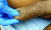
A 63-year-old male presented for evaluation of worsening genital lesions and associated swelling

Article Type
Changed
Mon, 06/19/2023 - 13:26

Elephantiasis nostras verrucosa (ENV) is a chronic, uncommon and progressively disfiguring disease most commonly presenting on the bilateral lower extremities in the setting of chronic nonfilarial lymphedema resulting in secondary dermatologic sequelae.1 Clinically, ENV presents as verrucous, hyperkeratotic, cobblestone-like patches, plaques, and nodules with associated nonpitting edema of the affected body area.1 Secondary bacterial infections are common and often worsen the clinical course. The etiology of ENV involves chronic lymphatic obstruction and venous insufficiency, with additional risk factors including obesity, chronic lymphedema, bacterial infection, surgery or trauma, neoplasia, radiation, congestive heart failure, or scleroderma.2,3 While most commonly presenting on the lower extremities, cases have been reported involving the abdomen, sacrum, ears, buttocks, and penoscrotal area.1,2

BiluMartin_Donna_FLORIDA_web.jpg
Dr. Donna Bilu Martin

Regardless of location, the pathogenesis of ENV remains the same. Chronic lymphatic obstruction results in accumulation and lymphostasis of protein-rich dermal fluid, which subsequently precipitates fibroblast proliferation and activation, suppression of the local immune response and development of recurrent lymphangitis, chronic inflammation, and potential secondary bacterial infection.2,4

There is no standard of care for the treatment and management of ENV and recurrence is common. Interventions often involve those used for chronic lymphedema – including leg elevation, compression stockings or devices, skin hygiene, and lymphatic pumping.2,3 Medical management with topical and oral retinoids has been reported, as well as emphasis on weight loss and infection control.1,4 Surgical intervention is often reserved for refractory cases that fail to respond to more conservative management, or severe presentations resulting in extensive functional and aesthetic impairment. Less commonly reported treatment modalities include lymphaticovenular anastomosis and ablative carbon dioxide laser use, although this latter intervention demonstrated minimal improvement in this patient.5,6

Penoscrotal ENV is a rare form of ENV affecting the genital region of males, often resulting in significant disfigurement, functional impairment, and psychosocial distress. Penoscrotal elephantiasis can be idiopathic, due to filarial infections, scleroinflammatory stricture of the urethra, Chlamydia trachomatis infection, and lymphostasis secondary to chronic inflammatory conditions such as streptococcal infections, radiotherapy, surgery, chronic venous stasis, or Kaposi sarcoma.7

In addition, hidradenitis suppurativa (HS) has been documented multiple times in the literature in association with the development of ENV, detailing lymphatic scarring secondary to chronic inguinal HS as the main pathogenic factor.8,9

Surgery is the mainstay of treatment for penoscrotal ENV, which not only improves functionality and cosmesis, but also aids in prevention of rare malignant sequelae, such as lymphangiosarcoma.10 Such interventions can involve lymphangioplasty to aid in lymphatic drainage or excision of the mass and subcutaneous tissue with full-thickness skin grafting for reconstruction.7 Collaboration between urology, plastic surgery, and dermatology is often essential to obtain adequate care with satisfactory outcomes and minimal recurrence for patients with this uncommon condition.

This case and photo were submitted by Marlee Hill, a medical student at the University of Oklahoma, Oklahoma City; and Michael Franzetti, MD, and Jeffrey McBride, MD, department of dermatology, University of Oklahoma Health Sciences Center. The column was edited by Donna Bilu Martin, MD.
 

Dr. Donna Bilu Martin is a board-certified dermatologist in private practice at Premier Dermatology, MD, in Aventura, Fla. More diagnostic cases are available at mdedge.com/dermatology. To submit a case for possible publication, send an email to dermnews@mdedge.com.

References

1. Hadian Y et al. Dermatol Online J. 2019 Dec 15;25(12):13030/qt6rn1s8ff.

2. Judge N and Kilic A. J Dermatol Case Rep. 2016 Nov 13;10(2):32-4.

3. Dean SM et al. J Am Acad Dermatol. 2011 Jun;64(6):1104-10.

4. Sisto K and Khachemoune A. Am J Clin Dermatol. 2008;9(3):141-6.

5. Motegi S et al. Dermatology. 2007;215(2):147-51.

6. Robinson CG et al. J Cutan Med Surg. 2018;22(6):611-3.

7. Koualla S et al. Ann Chir Plast Esthet. 2023 Apr 10;S0294-1260(23)00035-3.

8. Lelonek E et al. Acta Derm Venereol. 2021 Feb 11;101(2):adv00389.

9. Good LM et al. J Am Acad Dermatol. 2011 May;64(5):993-4.

10. Cerri A et al. Eur J Dermatol. 1998 Oct-Nov;8(7):511-4.

Publications
Topics
Sections

Elephantiasis nostras verrucosa (ENV) is a chronic, uncommon and progressively disfiguring disease most commonly presenting on the bilateral lower extremities in the setting of chronic nonfilarial lymphedema resulting in secondary dermatologic sequelae.1 Clinically, ENV presents as verrucous, hyperkeratotic, cobblestone-like patches, plaques, and nodules with associated nonpitting edema of the affected body area.1 Secondary bacterial infections are common and often worsen the clinical course. The etiology of ENV involves chronic lymphatic obstruction and venous insufficiency, with additional risk factors including obesity, chronic lymphedema, bacterial infection, surgery or trauma, neoplasia, radiation, congestive heart failure, or scleroderma.2,3 While most commonly presenting on the lower extremities, cases have been reported involving the abdomen, sacrum, ears, buttocks, and penoscrotal area.1,2

BiluMartin_Donna_FLORIDA_web.jpg
Dr. Donna Bilu Martin

Regardless of location, the pathogenesis of ENV remains the same. Chronic lymphatic obstruction results in accumulation and lymphostasis of protein-rich dermal fluid, which subsequently precipitates fibroblast proliferation and activation, suppression of the local immune response and development of recurrent lymphangitis, chronic inflammation, and potential secondary bacterial infection.2,4

There is no standard of care for the treatment and management of ENV and recurrence is common. Interventions often involve those used for chronic lymphedema – including leg elevation, compression stockings or devices, skin hygiene, and lymphatic pumping.2,3 Medical management with topical and oral retinoids has been reported, as well as emphasis on weight loss and infection control.1,4 Surgical intervention is often reserved for refractory cases that fail to respond to more conservative management, or severe presentations resulting in extensive functional and aesthetic impairment. Less commonly reported treatment modalities include lymphaticovenular anastomosis and ablative carbon dioxide laser use, although this latter intervention demonstrated minimal improvement in this patient.5,6

Penoscrotal ENV is a rare form of ENV affecting the genital region of males, often resulting in significant disfigurement, functional impairment, and psychosocial distress. Penoscrotal elephantiasis can be idiopathic, due to filarial infections, scleroinflammatory stricture of the urethra, Chlamydia trachomatis infection, and lymphostasis secondary to chronic inflammatory conditions such as streptococcal infections, radiotherapy, surgery, chronic venous stasis, or Kaposi sarcoma.7

In addition, hidradenitis suppurativa (HS) has been documented multiple times in the literature in association with the development of ENV, detailing lymphatic scarring secondary to chronic inguinal HS as the main pathogenic factor.8,9

Surgery is the mainstay of treatment for penoscrotal ENV, which not only improves functionality and cosmesis, but also aids in prevention of rare malignant sequelae, such as lymphangiosarcoma.10 Such interventions can involve lymphangioplasty to aid in lymphatic drainage or excision of the mass and subcutaneous tissue with full-thickness skin grafting for reconstruction.7 Collaboration between urology, plastic surgery, and dermatology is often essential to obtain adequate care with satisfactory outcomes and minimal recurrence for patients with this uncommon condition.

This case and photo were submitted by Marlee Hill, a medical student at the University of Oklahoma, Oklahoma City; and Michael Franzetti, MD, and Jeffrey McBride, MD, department of dermatology, University of Oklahoma Health Sciences Center. The column was edited by Donna Bilu Martin, MD.
 

Dr. Donna Bilu Martin is a board-certified dermatologist in private practice at Premier Dermatology, MD, in Aventura, Fla. More diagnostic cases are available at mdedge.com/dermatology. To submit a case for possible publication, send an email to dermnews@mdedge.com.

References

1. Hadian Y et al. Dermatol Online J. 2019 Dec 15;25(12):13030/qt6rn1s8ff.

2. Judge N and Kilic A. J Dermatol Case Rep. 2016 Nov 13;10(2):32-4.

3. Dean SM et al. J Am Acad Dermatol. 2011 Jun;64(6):1104-10.

4. Sisto K and Khachemoune A. Am J Clin Dermatol. 2008;9(3):141-6.

5. Motegi S et al. Dermatology. 2007;215(2):147-51.

6. Robinson CG et al. J Cutan Med Surg. 2018;22(6):611-3.

7. Koualla S et al. Ann Chir Plast Esthet. 2023 Apr 10;S0294-1260(23)00035-3.

8. Lelonek E et al. Acta Derm Venereol. 2021 Feb 11;101(2):adv00389.

9. Good LM et al. J Am Acad Dermatol. 2011 May;64(5):993-4.

10. Cerri A et al. Eur J Dermatol. 1998 Oct-Nov;8(7):511-4.

Publications
Publications
Topics
Article Type
Sections
Teambase XML
<?xml version="1.0" encoding="UTF-8"?>
<!--$RCSfile: InCopy_agile.xsl,v $ $Revision: 1.35 $-->
<!--$RCSfile: drupal.xsl,v $ $Revision: 1.7 $-->
<root generator="drupal.xsl" gversion="1.7"> <header> <fileName>163935</fileName> <TBEID>0C04AB18.SIG</TBEID> <TBUniqueIdentifier>MD_0C04AB18</TBUniqueIdentifier> <newsOrJournal>News</newsOrJournal> <publisherName>Frontline Medical Communications</publisherName> <storyname>July Make the Diagnosis</storyname> <articleType>353</articleType> <TBLocation>QC Done-All Pubs</TBLocation> <QCDate>20230615T163758</QCDate> <firstPublished>20230619T131537</firstPublished> <LastPublished>20230619T131537</LastPublished> <pubStatus qcode="stat:"/> <embargoDate/> <killDate/> <CMSDate>20230619T131537</CMSDate> <articleSource/> <facebookInfo/> <meetingNumber/> <byline>Donna Bilu Martin</byline> <bylineText>MARLEE HILL; MICHAEL FRANZETTI, MD; AND JEFFREY MCBRIDE, MD</bylineText> <bylineFull>MARLEE HILL; MICHAEL FRANZETTI, MD; AND JEFFREY MCBRIDE, MD</bylineFull> <bylineTitleText/> <USOrGlobal/> <wireDocType/> <newsDocType>Column</newsDocType> <journalDocType/> <linkLabel/> <pageRange/> <citation/> <quizID/> <indexIssueDate/> <itemClass qcode="ninat:text"/> <provider qcode="provider:imng"> <name>IMNG Medical Media</name> <rightsInfo> <copyrightHolder> <name>Frontline Medical News</name> </copyrightHolder> <copyrightNotice>Copyright (c) 2015 Frontline Medical News, a Frontline Medical Communications Inc. company. All rights reserved. This material may not be published, broadcast, copied, or otherwise reproduced or distributed without the prior written permission of Frontline Medical Communications Inc.</copyrightNotice> </rightsInfo> </provider> <abstract/> <metaDescription>Elephantiasis nostras verrucosa (ENV) is a chronic, uncommon and progressively disfiguring disease most commonly presenting on the bilateral lower extremities i</metaDescription> <articlePDF/> <teaserImage>271802</teaserImage> <title>Elephantiasis nostras verrucosa</title> <deck/> <disclaimer/> <AuthorList/> <articleURL/> <doi/> <pubMedID/> <publishXMLStatus/> <publishXMLVersion>1</publishXMLVersion> <useEISSN>0</useEISSN> <urgency/> <pubPubdateYear/> <pubPubdateMonth/> <pubPubdateDay/> <pubVolume/> <pubNumber/> <wireChannels/> <primaryCMSID/> <CMSIDs/> <keywords/> <seeAlsos/> <publications_g> <publicationData> <publicationCode>skin</publicationCode> <pubIssueName/> <pubArticleType/> <pubTopics/> <pubCategories/> <pubSections/> </publicationData> <publicationData> <publicationCode>fp</publicationCode> <pubIssueName/> <pubArticleType/> <pubTopics/> <pubCategories/> <pubSections/> </publicationData> <publicationData> <publicationCode>im</publicationCode> <pubIssueName/> <pubArticleType/> <pubTopics/> <pubCategories/> <pubSections/> </publicationData> </publications_g> <publications> <term canonical="true">13</term> <term>15</term> <term>21</term> </publications> <sections> <term>52</term> <term canonical="true">87</term> </sections> <topics> <term>27442</term> <term canonical="true">39212</term> <term>203</term> </topics> <links> <link> <itemClass qcode="ninat:picture"/> <altRep contenttype="image/jpeg">images/2400f1a6.jpg</altRep> <description role="drol:caption">Dr. Donna Bilu Martin</description> <description role="drol:credit"/> </link> </links> </header> <itemSet> <newsItem> <itemMeta> <itemRole>Main</itemRole> <itemClass>text</itemClass> <title>Elephantiasis nostras verrucosa</title> <deck/> </itemMeta> <itemContent> <p><span class="tag metaDescription">Elephantiasis nostras verrucosa (ENV) is a chronic, uncommon and progressively disfiguring disease most commonly presenting on the bilateral lower extremities in the setting of chronic nonfilarial lymphedema resulting in secondary dermatologic sequelae</span>.<sup>1</sup> Clinically, ENV presents as verrucous, hyperkeratotic, cobblestone-like patches, plaques, and nodules with associated nonpitting edema of the affected body area.<sup>1</sup> Secondary bacterial infections are common and often worsen the clinical course. The etiology of ENV involves chronic lymphatic obstruction and venous insufficiency, with additional risk factors including obesity, chronic lymphedema, bacterial infection, surgery or trauma, neoplasia, radiation, congestive heart failure, or scleroderma.<sup>2,3</sup> While most commonly presenting on the lower extremities, cases have been reported involving the abdomen, sacrum, ears, buttocks, and penoscrotal area.<sup>1,2</sup></p> <p>[[{"fid":"271802","view_mode":"medstat_image_flush_right","fields":{"format":"medstat_image_flush_right","field_file_image_alt_text[und][0][value]":"Dr. Donna Bilu Martin, Premier Dermatology, MD, Aventura, Fla.","field_file_image_credit[und][0][value]":"","field_file_image_caption[und][0][value]":"Dr. Donna Bilu Martin"},"type":"media","attributes":{"class":"media-element file-medstat_image_flush_right"}}]]Regardless of location, the pathogenesis of ENV remains the same. Chronic lymphatic obstruction results in accumulation and lymphostasis of protein-rich dermal fluid, which subsequently precipitates fibroblast proliferation and activation, suppression of the local immune response and development of recurrent lymphangitis, chronic inflammation, and potential secondary bacterial infection.<sup>2,4</sup> <br/><br/>There is no standard of care for the treatment and management of ENV and recurrence is common. Interventions often involve those used for chronic lymphedema – including leg elevation, compression stockings or devices, skin hygiene, and lymphatic pumping.<sup>2,3</sup> Medical management with topical and oral retinoids has been reported, as well as emphasis on weight loss and infection control.<sup>1,4</sup> Surgical intervention is often reserved for refractory cases that fail to respond to more conservative management, or severe presentations resulting in extensive functional and aesthetic impairment. Less commonly reported treatment modalities include lymphaticovenular anastomosis and ablative carbon dioxide laser use, although this latter intervention demonstrated minimal improvement in this patient.<sup>5,6</sup><br/><br/>Penoscrotal ENV is a rare form of ENV affecting the genital region of males, often resulting in significant disfigurement, functional impairment, and psychosocial distress. Penoscrotal elephantiasis can be idiopathic, due to filarial infections, scleroinflammatory stricture of the urethra, <em>Chlamydia trachomatis</em> infection, and lymphostasis secondary to chronic inflammatory conditions such as streptococcal infections, radiotherapy, surgery, chronic venous stasis, or Kaposi sarcoma.<sup>7</sup> <br/><br/>In addition, hidradenitis suppurativa (HS) has been documented multiple times in the literature in association with the development of ENV, detailing lymphatic scarring secondary to chronic inguinal HS as the main pathogenic factor.<sup>8,9<br/><br/></sup>Surgery is the mainstay of treatment for penoscrotal ENV, which not only improves functionality and cosmesis, but also aids in prevention of rare malignant sequelae, such as lymphangiosarcoma.<sup>10</sup> Such interventions can involve lymphangioplasty to aid in lymphatic drainage or excision of the mass and subcutaneous tissue with full-thickness skin grafting for reconstruction.<sup>7</sup> Collaboration between urology, plastic surgery, and dermatology is often essential to obtain adequate care with satisfactory outcomes and minimal recurrence for patients with this uncommon condition.<br/><br/>This case and photo were submitted by Marlee Hill, a medical student at the University of Oklahoma, Oklahoma City; and Michael Franzetti, MD, and Jeffrey McBride, MD, department of dermatology, University of Oklahoma Health Sciences Center. The column was edited by Donna Bilu Martin, MD.<br/><br/><br/><br/></p> <p> <em>Dr. Donna Bilu Martin is a board-certified dermatologist in private practice at Premier Dermatology, MD, in Aventura, Fla. More diagnostic cases are available at <span class="Hyperlink"><a href="http://mdedge.com/dermatology">mdedge.com/dermatology</a></span>. To submit a case for possible publication, send an email to d<span class="Hyperlink"><a href="mailto:ermnews%40mdedge.com?subject=">ermnews@mdedge.com</a></span>.</em> </p> <h2>References</h2> <p>1. Hadian Y et al. <span class="Hyperlink"><a href="https://escholarship.org/uc/item/6rn1s8ff">Dermatol Online J. 2019 Dec 15;25(12):13030/qt6rn1s8ff</a></span>.<br/><br/>2. Judge N and Kilic A. <span class="Hyperlink"><a href="https://www.ncbi.nlm.nih.gov/pmc/articles/PMC5124369/">J Dermatol Case Rep. 2016 Nov 13;10(2):32-4</a></span>.<br/><br/>3. Dean SM et al. <span class="Hyperlink"><a href="https://www.jaad.org/article/S0190-9622(10)00560-8/fulltext">J Am Acad Dermatol. 2011 Jun;64(6):1104-10</a></span>.<br/><br/>4. Sisto K and Khachemoune A. <span class="Hyperlink"><a href="https://link.springer.com/article/10.2165/00128071-200809030-00001">Am J Clin Dermatol. 2008;9(3):141-6</a></span>.<br/><br/>5. Motegi S et al. <span class="Hyperlink"><a href="https://karger.com/drm/article-abstract/215/2/147/112970/Successful-Treatment-with-Lymphaticovenular?redirectedFrom=fulltext">Dermatology. 2007;215(2):147-51</a></span>.<br/><br/>6. Robinson CG et al. <span class="Hyperlink"><a href="https://journals.sagepub.com/doi/10.1177/1203475418763548?url_ver=Z39.88-2003&amp;rfr_id=ori:rid:crossref.org&amp;rfr_dat=cr_pub%20%200pubmed">J Cutan Med Surg. 2018;22(6):611-3</a></span>. <br/><br/>7. Koualla S et al. <span class="Hyperlink"><a href="https://www.sciencedirect.com/science/article/pii/S0294126023000353?via%3Dihub">Ann Chir Plast Esthet. 2023 Apr 10;S0294-1260(23)00035-3</a></span>.<br/><br/>8. Lelonek E et al. <span class="Hyperlink"><a href="https://www.ncbi.nlm.nih.gov/pmc/articles/PMC9366703/">Acta Derm Venereol. 2021 Feb 11;101(2):adv00389</a></span>.<br/><br/>9. Good LM et al. <span class="Hyperlink"><a href="https://www.jaad.org/article/S0190-9622(09)00998-0/fulltext">J Am Acad Dermatol. 2011 May;64(5):993-4</a></span>.<br/><br/>10. Cerri A et al. <span class="Hyperlink"><a href="https://pubmed.ncbi.nlm.nih.gov/9854166/">Eur J Dermatol. 1998 Oct-Nov;8(7):511-4</a></span>.</p> </itemContent> </newsItem> <newsItem> <itemMeta> <itemRole>teaser</itemRole> <itemClass>text</itemClass> <title/> <deck/> </itemMeta> <itemContent> <p>Clinically, ENV presents as verrucous, hyperkeratotic, cobblestonelike patches, plaques, and nodules with associated nonpitting edema of the affected body area.</p> </itemContent> </newsItem> </itemSet></root>
Questionnaire Body

163935_photo_web.jpg
A 63-year-old male with a medical history of hidradenitis suppurativa (HS) and scrotoplasty presented for evaluation of worsening genital lesions and associated swelling despite treatment with imiquimod. HS had been treated in the past with antibiotics and infliximab infusions -  with clinical improvement - and control of HS was maintained control with adalimumab treatment for the past year. His refractory genital lesions had previously been treated with carbon dioxide laser. Physical exam of the penile shaft and scrotum was significant for smooth papules with a cobblestone-like appearance. Shave biopsy of the penile shaft demonstrated dermal interstitial edema with dilated thin-walled vessels and overlying acanthosis with mild spongiosis of the epidermis.

Disallow All Ads
Content Gating
No Gating (article Unlocked/Free)
Alternative CME
Disqus Comments
Default
Use ProPublica
Hide sidebar & use full width
render the right sidebar.
Conference Recap Checkbox
Not Conference Recap
Clinical Edge
Display the Slideshow in this Article
Medscape Article
Display survey writer
Reuters content
Disable Inline Native ads
WebMD Article