User login
Weight Gain After Age 20 May Raise Colon Adenoma Risk
Major Finding: Subjects who became overweight or obese after age 20 were 1.8 times more likely to have colon adenomas than were patients who maintained a normal weight.
Data Source: A prospective study of 1,865 people who underwent a screening colonoscopy.
Disclosures: None.
NEW ORLEANS — Adults who have been overweight since early adulthood are nearly twice as likely to have colon adenomas as those with a history of normal weight.
The findings, presented at the annual Digestive Disease Week, reinforce the benefit of maintaining a healthy weight throughout life, Dr. Fritz Francois of New York University wrote in a statement.
They “suggest that the chronicity of obesity is a significant risk factor for developing colon cancer. Given the continued rise in early-onset obesity, especially in minority populations, there is a need for interventions and lifestyle modifications earlier in life to help lessen this serious health risk later in life.”
Mr. Ian Fagan, a fourth-year medical student and protégé of Dr. Francois, presented the data during the meeting. The colleagues conducted a prospective study of 1,865 patients who were referred for a screening colonoscopy. Their mean age was 57 years. Body mass index was normal in 38%, whereas 39% were overweight and 23% were obese.
The patients provided information allowing the researchers to estimate their body mass index (BMI) and waist circumference at age 10 and age 20. The investigators compared the past weights to the finding of any adenoma, including advanced neoplasia, during the colonoscopy.
The subjects were divided into three groups: those who had normal weights at age 20 and at present; those who had a normal weight at 20 and were now overweight or obese; and those who had been overweight or obese since age 20.
“Race and ethnicity had a significant impact on weight change since early adulthood,” Mr. Fagan said. Sixty-one percent of Hispanics fell into the group that changed from normal weight to overweight or obese, as well as 50% of blacks, 46% of whites, and 7% of Asians.
Adenomas were significantly more common in patients who had been overweight or obese since age 10 (at a rate of 27%) and in those who went from normal weight to overweight (19%), compared with those who had maintained a normal weight (13%).
After controlling for age, gender, current BMI, U.S. birth, and red meat consumption, the investigators found that becoming overweight or obese in early adulthood almost doubled the risk of an adenoma on screening colonoscopy (odds ratio 1.8).
Disclosures: Neither Mr. Fagan nor Dr. Francois had any potential financial conflicts.
'Race and ethnicity had a significant impact on weight change since early adulthood.'
Source MR. FAGAN
From the annual Digestive Disease Week
Laparoscopic GI Surgery Safe for Octogenarians
Major Finding: Octogenarians who underwent elective laparoscopic colorectal surgery had significantly lower overall morbidity than did those who underwent open surgery (30% vs. 49%) and left the hospital earlier (6 vs. 8 days). Octogenarians who had laparoscopic repair of paraesophageal hernia had an 86% rate of symptom resolution and a 36% rate of major complications over the 30-day postoperative period.
Data Source: Two retrospective series comprising 258 patients.
Disclosures: Dr. Pinto and Dr. Fitzgerald had no disclosures. Dr. Wexner disclosed relationships with numerous medical equipment companies, including some that manufacture laparoscopic surgical instruments.
NATIONAL HARBOR, MD. — Advanced age need not be an impediment to laparoscopic surgery for colon resection or paraesophageal hernia.
Two retrospective studies found that octogenarians not only tolerated laparoscopic surgery, but came through both colorectal surgery and paraesophageal hernia repair with excellent outcomes.
A shortened hospital stay is one of the biggest benefits the elderly can reap from a laparoscopic procedure, said Dr. Steven Wexner, chair of the colorectal surgery department at the Cleveland Clinic, Weston, Fla.
“Leaving the hospital sooner is beneficial to older patients because it lessens their chances of a hospital-acquired infection, fall, or psychological changes,” said Dr. Wexner, lead author on one of the studies. “Unless there is a specific contraindication, these older patients [who need colorectal surgery] should be offered a laparoscopic procedure.”
Dr. Wexner and his colleague, Dr. Rodrigo Pinto, examined outcomes in 83 laparoscopic and 116 open colorectal resections among a group of 199 octogenarians. The patients' mean age was 84 years, and their mean American Society of Anesthesiologists class was 2.7. Cancer was the most common diagnosis, occurring in 86% of the open surgery group and 89% of the laparoscopic surgery group. Diverticular disease was present in 9% of the open group and 8% of the laparoscopic group. The remainder of the patients had other disorders.
The patients underwent a variety of surgical procedures including right, sigmoid, and transverse colectomy; sigmoid colectomy; low anterior resection; abdomino-perineal resection; left hemicolectomy; and proctacolectomy. Stomas were constructed in 47% of the open group and 10% of the laparoscopic group.
The mean operative time was not significantly different between the groups. However, the laparoscopic group lost significantly less blood than the open group (mean 100 vs. 200 mL), required significantly fewer intraoperative transfusions (3 vs. 19), and had a significantly shorter incision length (mean 9 vs. 23 cm).
The overall rate of major surgical complications was 5% in each group. Three patients in each group required reoperation. The rate of medical complications was lower—but not significantly lower—in the laparoscopic group, compared with the open group (25% vs. 39%). There was no significant difference in mortality.
The overall morbidity rate was 49% for open surgery and 30% for laparoscopic surgery, a significant difference. Patients who underwent laparoscopic surgery left the hospital a mean of 2 days earlier than did open surgery patients (6 vs. 8 days).
The open conversion rate was 25% (21 cases). The converted cases had a longer mean operative time than those completed laparoscopically (197 vs. 156 minutes), greater mean blood loss (220 vs. 129 mL), more surgical complications (96% vs. 5%), and more postsurgical medical complications (79% vs. 21%). All the differences were significant. Overall, however, “laparoscopic colorectal resection was very safe and effective for these patients,” Dr. Wexner said.
Dr. Heidi Fitzgerald of Dartmouth-Hitchcock Medical Center, Lebanon, N.H., reported on a series of 59 elderly patients (aged 80 or older) who underwent paraesophageal hernia repair. Based on her findings of a low mortality rate (two patients) and an 86% rate of symptom resolution, she and her colleagues recommended elective laparoscopic repair for octogenarians rather than watchful waiting.
The decision to repair electively or not is a controversial one, Dr. Fitzgerald said. “The major concern with paraesophageal hernias is their 10%–30% risk of complications, including hemorrhage, volvulus, strangulation, and perforation,” she said. Mortality rates in untreated patients undergoing emergent surgery have ranged from 5.4% to 26% in various studies. The only study to examine the issue in the very elderly found a 16% mortality rate associated with emergent repair, compared with a 2.5% rate in elective repair in patients 80 years and older.
The mean age of Dr. Fitzgerald's patients was 86 years, and 76% were women. All repairs were completed laparoscopically.
The mean operating time was 193 minutes. Five intraoperative complications occurred. They included three pneumothoraces, which were treated in the recovery unit with needle decompression, an esophageal perforation that was recognized and repaired intraoperatively, and an esophageal perforation that was noted on postoperative day 2 and required a reoperation. Major
Major complications occurred in 21 patients (36%) over the 30-day postoperative period. They included two cardiac arrhythmias; four cases of dysphagia, three of which required dilation; one empyema and one aspiration pneumonia that required admission to the intensive care unit; and four cases of anemia that required transfusion.
Two patients died in the hospital after surgery. One patient had an esophageal leak that was repaired, but resulted in a fatal sepsis. One patient needed a reoperation for bleeding and subsequently developed renal and cardiac failure; the family elected to withdraw life support.
Dr. Fitzgerald had complete 1-month follow-up data on 86% of the patients (51). Thirty-nine percent of the patients (23) reported complete symptom resolution, and 47% (28) reported partial resolution.
“This was a small sample size, but despite this, we feel that laparoscopic paraesophageal hernia repair can be performed safely with minimal perioperative morbidity in octogenarians. We now advocate this approach as opposed to watchful waiting in this subset of symptomatic patients.”
Support Grows for Earlier Colonoscopy in Blacks
NEW ORLEANS — Screening colonoscopy in asymptomatic black patients found a 4.5% rate of high-grade dysplastic polyps in those aged 45–49 years, according to the findings of a retrospective review.
The findings suggest that blacks should have their first colonoscopy before they reach age 50, said Dr. Frank Friedenberg, a professor of medicine at Temple University Hospital, Philadelphia.
About a month after the meeting, the American Society for Gastrointestinal Endoscopy issued separate guidelines suggesting that blacks begin screening colonoscopy at age 45. Developed by the ASGE's Standards of Practice Committee, the guidelines on ethnicity, gastrointestinal diseases, and endoscopic procedures appear in the June issue of GIE: Gastrointestinal Endoscopy (2010;71:1108–11). The document is available at www.giejournal.orgwww.asge.org
The guidelines, which are based on literature reviews, include “recommendations” as well as “suggestions,” with the latter term indicating that the quality of evidence is weaker.
The ASGE suggestion regarding screening for black patients reflects several risk factors: “African Americans with colon cancer have a 20% stage-adjusted increase in mortality risk, compared with European Americans,” are younger at presentation, have a higher proportion of cancers presenting before age 50, and generally more advanced stage at the time of diagnosis, the authors of the guidelines noted.
Many ethnic groups have low colon cancer screening rates; the guidelines support an increased emphasis on screening for those groups.
No studies have assessed the impact of modifying specific endoscopic standards based on ethnicity, but “it is logical to assume that increased awareness of differences in disease patterns and management among different ethnic groups could have beneficial impacts on the health-related quality of life of people in these groups,” said Dr. Jason A. Dominitz, chair of ASGE's Standards of Practice Committee.
“It is important to recognize that ethnic populations are not homogeneous and that additional factors, such as environment and behavior, also play important roles in disease,” Dr. Dominitz added in a statement.
In presenting his findings at Digestive Disease Week, Dr. Friedenberg noted that “several lines of evidence suggest we should be looking at screening blacks at an earlier age.”
“National data suggest that colorectal cancer is actually increasing in blacks, while it's declining in the general population. Blacks also seem to have a predilection for proximal colorectal neoplasia, suggesting that flexible sigmoidoscopy is not an appropriate screening tool.
“Finally, patients younger than 50 who develop colorectal cancer are twice as likely to be black as white. All these are very compelling reasons to think about earlier screening,” he said.
Dr. Douglas K. Rex, distinguished professor of medicine at Indiana University, Indianapolis, and director of endoscopy at Indiana University Hospital, commented in an interview that “the approach of screening colonoscopy starting at age 45 in African Americans was endorsed by the American College of Gastroenterology, but has been controversial. This study lends support to the ACG recommendation, but is unlikely to resolve the controversy.”
Dr. Friedenberg and his colleagues reviewed the charts of 109 black patients aged 45–49 years matched with 226 black patients aged 50–59 years, all of whom underwent a screening colonoscopy. All of the patients were asymptomatic, and none of them had a family history of colorectal cancer. The groups were well-matched in terms of gender and body mass index (mean, 31 kg/m
The older patients were significantly more likely than the younger patients to smoke (48% vs. 37%), drink alcohol (43% vs. 21%), and use aspirin regularly (26% vs. 15%).
Polyps were found in 22% of the younger group and 20% of the older group, not a significant difference. Thirty percent of each group had more than one polyp. The polyps were on the right side of the colon in about half of each group. “This is much more than you would expect, lending credence to the observation that blacks have a predilection for right-sided lesions,” Dr. Friedenberg said.
Significant differences emerged in polyp histology. High-grade dysplastic lesions occurred in 4.5% of the younger group and 0.4% of the older group. “Theoretically, this means that there were several younger individuals who, if they had waited until their 50s to have a colonoscopy, could have had a colon cancer by then,” Dr. Friedenberg said.
The significantly higher use of aspirin in the older group may have exerted a protective influence against high-grade lesions, he said, “although since that group smoked significantly more, it might have balanced out the risk.”
The study suggests that screening colonoscopy should begin earlier in black patients, Dr. Friedenberg said. “We are already swamped with colonoscopies, and to begin screening years earlier in this population would be a financial challenge,” he said. “Still, it is something that needs to be looked at on a larger scale.”
Dr. Rex added that “some have argued that an early screening recommendation for African Americans would have the added benefit of educating both patients and primary care physicians that there is higher risk in this group, but there are also lower rates of screening adherence, so special efforts are needed to ensure that screening in African Americans takes place.”
The ASGE guidelines also suggest a screening esophagogastroduodenoscopy (EGD) for gastric cancer in new immigrants from high-risk regions, such as Korea, Japan, China, Russia, and South America, in particular if there is a family history in a first-degree relative.
However, screening EGD for adenocarcinoma or squamous cell carcinoma of the esophagus should be based on clinical considerations and not on ethnicity, according to the guidelines.
Disclosures: Dr. Friedenberg had no relevant financial disclosures.
Alicia Ault contributed to this report.
Tanners Risk Sunbed Use Despite Knowing the Dangers
CANCUN, Mexico — Knowledge doesn't necessarily mean power when it comes to tanning beds.
The results of a small British survey show that the majority of patients in a dermatology clinic who used tanning beds for purely cosmetic reasons were well acquainted with the risks.

Ninety-four percent of the patients surveyed were aware of the link between tanning bed use and skin cancer, and even volunteered that they knew other risks, Dr. Tina Ninan said at Wonca 2010, the conference of the World Organization of Family Doctors. "Knowing the risks does not deter patients from engaging in this risky behavior," said Dr. Ninan, a general practice physician in Newcastle upon Tyne, England.
She distributed some simple surveys to 102 patients waiting in a dermatology clinic at the University Hospital of North Durham. Most of the respondents (65%) were women, and most (60%) were older than 40 years.
Thirty-four patients admitted to using tanning beds. Most of these (27) were women, meaning that 42% of the women surveyed admitted to using the devices. But they weren't alone: 18% of the men surveyed also said they used tanning beds.
Most of the users (88%) said that they had started visiting tanning salons before they turned 35 years old—a period considered crucial in developing an increased risk of skin cancers. "More disturbingly, 58% said they began tanning at age 17-25, and 18% of the users started at younger than 16 years," Dr. Ninan said.
Although most reported occasional use, 38% of the tanners said they visited a tanning bed weekly. Most used tanning beds at their local beauty salon, but 9% said they had the devices at home. "Six percent also said they tanned at their gym. Having [tanning] beds at gyms and other health centers really sends a mixed message about their health benefits," Dr. Ninan said.
Coin-operated tanning beds were also popular among the group, with 16% reporting use. This trend is particularly disturbing, Dr. Ninan said, because it circumvents what little control there may be over exposure. And although the United Kingdom recently passed a law forbidding teens to use tanning beds, no one can prevent them from accessing the coin-operated types, she added. Additionally, there is no guarantee that unsupervised tanners use the correct eye protection.
Looking good was the main motivation for tanning. Most tanners (64%) said that they just like the look of tanned skin, although 14% said they were trying to treat a skin condition, including acne, rosacea, and psoriasis. Seventeen percent of the tanners were getting ready for a beach vacation and thought a base tan would help protect them from sunburn. "Unfortunately, it's been shown that going away on vacation with a tan only offers an SPF of about 2-3," Dr. Ninan said.
The motivations for tanning were apparently stronger than worries about its risks.
Almost everyone (94%) was aware of the connection between tanning bed use and skin cancer. Respondents were also free to write in other risks they knew of— most frequently listed were wrinkles and eye damage.
Although she admitted that understanding risks won't necessarily change behavior, Dr. Ninan encouraged physicians to find "teachable moments" to educate their patients about tanning bed use. "For example, I frequently have patients asking me to check their moles. This is always the chance I take to ask about [tanning] bed use and its risks."
Dr. Ninan disclosed that she had no financial conflicts.
CANCUN, Mexico — Knowledge doesn't necessarily mean power when it comes to tanning beds.
The results of a small British survey show that the majority of patients in a dermatology clinic who used tanning beds for purely cosmetic reasons were well acquainted with the risks.
Ninety-four percent of the patients surveyed were aware of the link between tanning bed use and skin cancer, and even volunteered that they knew other risks, Dr. Tina Ninan said at Wonca 2010, the conference of the World Organization of Family Doctors. "Knowing the risks does not deter patients from engaging in this risky behavior," said Dr. Ninan, a general practice physician in Newcastle upon Tyne, England.
She distributed some simple surveys to 102 patients waiting in a dermatology clinic at the University Hospital of North Durham. Most of the respondents (65%) were women, and most (60%) were older than 40 years.
Thirty-four patients admitted to using tanning beds. Most of these (27) were women, meaning that 42% of the women surveyed admitted to using the devices. But they weren't alone: 18% of the men surveyed also said they used tanning beds.
Most of the users (88%) said that they had started visiting tanning salons before they turned 35 years old—a period considered crucial in developing an increased risk of skin cancers. "More disturbingly, 58% said they began tanning at age 17-25, and 18% of the users started at younger than 16 years," Dr. Ninan said.
Although most reported occasional use, 38% of the tanners said they visited a tanning bed weekly. Most used tanning beds at their local beauty salon, but 9% said they had the devices at home. "Six percent also said they tanned at their gym. Having [tanning] beds at gyms and other health centers really sends a mixed message about their health benefits," Dr. Ninan said.
Coin-operated tanning beds were also popular among the group, with 16% reporting use. This trend is particularly disturbing, Dr. Ninan said, because it circumvents what little control there may be over exposure. And although the United Kingdom recently passed a law forbidding teens to use tanning beds, no one can prevent them from accessing the coin-operated types, she added. Additionally, there is no guarantee that unsupervised tanners use the correct eye protection.
Looking good was the main motivation for tanning. Most tanners (64%) said that they just like the look of tanned skin, although 14% said they were trying to treat a skin condition, including acne, rosacea, and psoriasis. Seventeen percent of the tanners were getting ready for a beach vacation and thought a base tan would help protect them from sunburn. "Unfortunately, it's been shown that going away on vacation with a tan only offers an SPF of about 2-3," Dr. Ninan said.
The motivations for tanning were apparently stronger than worries about its risks.
Almost everyone (94%) was aware of the connection between tanning bed use and skin cancer. Respondents were also free to write in other risks they knew of— most frequently listed were wrinkles and eye damage.
Although she admitted that understanding risks won't necessarily change behavior, Dr. Ninan encouraged physicians to find "teachable moments" to educate their patients about tanning bed use. "For example, I frequently have patients asking me to check their moles. This is always the chance I take to ask about [tanning] bed use and its risks."
Dr. Ninan disclosed that she had no financial conflicts.
Treatments Minimize Infantile Hemangiomas
New medical and surgical treatments can minimize infantile hemangiomas in their proliferative phase and decrease residual scarring after they involute, according to Dr. Brandie J. Metz.
The first decision in treating a hemangioma is whether to use a surgical or medical approach, Dr. Metz said at a cosmetic dermatology seminar sponsored by Skin Disease Education Foundation in Santa Monica, Calif.

Medical treatment is usually a first-line choice, although surgery may be considered when the patient presents with severe refractory pain from ulceration or when residua after involution would likely require surgical treatment. "In that case, why wait?" said Dr. Metz, the director of pediatric dermatology at the University of California, Irvine.
In the past 2 years, propranolol has gained acceptance as an effective medical therapy. Its use was first noted in a case series published in 2008 (N. Engl. J. Med. 2008;358:2649-51). In this French report, 11 infants received propranolol for varying treatment periods. Within 24 hours, all of the hemangiomas responded with softening and a change in color from red to purple, indicating decreased vascularity.
The treatment is not without risks, Dr. Metz noted. "Possible side effects are bradycardia, hypotension, hypoglycemia, and bronchospasm. A gradual dose escalation with close monitoring is the best way to go."
She is an investigator for a large, double-blind, placebo-controlled trial of propranolol that is currently underway. Patients receive either placebo or propranolol at 3 mg/kg for 15 days, followed by another 15 days at 4 mg/kg. The primary outcome will be hemangioma thickness variation measured by ultrasonography from baseline to 1 month.
When a lesion is easily resectable and a patient has failed medical therapy, or if ulceration and pain are present, surgery can be considered. Generally, thin plaques with a gradual slope have a better surgical prognosis. A circular incision around the lesion, with purse string closure, is the usual technique. A 2002 study of 25 patients found that this method reduced hemangiomas by a mean of 73% in width and 45% in length (Plast. Reconstr. Surg. 2002;109:1544-54).
Lasers can treat superficial proliferating hemangiomas, as well as the residua left after involution, Dr. Metz said. The pulsed dye laser is one option. Short pulses of 0.45-1.5 millisecond are used at either a 585- or 595-nm wavelength. The concomitant use of a dynamic cooling device makes the treatment less painful and can allow the use of a higher fluence.
Several studies attest to the success of pulsed dye lasers for hemangiomas. A 2009 study of 90 patients with 105 hemangiomas reported complete or near-complete clearance of color for 81% and thickness for 64%. Although there was no scarring or atrophy, one patient did develop an ulceration that resolved. Hyperpigmentation occurred in 4% and hypopigmentation in 14% (Derm. Surg. 2009;35:1947-54).
A 2006 study noted complications related to pulsed dye laser treatment. Of 12 patients with segmental facial hemangiomas, there were 8 ulcerations and 4 with atrophic scarring (Lasers Surg. Med. 2006;38:116-23).
Two published reports detail the use of fractionated laser for residua. A 2008 report found that the laser effectively decreased fibrofatty tissue and redundant skin that resulted from an extensive mixed facial hemangioma (Derm. Surg. 2008;34:1112-4). A 2009 report found similar results on residua from another facial hemangioma (Arch. Dermatol. 2009;145:748-50).
Dr. Metz disclosed that she is an investigator on the current propranolol trial, sponsored by University Hospital, Bordeaux, France.
SDEF and this news organization are owned by Elsevier.
New medical and surgical treatments can minimize infantile hemangiomas in their proliferative phase and decrease residual scarring after they involute, according to Dr. Brandie J. Metz.
The first decision in treating a hemangioma is whether to use a surgical or medical approach, Dr. Metz said at a cosmetic dermatology seminar sponsored by Skin Disease Education Foundation in Santa Monica, Calif.
Medical treatment is usually a first-line choice, although surgery may be considered when the patient presents with severe refractory pain from ulceration or when residua after involution would likely require surgical treatment. "In that case, why wait?" said Dr. Metz, the director of pediatric dermatology at the University of California, Irvine.
In the past 2 years, propranolol has gained acceptance as an effective medical therapy. Its use was first noted in a case series published in 2008 (N. Engl. J. Med. 2008;358:2649-51). In this French report, 11 infants received propranolol for varying treatment periods. Within 24 hours, all of the hemangiomas responded with softening and a change in color from red to purple, indicating decreased vascularity.
The treatment is not without risks, Dr. Metz noted. "Possible side effects are bradycardia, hypotension, hypoglycemia, and bronchospasm. A gradual dose escalation with close monitoring is the best way to go."
She is an investigator for a large, double-blind, placebo-controlled trial of propranolol that is currently underway. Patients receive either placebo or propranolol at 3 mg/kg for 15 days, followed by another 15 days at 4 mg/kg. The primary outcome will be hemangioma thickness variation measured by ultrasonography from baseline to 1 month.
When a lesion is easily resectable and a patient has failed medical therapy, or if ulceration and pain are present, surgery can be considered. Generally, thin plaques with a gradual slope have a better surgical prognosis. A circular incision around the lesion, with purse string closure, is the usual technique. A 2002 study of 25 patients found that this method reduced hemangiomas by a mean of 73% in width and 45% in length (Plast. Reconstr. Surg. 2002;109:1544-54).
Lasers can treat superficial proliferating hemangiomas, as well as the residua left after involution, Dr. Metz said. The pulsed dye laser is one option. Short pulses of 0.45-1.5 millisecond are used at either a 585- or 595-nm wavelength. The concomitant use of a dynamic cooling device makes the treatment less painful and can allow the use of a higher fluence.
Several studies attest to the success of pulsed dye lasers for hemangiomas. A 2009 study of 90 patients with 105 hemangiomas reported complete or near-complete clearance of color for 81% and thickness for 64%. Although there was no scarring or atrophy, one patient did develop an ulceration that resolved. Hyperpigmentation occurred in 4% and hypopigmentation in 14% (Derm. Surg. 2009;35:1947-54).
A 2006 study noted complications related to pulsed dye laser treatment. Of 12 patients with segmental facial hemangiomas, there were 8 ulcerations and 4 with atrophic scarring (Lasers Surg. Med. 2006;38:116-23).
Two published reports detail the use of fractionated laser for residua. A 2008 report found that the laser effectively decreased fibrofatty tissue and redundant skin that resulted from an extensive mixed facial hemangioma (Derm. Surg. 2008;34:1112-4). A 2009 report found similar results on residua from another facial hemangioma (Arch. Dermatol. 2009;145:748-50).
Dr. Metz disclosed that she is an investigator on the current propranolol trial, sponsored by University Hospital, Bordeaux, France.
SDEF and this news organization are owned by Elsevier.
Isotretinoin-IBD Link Challenged by Two Studies
WAIKOLOA, HAWAII — Two studies—both conducted by gastroenterologists—dispute the notion that isotretinoin causes inflammatory bowel disease.
Neither found a basis for allegations of an increased risk of inflammatory bowel disease (IBD) in patients who were treated with isotretinoin.
“There has been a lot of concern about this. You can't say anything with certainty in life, but at least thus far the data we have are very reassuring that there is no association,” Dr. Sheila Fallon Friedlander said at the annual Hawaii Dermatology Seminar sponsored by Skin Disease Education Foundation.
She cited a retrospective, nested case-control study by investigators at the University of Manitoba, Winnipeg, who used the comprehensive provincial IBD database to demonstrate that patients with IBD were no more likely to have used isotretinoin before diagnosis than were matched controls. “Although there may be anecdotes of isotretinoin causing acute colitis, our data suggest that isotretinoin is not likely to cause chronic IBD,” the investigators concluded (Am. J. Gastroenterol. 2009;104:2774-8).
Dr. J. Mark Jackson of the University of Louisville (Ky.) characterized the Manitoba study as “a really well-done study coming at a critical time,” conducted by physicians who deal with IBD and therefore have no stake in protecting a drug that could cause it.
The second study was a seven-country, systematic data search led by gastroenterologists at the University of North Carolina at Chapel Hill, who found “no clear relationship” between the use of isotretinoin and IBD. Unlike the earlier study, this analysis used the rigorous Chapel Hill criteria designed to weigh the strength, consistency, specificity, and plausibility of scientific evidence, and on that basis, the investigators determined no causal association had been established (Am. J. Gastroenterol. 2009;104:2387-93).
“We now have some very good data reviews showing that IBD is not overrepresented in patients who use isotretinoin,” Dr. Jackson said in an interview. “When this issue comes up [in prescribing], we need to make people aware that this rumor has not been validated.”
Personal injury lawyers seized on an earlier study that concluded it was “highly probable” that isotretinoin was the cause of four cases of IBD reported to the Food and Drug Administration's MedWatch program, with the oral retinoid being deemed the “probable” cause of another 58 (Am. J. Gastroenterol. 2006;101:1569-73). Trial investigator Dr. Sunanda Kane of the University of Chicago was contacted for an interview, but was unable to comment because of pending litigation. The other three study investigators never responded to interview requests.
“This is a situation where there are conflicting data, but a bottom line of interest to all of us is that a New Jersey jury has awarded $12.9 million to patients who have taken isotretinoin and developed IBD,” said Dr. Friedlander, a dermatologist who is a clinical professor of pediatrics and medicine at the University of California, San Diego.
Lawyers shooting for claims have found isotretinoin to be an easy mark for years. The finding that it could increase birth defects by up to 30% left it “an open target,” Dr. Jackson said. “It's always been on the legal radar because of this past issue, and it tends to get put back in the forefront frequently—it's an easy medicine to beat up.”
He said he makes it a practice to discuss reports of these issues with patients before prescribing isotretinoin. “I bring it up with my patients because I don't want them hearing it from some other source first,” he said.
At the meeting, when Dr. Jackson asked for a show of hands as to how many audience members have fielded questions from their patients regarding a putative isotretinoin-IBD link, the majority of dermatologists' hands shot up. “It's amazing how isotretinoin continuously gets brought to the forefront of the legal realm. There's always something going on,” he said.
Dr. Seth D. Crockett, a coauthor of the University of North Carolina study, tried to bring some perspective to the issue in an interview with this news organization. “Our study was a critical appraisal of the literature and an assessment of causality. Basically we found that the only published evidence is case reports, which generally is considered poor evidence to establish causality. The best evidence is from epidemiologic studies such as the University of Manitoba study,” he said.
“It's important to recognize that the absence of published evidence does not mean the absence of an association; it just means that there's insufficient evidence in the scientific literature thus far to support a causal connection between isotretinoin and IBD,” added Dr. Crockett of the division of gastroenterology and hepatology at the University of North Carolina.
Isotretinoin is now available only in generic form.
Disclosures: Dr. Jackson and Dr. Friedlander indicated that they are longtime prescribers of isotretinoin in severe cases of acne. Dr. Jackson has received support from Roche Pharmaceuticals (manufacturer of Accutane). Dr. Crockett had no relevant conflicts of interest. SDEF and this news organization are owned by Elsevier.
“This rumor has not been validated,” Dr. J. Mark Jackson said.
Source ©Tom Fougerousse/University of Louisville
Abdominoplasty Plication Technique Could Revolutionize Facelifts
ORLANDO - By adopting an abdominoplasty plication technique, a California facial plastic and cosmetic surgeon is pioneering a minimally invasive facelift.
The revision rate among 1,535 patients was less than 1% over an average 1.5-year follow-up, Dr. Kevin Sadati said at the annual meeting of the American Academy of Cosmetic Surgery.
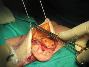
Initial anchoring of the suture at 45 degrees. (Photo courtesy: Dr. Kevin Sadati)
Using a double C-plication technique in the face offers several advantages over the traditional method, he said in an interview. "I use a double-layered interlocking suture, which gives you multiple fixation points and ensures that, over time, the tissue will stay in place and not lead to early tissue laxity," said Dr. Sadati of Newport Beach, Calif.
The technique also provides tissue traction along multiple vectors, he said in an interview. "It creates a more uniform suspension with less bunching compared to traditional plication sutures, which only provide point-specific tension," he said.
Dr. Sadati developed this technique after a number of his early facelift patients came back for revisions due to tissue laxity or bunching. "I decided to change my technique to something that would avoid failure of the plication. In my abdominoplasties, I was using a double-layer plication and, despite great forces exerted in the abdomen, the plication did not fail. I thought perhaps I could apply that same technique to the face," he said.
Using cadaver studies, Dr. Sadati determined that it was possible to adapt the same plication technique used in abdominoplasty to the facial superficial musculoaponeurotic system (SMAS). "I apply the first anchoring suture at the angle of the mandible, followed by double-layer interlocking sutures. In order to create a natural look without the 'pulled look,' I apply gradual tension with various vectors on the SMAS," he explained.
At the superior portions, he only tightens the SMAS 20%-30%. As he goes down toward the mandible, he increases the tension so that at the jaw lines, the SMAS is 100% tightened. "This creates a uniform, natural tension on the SMAS and a smooth look without pulled mouth or tissue bunching by the hairline," Dr. Sadati said.
The procedure lifts the jowls and provides excellent lift to the neck. "It improves any kind of neck, from neck with minimal laxity to the heaviest and the most difficult neck," he said.
He performs the surgery while patients are awake, using 1% lidocaine with epinephrine and tumescent infiltration to the submental and jowl area. Patients also receive oral sedation with diazepam and diphenhydramine. No drain is necessary, and the exterior sutures are absorbable.
He presented data on 1,532 patients he has followed for an average of 1.5 years (maximum 3 years). Of these, 110 had undergone previous facelifts by other surgeons. The postsurgical complication rate was low, with no facial nerve injuries and two greater auricular nerve injuries (some numbness behind ear).
There were 11 revisions (10 women and one man), for a total revision rate of 0.7%. The low revision rate is the best indication of the technique's success.
"The most important result is the high patient satisfaction rate," he noted.
Dr. Sadati had no financial disclosures relevant to his presentation. He teaches the technique but does not receive remuneration for that.
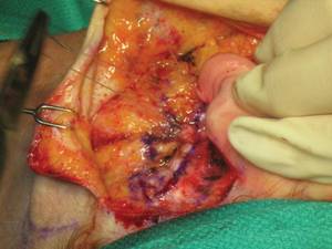
Double layered interlocking plication of the neck is shown. (Photo courtesy: Dr. Kevin Sadati)
ORLANDO - By adopting an abdominoplasty plication technique, a California facial plastic and cosmetic surgeon is pioneering a minimally invasive facelift.
The revision rate among 1,535 patients was less than 1% over an average 1.5-year follow-up, Dr. Kevin Sadati said at the annual meeting of the American Academy of Cosmetic Surgery.
Initial anchoring of the suture at 45 degrees. (Photo courtesy: Dr. Kevin Sadati)
Using a double C-plication technique in the face offers several advantages over the traditional method, he said in an interview. "I use a double-layered interlocking suture, which gives you multiple fixation points and ensures that, over time, the tissue will stay in place and not lead to early tissue laxity," said Dr. Sadati of Newport Beach, Calif.
The technique also provides tissue traction along multiple vectors, he said in an interview. "It creates a more uniform suspension with less bunching compared to traditional plication sutures, which only provide point-specific tension," he said.
Dr. Sadati developed this technique after a number of his early facelift patients came back for revisions due to tissue laxity or bunching. "I decided to change my technique to something that would avoid failure of the plication. In my abdominoplasties, I was using a double-layer plication and, despite great forces exerted in the abdomen, the plication did not fail. I thought perhaps I could apply that same technique to the face," he said.
Using cadaver studies, Dr. Sadati determined that it was possible to adapt the same plication technique used in abdominoplasty to the facial superficial musculoaponeurotic system (SMAS). "I apply the first anchoring suture at the angle of the mandible, followed by double-layer interlocking sutures. In order to create a natural look without the 'pulled look,' I apply gradual tension with various vectors on the SMAS," he explained.
At the superior portions, he only tightens the SMAS 20%-30%. As he goes down toward the mandible, he increases the tension so that at the jaw lines, the SMAS is 100% tightened. "This creates a uniform, natural tension on the SMAS and a smooth look without pulled mouth or tissue bunching by the hairline," Dr. Sadati said.
The procedure lifts the jowls and provides excellent lift to the neck. "It improves any kind of neck, from neck with minimal laxity to the heaviest and the most difficult neck," he said.
He performs the surgery while patients are awake, using 1% lidocaine with epinephrine and tumescent infiltration to the submental and jowl area. Patients also receive oral sedation with diazepam and diphenhydramine. No drain is necessary, and the exterior sutures are absorbable.
He presented data on 1,532 patients he has followed for an average of 1.5 years (maximum 3 years). Of these, 110 had undergone previous facelifts by other surgeons. The postsurgical complication rate was low, with no facial nerve injuries and two greater auricular nerve injuries (some numbness behind ear).
There were 11 revisions (10 women and one man), for a total revision rate of 0.7%. The low revision rate is the best indication of the technique's success.
"The most important result is the high patient satisfaction rate," he noted.
Dr. Sadati had no financial disclosures relevant to his presentation. He teaches the technique but does not receive remuneration for that.
Double layered interlocking plication of the neck is shown. (Photo courtesy: Dr. Kevin Sadati)
Imaging Sheds Light on Lasting Effects of TBI
Major Finding: Various functional and structural imaging techniques can provide a prognosis for TBI patients and objective evidence to insurers to cover rehabilitative services.
Data Source: A review of recent imaging studies of traumatic brain injury.
Disclosures: None
BANGKOK, THAILAND — New imaging techniques may help to explain the disabling symptoms that can plague patients with traumatic brain injury long after their acute problems have resolved, and eventually guide the best choice for medical therapy.
Survivors of traumatic brain injury who complain of depression, irritability, and memory or cognitive problems are often written off as psychiatric cases or malingerers, Dr. Ramon Diaz-Arrastia said at the World Congress of Neurology. “This is the frustrating thing about TBI. The patients might look OK—they don't have paralysis; they are walking around. But their cognitive and behavioral problems are real.”
Imaging techniques that are now well established in other areas of neurology—such as diffusion-weighted and susceptibility-weighted magnetic resonance imaging—are now being used to show that brain injuries leave permanent, life-altering marks behind after the contusions and hematomas have healed.
These findings may have both immediate and long-range benefits, said Dr. Diaz-Arrastia, a professor of neurology at the University of Texas Southwestern Medical Center, Dallas. “Right now, imaging these patients has the primary value of providing a prognosis and perhaps helping them obtain the care that they need. In terms of reimbursement, it's useful to have an objective documentation of the injury when trying to convince insurers to cover rehabilitative service.”
In the future, imaging the post-TBI brain may help guide medical treatment choices and monitor drugs' effectiveness.
So far, nearly 30 drugs have provided effective neuroprotection in animal models of TBI, he said. However, none that has undergone testing in well-designed phase III trials has proven beneficial to humans.
Part of the problem may be the heterogeneity of human brain injury, Dr. Diaz-Arrastia said. There are many subtypes of TBI, yet “from the point of view of the clinical trials, all patients who present in a coma [after a brain injury] are treated the same way, even though the injuries can be very different, with very different prognoses.”
Susceptibility-weighted imaging (SWI) is one technique being studied in TBI patients. It measures the paramagnetic shift of intravascular deoxyhemoglobin and methemoglobin, amplifying the appearance of microhemorrhages and making them much easier to identify. “SWI picks up 640% more lesions and 200% more lesion volume than does gradient-recall echo,” Dr. Diaz-Arrastia said, referring to work by Dr. Karen Tong from Loma Linda (California) University.
SWI is very good at identifying diffuse microvascular injury, a marker for diffuse axonal injury that is usually invisible on computed axial tomography. “The only problem is that SWI may be overly sensitive,” he said. “One patient with a lot of microhemorrhages might be complaining only of headache and dizziness, whereas another with a similar volume might have a lot more problems.”
Diffusion-weighted imaging (DWI), which is well established in the stroke world, is understudied in TBI, probably because it's a challenge to perform magnetic resonance imaging on these acutely ill patients. But this technique provides detailed information about the makeup of lesions, showing vasogenic and cytotoxic edema, as well as location in the superficial or deep structures in both gray and white matter.
Dr. Diaz-Arrastia and his colleagues performed DWI on 99 patients with TBI. Of these, the study identified corpus callosum lesions in 84%. It was able to differentiate between patients with primarily cytotoxic lesions (54%) and those with vasogenic lesions (46%). “We also found that the volume of these brain lesions, irrespective of location, explained about 28% of the variance in outcome among these patients,” he said. “It's a relatively modest correlation with outcome, but it shows that what we are measuring is something that is functionally important and affects outcome.”
Diffusion tensor imaging [DTI] shows how water tracks along the axons, giving a good view of white matter lesions. Follow-up scans on TBI patients have shown tantalizing clues to the possible causes of their long-term problems.
“When we scan patients within a day or two of injury, we may see subtle changes in the parameters. But if we come back 6 months later and rescan, we see much greater dropout of axons. Initially, the patient may be in a coma, and when they are scanned later they are usually much improved and walking around, albeit with problems with memory or executive function. So this tells us that something is happening weeks or months after the injury that results in white matter dropout.”
Another technique moving into trauma field is quantitative volumetric assessment of the cortical field. This technique measures the thickness and volume of different cortical and subcortical regions, Dr. Diaz-Arrastia said. “In our patients, we have found that the brain shrinks overall after a severe traumatic injury, but that not all structures shrink at the same rate. Some cortical regions shrink very little, while others, like the hippocampus, appear particularly sensitive to injury.” This finding makes sense given the cognitive and mood issues that TBI patients can experience, he said.
On average, whole brain volume and both grey and white matter volumes decreased by 3%–10% in the first few months after severe TBI. In comparison, Dr. Diaz-Arrastia said, the rate of atrophy for patients with Alzheimer's disease is about 1%–2% a year.
Functional MRIs also provide some clues that the injured brain sustains long-term problems. In the resting state, the blood oxygen level dependent signal typically shows seemingly random fluctuations when the brain is at rest. But recent studies have shown that these fluctuations actually represent the communication between brain regions that work together.
“In our patients, we found a very high indication that the functional connectivity between the hippocampi was greatly reduced,” compared with controls, he said.
Imaging provides objective documentation to help convince insurers to cover rehabilitative services.
Source DR. DIAZ-ARRASTIA
New Imaging Techniques Show Longer-Term Effects of TBI
BANGKOK, THAILAND – New imaging techniques might help to explain the disabling symptoms that can plague patients with traumatic brain injury long after their acute problems have resolved, and eventually guide the best choice for medical therapy, Dr. Ramon Diaz-Arrastia said at the World Congress of Neurology.
Imaging techniques that are now well established in other areas of neurology–such as diffusion-weighted and susceptibility-weighted MR imaging–are now being used to show that brain injuries leave permanent, life-altering marks behind after the contusions and hematomas have healed.
These findings might have both immediate and long-range benefits, said Dr. Diaz-Arrastia, a professor of neurology at the University of Texas Southwestern Medical Center, Dallas.
In the future, imaging the post-TBI brain might help guide medical treatment choices and monitor drugs' effectiveness.
So far, nearly 30 drugs have provided effective neuroprotection in animal models of TBI, he said. However, none that has undergone testing in well-designed phase III trials has proven beneficial to humans.
Part of the problem might be the heterogeneity of human brain injury, Dr. Diaz-Arrastia said. There are many subtypes of TBI, yet “from the point of view of the clinical trials, all patients who present in a coma [after a brain injury] are treated the same way, even though the injuries can be very different, with very different prognoses.”
Susceptibility-weighted imaging (SWI) is one technique being studied in TBI patients. It measures the paramagnetic shift of intravascular deoxyhemoglobin and methemoglobin, amplifying the appearance of microhemorrhages and making them much easier to identify. “SWI picks up 640% more lesions and 200% more lesion volume than does gradient-recall echo,” Dr. Diaz-Arrastia said, referring to work by Dr. Karen Tong from Loma Linda (Calif.) University.
SWI is very good at identifying diffuse microvascular injury, a marker for diffuse axonal injury that is usually invisible on computed axial tomography. “The only problem is that SWI may be overly sensitive,” he said.
Diffusion-weighted imaging (DWI), which is well established in the stroke world, is understudied in TBI, probably because it's a challenge to perform magnetic resonance imaging on these acutely ill patients. But this technique provides detailed information about the makeup of lesions.
Diffusion tensor imaging shows how water tracks along the axons, giving a good view of white matter lesions. Follow-up scans on TBI patients have shown tantalizing clues to the possible causes of their long-term problems.
Another technique moving into trauma field is quantitative volumetric assessment of the cortical field. “We have found that the brain shrinks overall after a severe traumatic injury, but that not all structures shrink at the same rate. Some cortical regions shrink very little, while others appear particularly sensitive to injury.” This finding makes sense given the cognitive and mood issues that TBI patients can experience, he said.
Susceptibility-weighted imaging picks up 640% more lesions than does gradient-recall echo.
Source DR. DIAZ-ARRASTIA
Imaging Sheds Light on Lasting Effects of TBI
BANGKOK, THAILAND — New imaging techniques may help to explain the disabling symptoms that can plague patients with traumatic brain injury long after their acute problems have resolved, and eventually guide the best choice for medical therapy.
Survivors of traumatic brain injury who complain of depression, irritability, and memory or cognitive problems may be written off as psychiatric cases or malingerers, Dr. Ramon Diaz-Arrastia said at the World Congress of Neurology. “This is the frustrating thing about TBI. The patients might look OK—they don't have paralysis, they are walking around. But their cognitive and behavioral problems are real.”
Imaging techniques that are now well established in other areas of neurology—such as diffusion-weighted and susceptibility-weighted MRI—are now being used to show that brain injuries leave permanent, life-altering marks behind after the contusions and hematomas have healed.
These findings may have both immediate and long-range benefits, said Dr. Diaz-Arrastia, a professor of neurology at the University of Texas Southwestern Medical Center, Dallas. “Right now, imaging these patients has the primary value of providing a prognosis and perhaps helping them obtain the care that they need. In terms of reimbursement, it's useful to have an objective documentation of the injury when trying to convince insurers to cover rehabilitative service.”
In the future, imaging the post-TBI brain may help guide medical treatment choices and monitor drugs' effectiveness. So far, nearly 30 drugs have provided effective neuroprotection in animal models of TBI, he said. However, no drug that has undergone testing in well-designed phase III trials has proven beneficial to humans.
Part of the problem may be the heterogeneity of human brain injury, Dr. Diaz-Arrastia said. There are many subtypes of TBI, yet “from the point of view of the clinical trials, all patients who present in a coma [after a brain injury] are treated the same way, even though the injuries can be very different, with very different prognoses.”
Susceptibility-weighted imaging (SWI) is one technique being studied in TBI patients. It measures the paramagnetic shift of intravascular deoxyhemoglobin and methemoglobin, amplifying the appearance of microhemorrhages and making them much easier to identify. “SWI picks up 640% more lesions and 200% more lesion volume than does gradient-recall echo,” Dr. Diaz-Arrastia said, referring to work by Dr. Karen Tong from Loma Linda (Calif.) University.
SWI is very good at identifying diffuse microvascular injury, a marker for diffuse axonal injury that is usually invisible on computed axial tomography. “The only problem is that SWI may be overly sensitive,” he said. “One patient with a lot of microhemorrhages might be complaining only of headache and dizziness, whereas another with a similar volume might have a lot more problems.”
Diffusion-weighted imaging (DWI), which is well established in the stroke world, is understudied in TBI, probably because it's a challenge to perform MRI on these acutely ill patients. But this technique provides detailed information about the makeup of lesions, showing vasogenic and cytotoxic edema, as well as location in the superficial or deep structures in both gray and white matter.
Dr. Diaz-Arrastia and his colleagues performed DWI on 99 patients with TBI. Of these, the study identified corpus callosum lesions in 84%. It was able to differentiate between patients with primarily cytotoxic lesions (54%) and those with vasogenic lesions (46%). “We also found that the volume of these brain lesions, irrespective of location, explained about 28% of the variance in outcome among these patients,” he said. “It's a relatively modest correlation with outcome, but it shows that what we are measuring is something that is functionally important and affects outcome.”
Diffusion tensor imaging shows how water tracks along the axons, giving a good view of white matter lesions. Follow-up scans on TBI patients have shown tantalizing clues to the possible causes of their long-term problems.
“When we scan patients within a day or two of injury, we may see subtle changes in the parameters. But if we come back 6 months later and rescan, we see much greater dropout of axons. Initially, the patient may be in a coma, and when they are scanned later they are usually much improved and walking around, albeit with problems with memory or executive function. So this tells us that something is happening weeks or months after the injury that results in white matter dropout,” he said.
Quantitative volumetric assessment of the cortical field measures the thickness and volume of different regions, Dr. Diaz-Arrastia said. “In our patients, we have found that the brain shrinks overall after a severe traumatic injury, but that not all structures shrink at the same rate. Some cortical regions shrink very little, while others, like the hippocampus, appear particularly sensitive to injury.” This makes sense given the cognitive and mood issues that TBI patients can experience.
On average, whole brain volume and both gray and white matter volumes decreased by 3%-10% in the first few months after severe TBI. In comparison, Dr. Diaz-Arrastia said, the rate of atrophy for patients with Alzheimer's disease is about 1%-2% a year.
Functional MRIs also provide some clues that the injured brain sustains long-term problems. The blood oxygen level dependent signal typically shows seemingly random fluctuations when the brain is at rest. But recent studies have shown that these fluctuations actually represent the communication between brain regions that work together. “In our patients, we found a very high indication that the functional connectivity between the hippocampi was greatly reduced,” compared with controls, he said.
TBI patients 'might look OK … but their cognitive and behavioral problems are real.'
Source DR. DIAZ-ARRASTIA