Article

Elephantiasis Nostras Verrucosa Secondary to Scleroderma
Scleroderma rarely may lead to elephantiasis nostras verrucosa (ENV) of the upper extremities. This report presents an unusual case of lymphedema...
Article
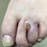
Nonhealing Eroded Plaque on an Interdigital Web Space of the Foot
A 53-year-old man with a history of numerous basal cell carcinomas and odontogenic keratocysts presented with a nonhealing erosion between the...